Application
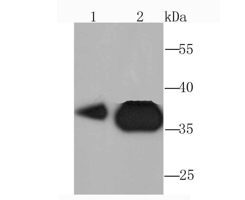
Fig1: Western blot analysis of Pyruvate Dehydrogenase E1 beta subunit on HepG2 (1) and Hela (2) cell lysate using anti-Pyruvate Dehydrogenase E1 beta subunit antibody at 1/1,000 dilution.Application
Fig2: Western blot analysis of Pyruvate Dehydrogenase E1 beta subunit on mouse kidney tissue (1) and PC-12 (2) cell lysate using anti-Pyruvate Dehydrogenase E1 beta subunit antibody at 1/1,000 dilution.Application
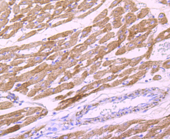
Fig3: Immunohistochemical analysis of paraffin-embedded rat heart tissue using anti-Pyruvate Dehydrogenase E1 beta subunit antibody. Counter stained with hematoxylin.Application
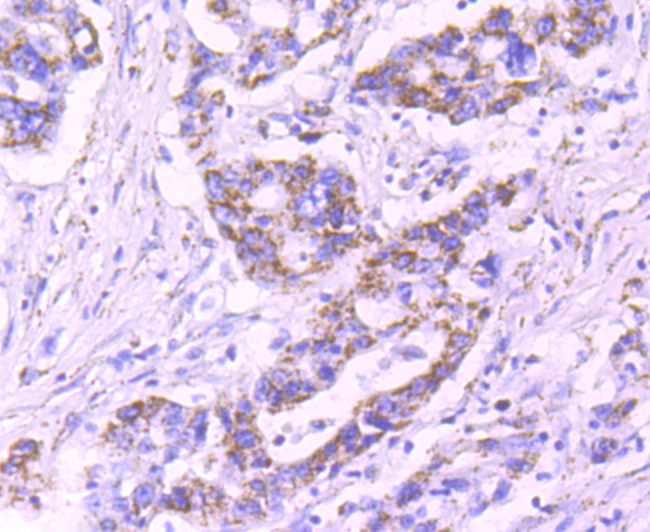
Fig4: Immunohistochemical analysis of paraffin-embedded human stomach cancer tissue using anti-Pyruvate Dehydrogenase E1 beta subunit antibody. Counter stained with hematoxylin.Application
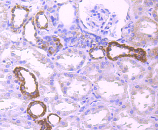
Fig5: Immunohistochemical analysis of paraffin-embedded human kidney tissue using anti-Pyruvate Dehydrogenase E1 beta subunit antibody. Counter stained with hematoxylin.Application
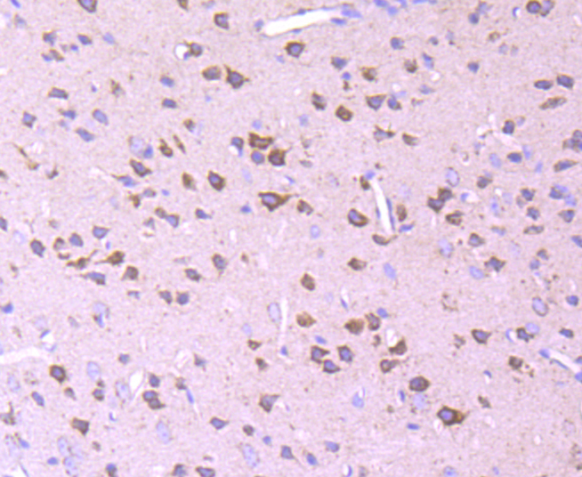
Fig6: Immunohistochemical analysis of paraffin-embedded mouse brain tissue using anti-Pyruvate Dehydrogenase E1 beta subunit antibody. Counter stained with hematoxylin.Application
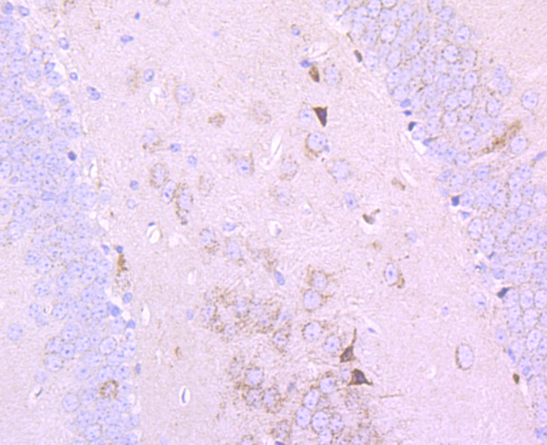
Fig7: Immunohistochemical analysis of paraffin-embedded mouse hippocampus tissue using anti-Pyruvate Dehydrogenase E1 beta subunit antibody. Counter stained with hematoxylin.Application
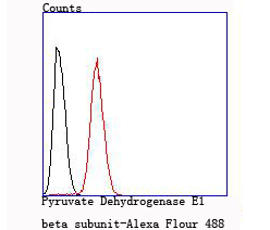
Fig8: Flow cytometric analysis of Hela cells with Pyruvate Dehydrogenase E1 beta subunit antibody at 1/100 dilution (red) compared with an unlabelled control (cells without incubation with primary antibody; black).| Product Name | Pyruvate Dehydrogenase E1 beta subunit |
|---|---|
| Antibody Type | Primary Antibodies |
| Antigen Alias | DKFZp564K0164 antibody mitochondrial antibody ODPB_HUMAN antibody pdhB antibody PDHBD antibody PDHE1 B antibody PDHE1-B antibody PHE1B antibody Pyruvate dehydrogenase (lipoamide) beta antibody Pyruvate dehydrogenase E1 beta polypeptide antibody Pyruvate dehydrogenase E1 component subunit beta antibody Pyruvate dehydrogenase E1 component subunit beta mitochondrial antibody |
| Product description | The pyruvate dehydrogenase complex catalyzes the overall conversion of pyruvate to acetyl-CoA and CO. It contains multiple copies of three enzymatic components: pyruvate dehydrogenase (E1), dihydrolipoamide acetyltransferase (E2) and lipoamide dehydrogenase (E3). Defects in PDHB are a cause of pyruvate dehydrogenase E1 component deficiency (PDHE1 deficiency). PDHE1 deficiency is the most common enzyme defect in patients with primary lactic acidosis. It is associated with variable clinical phenotypes ranging from neonatal death to prolonged survival complicated by developmental delay, seizures, ataxia, apnea, and in some cases to an X-linked form of Leigh syndrome (LS) (Leigh encephalomyelopathy). |
| Immunogen | Recombinant protein |
| Clonality | Monoclonal |
|---|---|
| Isotype | IgG |
| Host Species | Recombinant rabbit |
| Tested Applications | WBIHCIPFC |
| WB:1:500-1:2,000 IHC:1:50-1:200 FC:1:50-1:100 |
|
| Species Reactivity | HumanMouseRat |
| Concentration | 1mg/ml |
| Alternative Names | DKFZp564K0164 antibody mitochondrial antibody ODPB_HUMAN antibody pdhB antibody PDHBD antibody PDHE1 B antibody PDHE1-B antibody PHE1B antibody Pyruvate dehydrogenase (lipoamide) beta antibody Pyruvate dehydrogenase E1 beta polypeptide antibody Pyruvate dehydrogenase E1 component subunit beta antibody Pyruvate dehydrogenase E1 component subunit beta mitochondrial antibody |
|---|---|
| Cellular Localization | Mitochondrion matrix. |
Application
Fig1: Western blot analysis of Pyruvate Dehydrogenase E1 beta subunit on HepG2 (1) and Hela (2) cell lysate using anti-Pyruvate Dehydrogenase E1 beta subunit antibody at 1/1,000 dilution.
Application
Fig2: Western blot analysis of Pyruvate Dehydrogenase E1 beta subunit on mouse kidney tissue (1) and PC-12 (2) cell lysate using anti-Pyruvate Dehydrogenase E1 beta subunit antibody at 1/1,000 dilution.
Application
Fig3: Immunohistochemical analysis of paraffin-embedded rat heart tissue using anti-Pyruvate Dehydrogenase E1 beta subunit antibody. Counter stained with hematoxylin.
Application
Fig4: Immunohistochemical analysis of paraffin-embedded human stomach cancer tissue using anti-Pyruvate Dehydrogenase E1 beta subunit antibody. Counter stained with hematoxylin.
Application
Fig5: Immunohistochemical analysis of paraffin-embedded human kidney tissue using anti-Pyruvate Dehydrogenase E1 beta subunit antibody. Counter stained with hematoxylin.
Application
Fig6: Immunohistochemical analysis of paraffin-embedded mouse brain tissue using anti-Pyruvate Dehydrogenase E1 beta subunit antibody. Counter stained with hematoxylin.
Application
Fig7: Immunohistochemical analysis of paraffin-embedded mouse hippocampus tissue using anti-Pyruvate Dehydrogenase E1 beta subunit antibody. Counter stained with hematoxylin.
Application
Fig8: Flow cytometric analysis of Hela cells with Pyruvate Dehydrogenase E1 beta subunit antibody at 1/100 dilution (red) compared with an unlabelled control (cells without incubation with primary antibody; black).| Positive Control | HepG2, Hela, PC-12, mouse kidney, rat heart tissue, human kidney tissue, human stomach cancer tissue, mouse hippocampus tissue, mouse brain tissue. |
|---|---|
| Application Notes | WB:1:500-1:2,000 IHC:1:50-1:200 FC:1:50-1:100 |
| Form | Liquid |
|---|---|
| Storage Instructions | Store at +4℃ after thawing. Aliquot store at -20℃ or -80℃. Avoid repeated freeze / thaw cycles. |
| Storage Buffer | 1*TBS (pH7.4), 1%BSA, 40%Glycerol. Preservative: 0.05% Sodium Azide. |


