Application
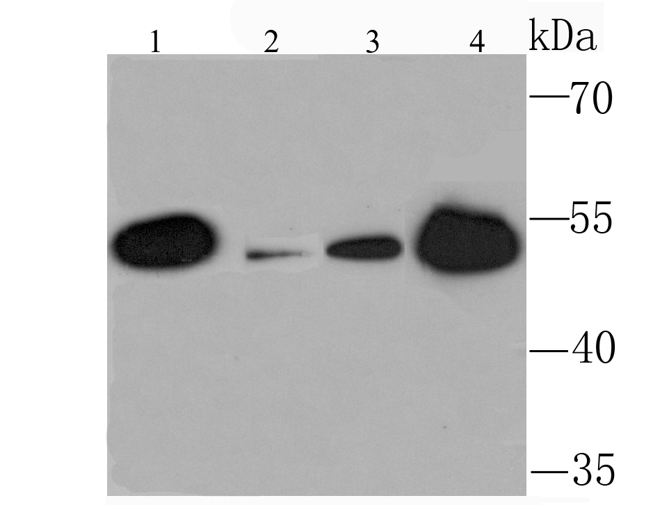
Fig1: Western blot analysis of KMT5A on different lysates using anti-KMT5A antibody at 1/1,000 dilution. Positive control: Lane 1: Mouse kidney Lane 2: Mouse liver Lane 3: 293T Lane 4: 293Application
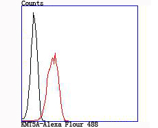
Fig2: Flow cytometric analysis of 293T cells with KMT5A antibody at 1/100 dilution (red) compared with an unlabelled control (cells without incubation with primary antibody; black).| Product Name | KMT5A/SETD8/Pr-SET7 |
|---|---|
| Antibody Type | Primary Antibodies |
| Antigen Alias | H4 K20 HMTase antibody H4 K20 specific histone methyltransferase antibody H4-K20-HMTase SETD8 antibody Histone H4 K20 methyltransferase antibody Histone lysine N methyltransferase H4 lysine 20 specific antibody Histone-lysine N-methyltransferase SETD8 antibody Lysine N-methyltransferase 5A antibody N-lysine methyltransferase SETD8 antibody Pr SET 7 antibody Pr SET7 antibody PR-Set7 antibody PR/SET domain containing protein 07 antibody PR/SET domain containing protein 8 antibody PR/SET domain-containing protein 07 antibody PR/SET07 antibody PrSET7 antibody SET 07 antibody SET 8 antibody SET domain containing (lysine methyltransferase) 8 antibody SET domain containing 8 antibody SET domain containing lysine methyltransferase 8 antibody SET domain containing protein 8 antibody SET domain-containing protein 8 antibody SET07 antibody SET8 antibody SETD 8 antibody setd8 antibody SETD8_HUMAN antibody |
| Product description | Protein-lysine N-methyltransferase that monomethylates both histones and non-histone proteins. All monomethylates 'Lys-20' of histone H4 (H4K20me1). H4K20me1 is enriched during mitosis and represents a specific tag for epigenetic transcriptional repression. Mainly functions in euchromatin regions , benef playing a central role in the silencing of euchromatic genes. Required for cell proliferation, probably by contribute to the maintenance of proper higher order structure of DNA during mitosis. Involved in chromosome condensation and proper cytokinesis. Nucleosomes are preferred as substrate compared to free histones. Mediates monomethylation of p53 / TP53 at 'Lys-382', leading to repress p53 / TP53-target genes. |
| Immunogen | Recombinant protein |
| Clonality | Monoclonal |
|---|---|
| Isotype | IgG |
| Host Species | Recombinant rabbit |
| Tested Applications | WBFC |
| WB:1:500-1:2,000 FC:1:50-1:100 |
|
| Species Reactivity | HumanMouse |
| Concentration | 1mg/ml |
| Alternative Names | H4 K20 HMTase antibody H4 K20 specific histone methyltransferase antibody H4-K20-HMTase SETD8 antibody Histone H4 K20 methyltransferase antibody Histone lysine N methyltransferase H4 lysine 20 specific antibody Histone-lysine N-methyltransferase SETD8 antibody Lysine N-methyltransferase 5A antibody N-lysine methyltransferase SETD8 antibody Pr SET 7 antibody Pr SET7 antibody PR-Set7 antibody PR/SET domain containing protein 07 antibody PR/SET domain containing protein 8 antibody PR/SET domain-containing protein 07 antibody PR/SET07 antibody PrSET7 antibody SET 07 antibody SET 8 antibody SET domain containing (lysine methyltransferase) 8 antibody SET domain containing 8 antibody SET domain containing lysine methyltransferase 8 antibody SET domain containing protein 8 antibody SET domain-containing protein 8 antibody SET07 antibody SET8 antibody SETD 8 antibody setd8 antibody SETD8_HUMAN antibody |
|---|---|
| Cellular Localization | Nucleus. |
Application
Fig1: Western blot analysis of KMT5A on different lysates using anti-KMT5A antibody at 1/1,000 dilution. Positive control: Lane 1: Mouse kidney Lane 2: Mouse liver Lane 3: 293T Lane 4: 293
Application
Fig2: Flow cytometric analysis of 293T cells with KMT5A antibody at 1/100 dilution (red) compared with an unlabelled control (cells without incubation with primary antibody; black).| Positive Control | Mouse kidney tissue, mouse liver tissue, 293T, 293. |
|---|---|
| Application Notes | WB:1:500-1:2,000 FC:1:50-1:100 |
| Form | Liquid |
|---|---|
| Storage Instructions | Store at +4℃ after thawing. Aliquot store at -20℃ or -80℃. Avoid repeated freeze / thaw cycles. |
| Storage Buffer | 1*TBS (pH7.4), 1%BSA, 40%Glycerol. Preservative: 0.05% Sodium Azide. |


