Application
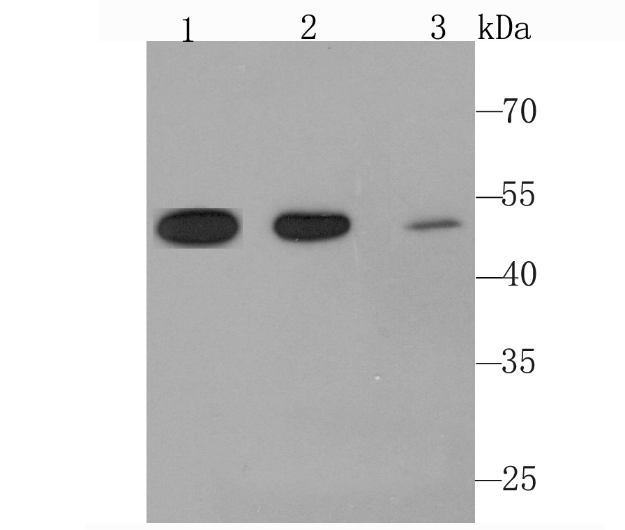
Fig1: Western blot analysis of Angiopoietin 2 on different cell lysate using anti-Angiopoietin 2 antibody at 1/1,000 dilution. Positive control: Lane 1: TF-1 Lane 2: Human liver tissue Lane 3: Human placenta tissueApplication
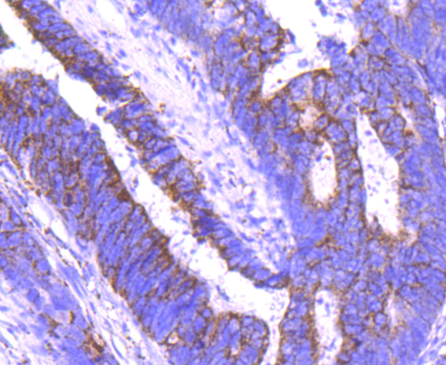
Fig2: Immunohistochemical analysis of paraffin-embedded human colon cancer tissue using anti-Angiopoietin 2 antibody. Counter stained with hematoxylin.Application
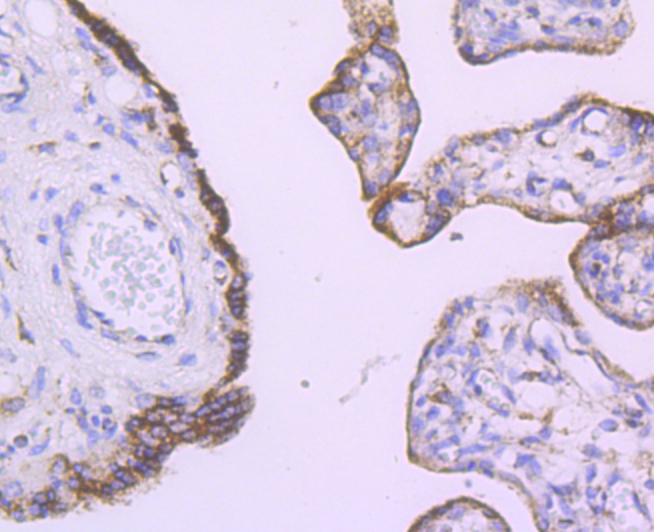
Fig3: Immunohistochemical analysis of paraffin-embedded human placenta tissue using anti-Angiopoietin 2 antibody. Counter stained with hematoxylin.Application
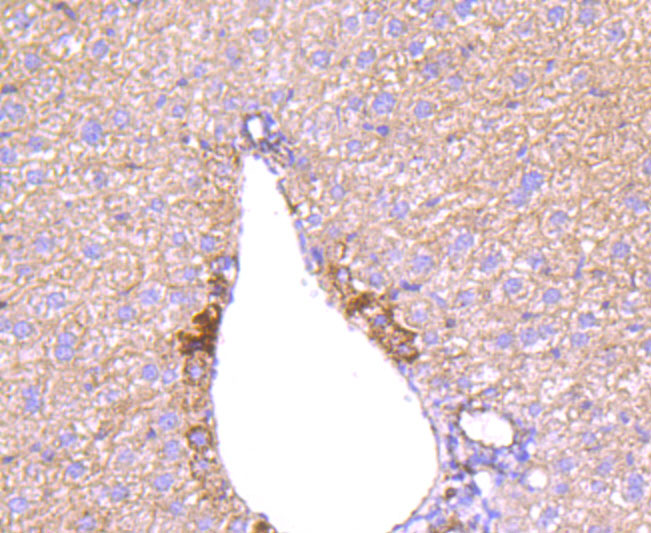
Fig4: Immunohistochemical analysis of paraffin-embedded mouse liver tissue using anti-Angiopoietin 2 antibody. Counter stained with hematoxylin.| Product Name | Angiopoietin 2 [JM71-34] |
|---|---|
| Antibody Type | Primary Antibodies |
| Antigen Alias | AGPT 2 antibody Agpt2 antibody ANG 2 antibody ANG-2 antibody ANG2 antibody Angiopoietin 2a antibody Angiopoietin 2B antibody Angiopoietin-2 antibody Angiopoietin2 antibody ANGP2_HUMAN antibody ANGPT 2 antibody Angpt2 antibody Tie2 ligand antibody |
| Product description | Tie-1 and Tie-2 (also designated Tek) are novel cell surface receptor tyrosine kinases. The extracellular domain of Tie-1 has an unusual multidomain structure consisting of a cluster of three epidermal growth factor homology motifs localized between two immunoglobulin-like loops, which are followed by three fibronectin type III repeats next to the transmembrane region. Angiopoietin-1 (Ang-1) is a secreted ligand for Tie-2. Preliminary biochemical analyses of Ang-1 reveal a potential fibrinogen-like domain at the carboxy terminus and coiled-coil regions in the amino terminus. Ang-1 is an angiogenic factor that is thought to be involved in endothelial development. A related protein, angiopoietin-2 (Ang-2), has been identified as a naturally occurring antagonist of Ang-1 activation of Tie-2. In adult tissue, Ang-2 expression seems to be restricted to sites of vascular remodeling. |
| Immunogen | recombinant protein |
| Clonality | Monoclonal |
|---|---|
| Isotype | IgG |
| Host Species | Recombinant rabbit |
| Tested Applications | WBIHC |
| WB:1:500-1:2,000 IHC:1:50-1:200 |
|
| Species Reactivity | HumanMouseRat |
| Concentration | 1mg/ml |
| Alternative Names | AGPT 2 antibody Agpt2 antibody ANG 2 antibody ANG-2 antibody ANG2 antibody Angiopoietin 2a antibody Angiopoietin 2B antibody Angiopoietin-2 antibody Angiopoietin2 antibody ANGP2_HUMAN antibody ANGPT 2 antibody Angpt2 antibody Tie2 ligand antibody |
|---|---|
| Molecular Weight(MW) | 50 kDa |
| Cellular Localization | Secreted. |
Application
Fig1: Western blot analysis of Angiopoietin 2 on different cell lysate using anti-Angiopoietin 2 antibody at 1/1,000 dilution. Positive control: Lane 1: TF-1 Lane 2: Human liver tissue Lane 3: Human placenta tissue
Application
Fig2: Immunohistochemical analysis of paraffin-embedded human colon cancer tissue using anti-Angiopoietin 2 antibody. Counter stained with hematoxylin.
Application
Fig3: Immunohistochemical analysis of paraffin-embedded human placenta tissue using anti-Angiopoietin 2 antibody. Counter stained with hematoxylin.
Application
Fig4: Immunohistochemical analysis of paraffin-embedded mouse liver tissue using anti-Angiopoietin 2 antibody. Counter stained with hematoxylin.| Positive Control | TF-1 cell lysate, human liver tissue lysate, human placenta tissue lysate, human colon tissue, human placenta tissue. |
|---|---|
| Application Notes | WB:1:500-1:2,000 IHC:1:50-1:200 |
| Form | Liquid |
|---|---|
| Storage Instructions | Store at +4℃ after thawing. Aliquot store at -20℃ or -80℃. Avoid repeated freeze / thaw cycles. |
| Storage Buffer | 1*TBS (pH7.4), 1%BSA, 40%Glycerol. Preservative: 0.05% Sodium Azide. |


