Application
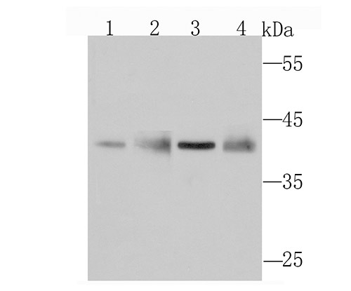
Fig1: Western blot analysis of WDR5 on different cell lysates using anti-WDR5 antibody at 1/1,000 dilution. Positive control:Lane 1: HelaLane 2: A549Lane 3: NIH-3T3Lane 4: PC-12Application
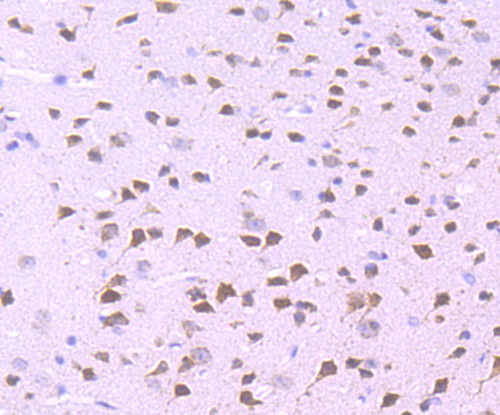
Fig2: Immunohistochemical analysis of paraffin-embedded mouse brain tissue using anti-WDR5 antibody. Counter stained with hematoxylin.Application
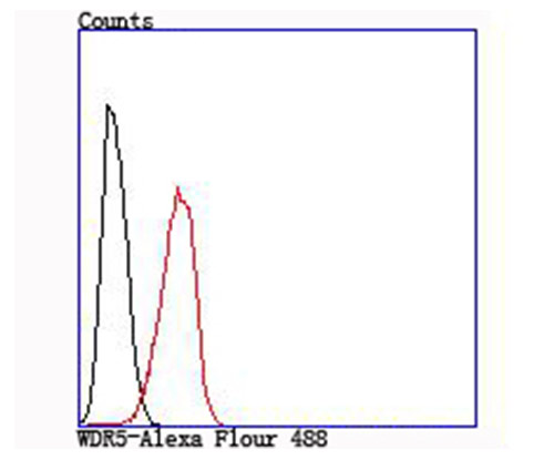
Fig3: Flow cytometric analysis of Hela cells with WDR5 antibody at 1/100 dilution (red) compared with an unlabelled control (cells without incubation with primary antibody; black).| Product Name | WDR5 [JM73-53] |
|---|---|
| Antibody Type | Primary Antibodies |
| Antigen Alias | 2410008O07Rik antibody AA408785 antibody AA960360 antibody BIG 3 antibody Big antibody BIG3 antibody BMP2 induced 3 kb gene protein antibody BMP2 induced gene, 3-KB antibody BMP2-induced 3-kb gene protein antibody MGC114572 antibody OTTHUMP00000162494 antibody SWD 3 antibody SWD3 antibody SWD3 Set1c WD40 repeat protein homolog antibody WD repeat containing protein 5 antibody WD repeat domain 5 antibody WD repeat protein 5 antibody WD repeat-containing protein 5 antibody WD repeat-containing protein BIG-3 antibody WDR 5 antibody Wdr5 antibody WDR5_HUMAN antibody |
| Product description | Contributes to histone modification. May position the N-terminus of histone H3 for efficient trimethylation at 'Lys-4'. As part of the MLL1/MLL complex it is involved in methylation and dimethylation at 'Lys-4' of histone H3. H3 'Lys-4' methylation represents a specific tag for epigenetic transcriptional activation. As part of the NSL complex it may be involved in acetylation of nucleosomal histone H4 on several lysine residues. May regulate osteoblasts differentiation. |
| Immunogen | Recombinant protein |
| Clonality | Monoclonal |
|---|---|
| Isotype | IgG |
| Host Species | Recombinant rabbit |
| Tested Applications | WBIHCFC |
| WB:1:500~1:2000 IHC:1:50~1:200 FC:1:200~1:500 Notes:Optimal dilutions/concentrations should be determined by the researcher. |
|
| Species Reactivity | HumanMouseRat |
| Concentration | 1mg/ml |
| Alternative Names | 2410008O07Rik antibody
AA408785 antibody
AA960360 antibody
BIG 3 antibody
Big antibody
BIG3 antibody
BMP2 induced 3 kb gene protein antibody
BMP2 induced gene 3-KB antibody BMP2-induced 3-kb gene protein antibody MGC114572 antibody OTTHUMP00000162494 antibody SWD 3 antibody SWD3 antibody SWD3 Set1c WD40 repeat protein homolog antibody WD repeat containing protein 5 antibody WD repeat domain 5 antibody WD repeat protein 5 antibody WD repeat-containing protein 5 antibody WD repeat-containing protein BIG-3 antibody WDR 5 antibody Wdr5 antibody WDR5_HUMAN antibody |
|---|---|
| Molecular Weight(MW) | 36 kDa |
| Cellular Localization | Nucleus. |
Application
Fig1: Western blot analysis of WDR5 on different cell lysates using anti-WDR5 antibody at 1/1,000 dilution. Positive control:Lane 1: HelaLane 2: A549Lane 3: NIH-3T3Lane 4: PC-12
Application
Fig2: Immunohistochemical analysis of paraffin-embedded mouse brain tissue using anti-WDR5 antibody. Counter stained with hematoxylin.
Application
Fig3: Flow cytometric analysis of Hela cells with WDR5 antibody at 1/100 dilution (red) compared with an unlabelled control (cells without incubation with primary antibody; black).| Positive Control | Hela, A549, NIH-3T3, PC-12, mouse brain tissue. |
|---|---|
| Application Notes | WB:1:500~1:2000 IHC:1:50~1:200 FC:1:200~1:500 Notes:Optimal dilutions/concentrations should be determined by the researcher. |
| Form | Liquid |
|---|---|
| Storage Instructions | Store at +4℃ after thawing. Aliquot store at -20℃ or -80℃. Avoid repeated freeze / thaw cycles. |
| Storage Buffer | 1*TBS (pH7.4), 1%BSA, 40%Glycerol. Preservative: 0.05% Sodium Azide. |


