WB
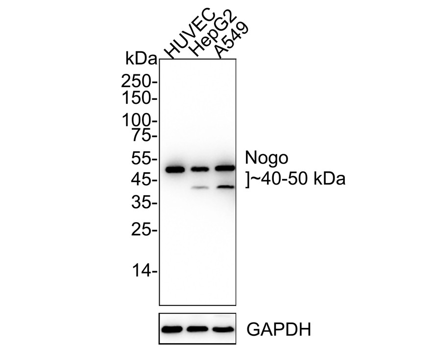
Western blot analysis of Nogo on different lysates with Rabbit anti-Nogo antibody at 1/1,000 dilution. Lane 1: HUVEC cell lysate, Lane 2: HepG2 cell lysate, Lane 3: A549 cell lysate, Lysates/proteins at 10 µg/Lane. Exposure time: 6 seconds; 4-20% SDS-PAGE gel. Proteins were transferred to a PVDF membrane and blocked with 5% NFDM/TBST for 1 hour at room temperature. The primary antibody at 1/1,000 dilution was used in 5% NFDM/TBST at 4℃ overnight. Goat Anti-Rabbit IgG - HRP Secondary Antibody at 1/50,000 dilution was used for 1 hour at room temperature.IHC
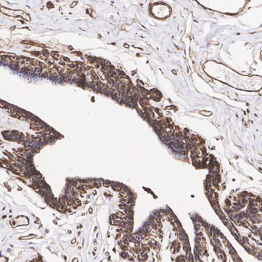
Immunohistochemical analysis of paraffin-embedded human breast tissue with Rabbit anti-Nogo antibody at 1/1,000 dilution. The section was pre-treated using heat mediated antigen retrieval with Tris-EDTA buffer (pH 9.0) for 20 minutes. The tissues were blocked in 1% BSA for 20 minutes at room temperature, washed with ddH2O and PBS, and then probed with the primary antibody at 1/1,000 dilution for 1 hour at room temperature. The detection was performed using an HRP conjugated compact polymer system. DAB was used as the chromogen. Tissues were counterstained with hematoxylin and mounted with DPX.IHC
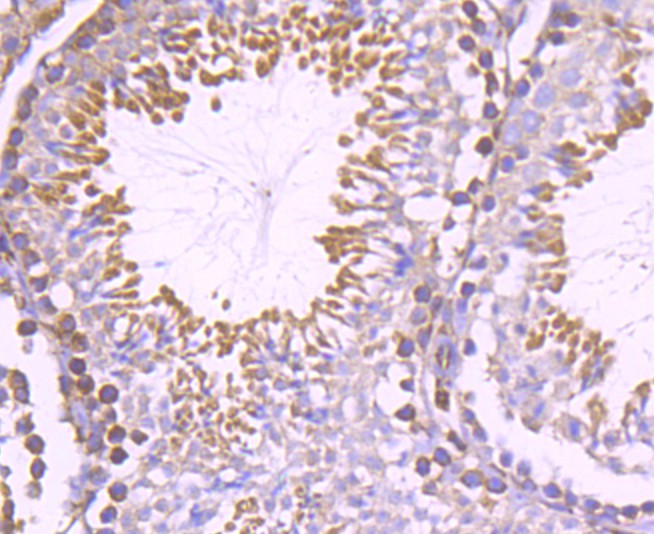
Immunohistochemical analysis of paraffin-embedded mouse testis tissue using anti-Nogo antibody. The section was pre-treated using heat mediated antigen retrieval with Tris-EDTA buffer (pH 9.0) for 20 minutes.The tissues were blocked in 1% BSA for 30 minutes at room temperature, washed with ddH2O and PBS, and then probed with the primary antibody (1/500) for 30 minutes at room temperature. The detection was performed using an HRP conjugated compact polymer system. DAB was used as the chromogen. Tissues were counterstained with hematoxylin and mounted with DPX.ICC/IF
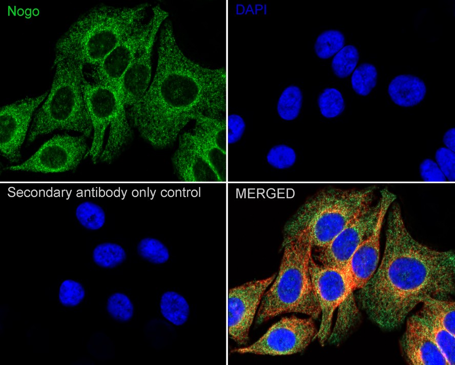
Immunocytochemistry analysis of A549 cells labeling Nogo with Rabbit anti-Nogo antibody at 1/250 dilution. Cells were fixed in 4% paraformaldehyde for 20 minutes at room temperature, permeabilized with 0.1% Triton X-100 in PBS for 5 minutes at room temperature, then blocked with 1% BSA in 10% negative goat serum for 1 hour at room temperature. Cells were then incubated with Rabbit anti-Nogo antibody at 1/250 dilution in 1% BSA in PBST overnight at 4 ℃. Goat Anti-Rabbit IgG H&L (488) was used as the secondary antibody at 1/1,000 dilution. PBS instead of the primary antibody was used as the secondary antibody only control. Nuclear DNA was labelled in blue with DAPI. Beta tubulin (red) was stained at 1/100 dilution overnight at +4℃. Goat Anti-Mouse IgG H&L (594) was used as the secondary antibody at 1/1,000 dilution.ICC/IF
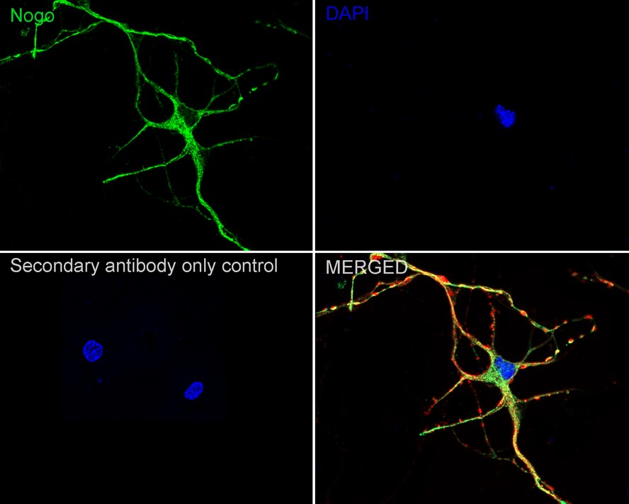
Immunocytochemistry analysis of mouse neuron cells labeling Nogo with Rabbit anti-Nogo antibody at 1/250 dilution. Cells were fixed in 4% paraformaldehyde for 20 minutes at room temperature, permeabilized with 0.1% Triton X-100 in PBS for 5 minutes at room temperature, then blocked with 1% BSA in 10% negative goat serum for 1 hour at room temperature. Cells were then incubated with Rabbit anti-Nogo antibody at 1/250 dilution in 1% BSA in PBST overnight at 4 ℃. Goat Anti-Rabbit IgG H&L (488) was used as the secondary antibody at 1/1,000 dilution. PBS instead of the primary antibody was used as the secondary antibody only control. Nuclear DNA was labelled in blue with DAPI. Beta tubulin (red) was stained at 1/100 dilution overnight at +4℃. Goat Anti-Mouse IgG H&L (594) was used as the secondary antibody at 1/1,000 dilution.FC
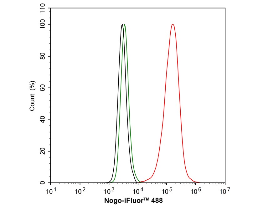
Flow cytometric analysis of A549 cells labeling Nogo. Cells were fixed and permeabilized. Then stained with the primary antibody (1μg/mL) (red) compared with Rabbit IgG Isotype Control (green). After incubation of the primary antibody at +4℃ for an hour, the cells were stained with a 488 conjugate-Goat anti-Rabbit IgG Secondary antibody at 1/1,000 dilution for 30 minutes at +4℃. Unlabelled sample was used as a control (cells without incubation with primary antibody; black).| Product Name | Nogo Recombinant Rabbit Monoclonal Antibody |
|---|---|
| Antibody Type | Primary Antibodies |
| Product description | Developmental neurite growth regulatory factor with a role as a negative regulator of axon-axon adhesion and growth, and as a facilitator of neurite branching. Regulates neurite fasciculation, branching and extension in the developing nervous system. Involved in down-regulation of growth, stabilization of wiring and restriction of plasticity in the adult CNS. Regulates the radial migration of cortical neurons via an RTN4R-LINGO1 containing receptor complex. Isoform 2 reduces the anti-apoptotic activity of Bcl-xl and Bcl-2. This is likely consecutive to their change in subcellular location, from the mitochondria to the endoplasmic reticulum, after binding and sequestration. Isoform 2 and isoform 3 inhibit BACE1 activity and amyloid precursor protein processing. |
| Immunogen | Synthetic peptide within Human Nogo aa 111-160 / 1192. |
| Clonality | Monoclonal |
|---|---|
| Isotype | IgG |
| Host Species | Recombinant rabbit |
| Tested Applications | FCICC/IFIHCWB |
| WB:1:1:000-1:2000 IHC1:200-1:1000ICC/IF:1:50-1:200 FC:1:1000 |
|
| Species Reactivity | HumanMouse |
| Concentration | 1mg/ml |
| Purification | Protein A |
| Alternative Names | 1110020G17Rik antibody
AA407876 antibody
AA409940 antibody
AA960376 antibody
ASY antibody
C130026I10Rik antibody
Foocen antibody
Glut4 vesicle 20 kDa protein antibody
Human NogoA antibody
Kiaa0886 antibody
KIAA4153 antibody
MGC116054 antibody
MGC139261 antibody
mKIAA0886 antibody
mKIAA4153 antibody
My043 protein antibody
Nbla00271 antibody
Nbla10545 antibody
Neurite growth inhibitor 220 antibody
Neurite Growth Inhibitor 220 included antibody Neurite outgrowth inhibitor antibody Neuroendocrine-specific protein antibody Neuroendocrine-specific protein C homolog antibody NI-250 antibody NI220/250 antibody Nogo A antibody NOGO antibody Nogo B antibody Nogo C antibody Nogo protein antibody NOGOC antibody NSP antibody NSP-CL antibody rat N antibody Reticulon 4 antibody Reticulon 5 antibody Reticulon-4 antibody Reticulon-5 antibody RTN X antibody RTN-x antibody Rtn4 antibody Rtn4 reticulon 4 antibody RTN4-A antibody RTN4-B1 antibody RTN4-B2 antibody RTN4-C antibody RTN4_HUMAN antibody Vp20 antibody |
|---|---|
| Molecular Weight(MW) | 130 kDa(Observed band size: 40-50 kDa) |
| Cellular Localization | Endoplasmic reticulum membrane. |
WB
Western blot analysis of Nogo on different lysates with Rabbit anti-Nogo antibody at 1/1,000 dilution. Lane 1: HUVEC cell lysate, Lane 2: HepG2 cell lysate, Lane 3: A549 cell lysate, Lysates/proteins at 10 µg/Lane. Exposure time: 6 seconds; 4-20% SDS-PAGE gel. Proteins were transferred to a PVDF membrane and blocked with 5% NFDM/TBST for 1 hour at room temperature. The primary antibody at 1/1,000 dilution was used in 5% NFDM/TBST at 4℃ overnight. Goat Anti-Rabbit IgG - HRP Secondary Antibody at 1/50,000 dilution was used for 1 hour at room temperature.
IHC
Immunohistochemical analysis of paraffin-embedded human breast tissue with Rabbit anti-Nogo antibody at 1/1,000 dilution. The section was pre-treated using heat mediated antigen retrieval with Tris-EDTA buffer (pH 9.0) for 20 minutes. The tissues were blocked in 1% BSA for 20 minutes at room temperature, washed with ddH2O and PBS, and then probed with the primary antibody at 1/1,000 dilution for 1 hour at room temperature. The detection was performed using an HRP conjugated compact polymer system. DAB was used as the chromogen. Tissues were counterstained with hematoxylin and mounted with DPX.
IHC
Immunohistochemical analysis of paraffin-embedded mouse testis tissue using anti-Nogo antibody. The section was pre-treated using heat mediated antigen retrieval with Tris-EDTA buffer (pH 9.0) for 20 minutes.The tissues were blocked in 1% BSA for 30 minutes at room temperature, washed with ddH2O and PBS, and then probed with the primary antibody (1/500) for 30 minutes at room temperature. The detection was performed using an HRP conjugated compact polymer system. DAB was used as the chromogen. Tissues were counterstained with hematoxylin and mounted with DPX.
ICC/IF
Immunocytochemistry analysis of A549 cells labeling Nogo with Rabbit anti-Nogo antibody at 1/250 dilution. Cells were fixed in 4% paraformaldehyde for 20 minutes at room temperature, permeabilized with 0.1% Triton X-100 in PBS for 5 minutes at room temperature, then blocked with 1% BSA in 10% negative goat serum for 1 hour at room temperature. Cells were then incubated with Rabbit anti-Nogo antibody at 1/250 dilution in 1% BSA in PBST overnight at 4 ℃. Goat Anti-Rabbit IgG H&L (488) was used as the secondary antibody at 1/1,000 dilution. PBS instead of the primary antibody was used as the secondary antibody only control. Nuclear DNA was labelled in blue with DAPI. Beta tubulin (red) was stained at 1/100 dilution overnight at +4℃. Goat Anti-Mouse IgG H&L (594) was used as the secondary antibody at 1/1,000 dilution.
ICC/IF
Immunocytochemistry analysis of mouse neuron cells labeling Nogo with Rabbit anti-Nogo antibody at 1/250 dilution. Cells were fixed in 4% paraformaldehyde for 20 minutes at room temperature, permeabilized with 0.1% Triton X-100 in PBS for 5 minutes at room temperature, then blocked with 1% BSA in 10% negative goat serum for 1 hour at room temperature. Cells were then incubated with Rabbit anti-Nogo antibody at 1/250 dilution in 1% BSA in PBST overnight at 4 ℃. Goat Anti-Rabbit IgG H&L (488) was used as the secondary antibody at 1/1,000 dilution. PBS instead of the primary antibody was used as the secondary antibody only control. Nuclear DNA was labelled in blue with DAPI. Beta tubulin (red) was stained at 1/100 dilution overnight at +4℃. Goat Anti-Mouse IgG H&L (594) was used as the secondary antibody at 1/1,000 dilution.
FC
Flow cytometric analysis of A549 cells labeling Nogo. Cells were fixed and permeabilized. Then stained with the primary antibody (1μg/mL) (red) compared with Rabbit IgG Isotype Control (green). After incubation of the primary antibody at +4℃ for an hour, the cells were stained with a 488 conjugate-Goat anti-Rabbit IgG Secondary antibody at 1/1,000 dilution for 30 minutes at +4℃. Unlabelled sample was used as a control (cells without incubation with primary antibody; black).| Application Notes | WB:1:1:000-1:2000 IHC1:200-1:1000ICC/IF:1:50-1:200 FC:1:1000 |
|---|
| Form | Liquid |
|---|---|
| Storage Instructions | Store at +4℃ after thawing. Aliquot store at -20℃ or -80℃. Avoid repeated freeze / thaw cycles. |
| Storage Buffer | 1*TBS (pH7.4), 1%BSA, 40%Glycerol. Preservative: 0.05% Sodium Azide. |


