Application
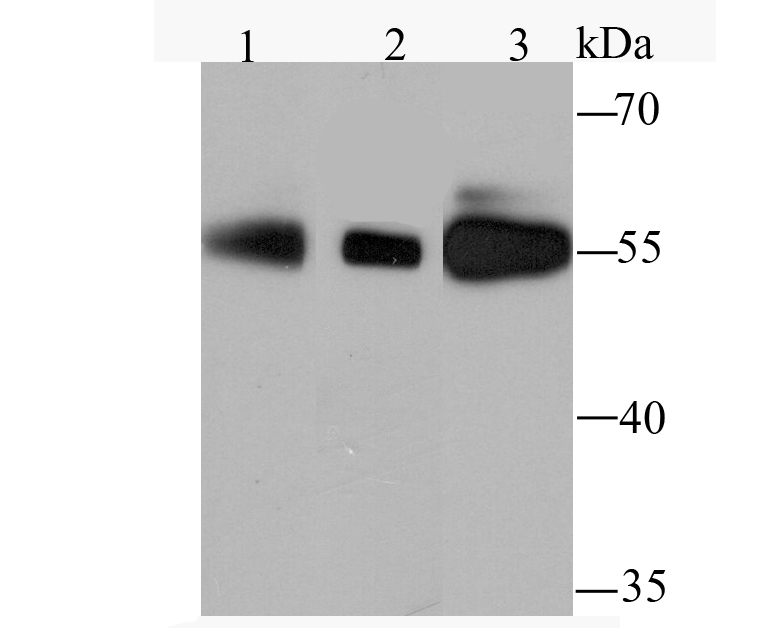
Fig1: Western blot analysis of PFKFB3 on different cell lysates using anti-PFKFB3 antibody at 1/1,000 dilution. Positive control: Lane 1: Hela Lane 2: PC-12 Lane 3: A431Application
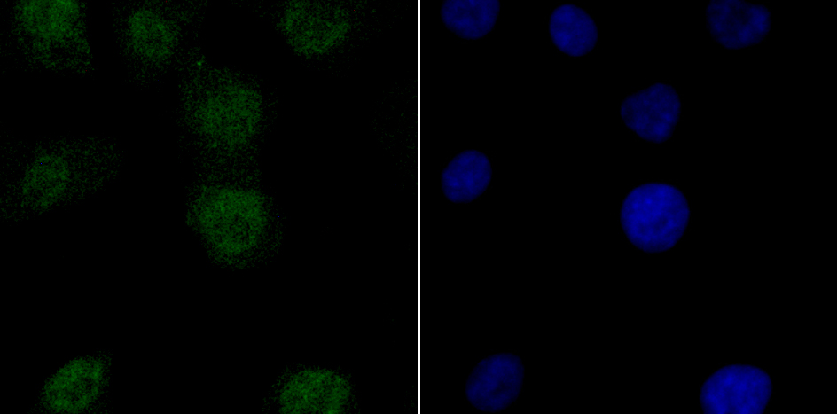
Fig2: ICC staining PFKFB3 in A549 cells (green). The nuclear counter stain is DAPI (blue). Cells were fixed in paraformaldehyde, permeabilised with 0.25% Triton X100/PBS.Application
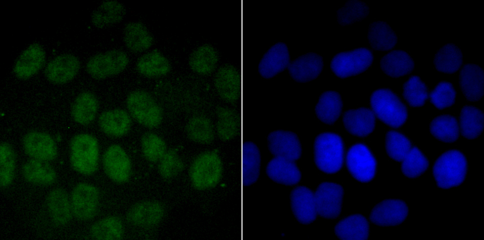
Fig3: ICC staining PFKFB3 in Hela cells (green). The nuclear counter stain is DAPI (blue). Cells were fixed in paraformaldehyde, permeabilised with 0.25% Triton X100/PBS.Application
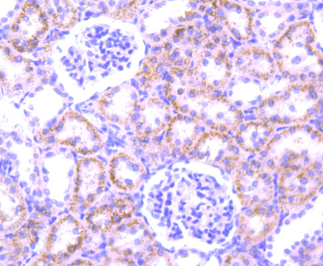
Fig4: Immunohistochemical analysis of paraffin-embedded rat kidney tissue using anti-PFKFB3 antibody. Counter stained with hematoxylin.Application
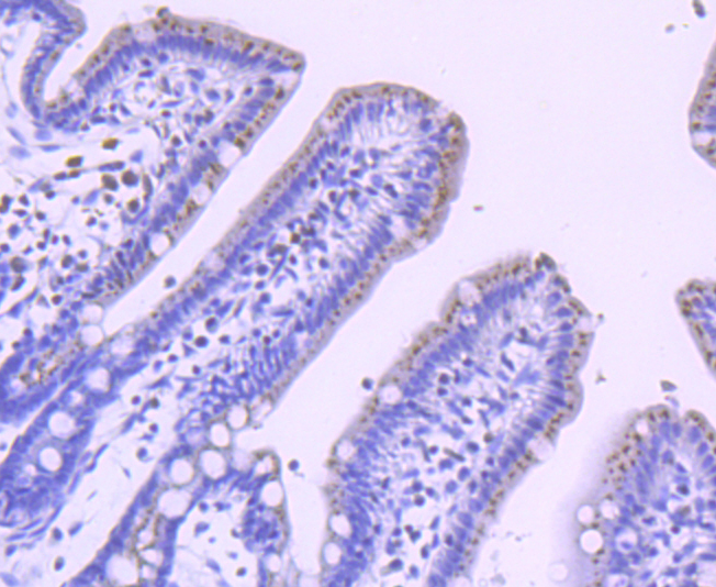
Fig5: Immunohistochemical analysis of paraffin-embedded mouse colon tissue using anti-PFKFB3 antibody. Counter stained with hematoxylin.Application
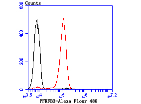
Fig6: Flow cytometric analysis of A431 cells with PFKFB3 antibody at 1/100 dilution (red) compared with an unlabelled control (cells without incubation with primary antibody; black).| Product Name | PFKFB3 |
|---|---|
| Antibody Type | Primary Antibodies |
| Antigen Alias | 6 phosphofructo 2 kinase/ fructose 2,6 bisphosphatase antibody 6 phosphofructo 2 kinase/fructose 2,6 biphosphatase 3 antibody 6-bisphosphatase antibody 6-P2ase 3 antibody 6-P2ASE brain/placenta-type isozyme antibody 6PF 2 K/Fru 2,6 P2ASE brain/placenta type isozyme antibody 6PF 2-K/Fru 2,6 P2ase 3 antibody 6PF-2-K/Fru-2 antibody F263_HUMAN antibody fructose 6 phosphate,2 kinase/fructose 2, 6 bisphosphatase antibody Fructose-2 antibody Inducible 6 phosphofructo 2 kinase/fructose 2,6 bisphosphatase antibody iPFK 2 antibody iPFK-2 antibody IPFK2 antibody PFK/FBPase 3 antibody PFK2 antibody PFKFB3 antibody Renal carcinoma antigen NY REN 56 antibody Renal carcinoma antigen NY-REN-56 antibody uPFK 2 antibody |
| Product description | Among the enzymes playing role in glycolysis,four allosteric PFKFB enzymes 1–4 expressed by four independent PFKFB genes, catalyze the rate-limiting phosphorylation of fructose-6-phosphate to fructose-1, 6-bisphosphate, using ATP as the energy source in the glycolysis pathway. Among these four allosteric enzymes, PFKFB3 enzyme retains the highest Kinase/Biphosphatase activity ratio and is expressed by PFKFB3 gene which has been demonstrated to be highly expressed in leukemic cells and in solid tumors. Moreover, mitogenic, hypoxic and inflammatory conditions have an inductive effect on the expression of PFKFB3. Hence upregulation of PFKFB genes specific to cancer cells compared to their normal counterparts (from the same patients) with more robust over-expression in breast and lung cancer make it a more appropriate target. |
| Immunogen | Recombinant protein |
| Clonality | Monoclonal |
|---|---|
| Isotype | IgG |
| Host Species | Recombinant rabbit |
| Tested Applications | WBICCIHCFCIP |
| WB:1:500-1:1,000 ICC:1:50-1:200 IHC:1:50-1:200 FC:1:50-1:100 |
|
| Species Reactivity | HumanMouseRat |
| Concentration | 1mg/ml |
| Alternative Names | 6 phosphofructo 2 kinase/ fructose 2 6 bisphosphatase antibody 6 phosphofructo 2 kinase/fructose 2 6 biphosphatase 3 antibody 6-bisphosphatase antibody 6-P2ase 3 antibody 6-P2ASE brain/placenta-type isozyme antibody 6PF 2 K/Fru 2 6 P2ASE brain/placenta type isozyme antibody 6PF 2-K/Fru 2 6 P2ase 3 antibody 6PF-2-K/Fru-2 antibody F263_HUMAN antibody fructose 6 phosphate 2 kinase/fructose 2 6 bisphosphatase antibody Fructose-2 antibody Inducible 6 phosphofructo 2 kinase/fructose 2 6 bisphosphatase antibody iPFK 2 antibody iPFK-2 antibody IPFK2 antibody PFK/FBPase 3 antibody PFK2 antibody PFKFB3 antibody Renal carcinoma antigen NY REN 56 antibody Renal carcinoma antigen NY-REN-56 antibody uPFK 2 antibody |
|---|---|
| Cellular Localization | Cytosol. Nucleoplasm. |
Application
Fig1: Western blot analysis of PFKFB3 on different cell lysates using anti-PFKFB3 antibody at 1/1,000 dilution. Positive control: Lane 1: Hela Lane 2: PC-12 Lane 3: A431
Application
Fig2: ICC staining PFKFB3 in A549 cells (green). The nuclear counter stain is DAPI (blue). Cells were fixed in paraformaldehyde, permeabilised with 0.25% Triton X100/PBS.
Application
Fig3: ICC staining PFKFB3 in Hela cells (green). The nuclear counter stain is DAPI (blue). Cells were fixed in paraformaldehyde, permeabilised with 0.25% Triton X100/PBS.
Application
Fig4: Immunohistochemical analysis of paraffin-embedded rat kidney tissue using anti-PFKFB3 antibody. Counter stained with hematoxylin.
Application
Fig5: Immunohistochemical analysis of paraffin-embedded mouse colon tissue using anti-PFKFB3 antibody. Counter stained with hematoxylin.
Application
Fig6: Flow cytometric analysis of A431 cells with PFKFB3 antibody at 1/100 dilution (red) compared with an unlabelled control (cells without incubation with primary antibody; black).| Positive Control | A431, PC-12, Hela, A549, rat kidney tissue, mouse colon tissue. |
|---|---|
| Application Notes | WB:1:500-1:1,000 ICC:1:50-1:200 IHC:1:50-1:200 FC:1:50-1:100 |
| Form | Liquid |
|---|---|
| Storage Instructions | Store at +4℃ after thawing. Aliquot store at -20℃ or -80℃. Avoid repeated freeze / thaw cycles. |
| Storage Buffer | 1*TBS (pH7.4), 1%BSA, 40%Glycerol. Preservative: 0.05% Sodium Azide. |


