Application
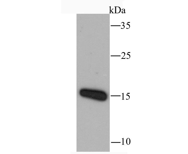
Fig1: Western blot analysis of RANTES on mouse thymus tissue lysate using anti-RANTES antibody at 1/1,000 dilution.Application
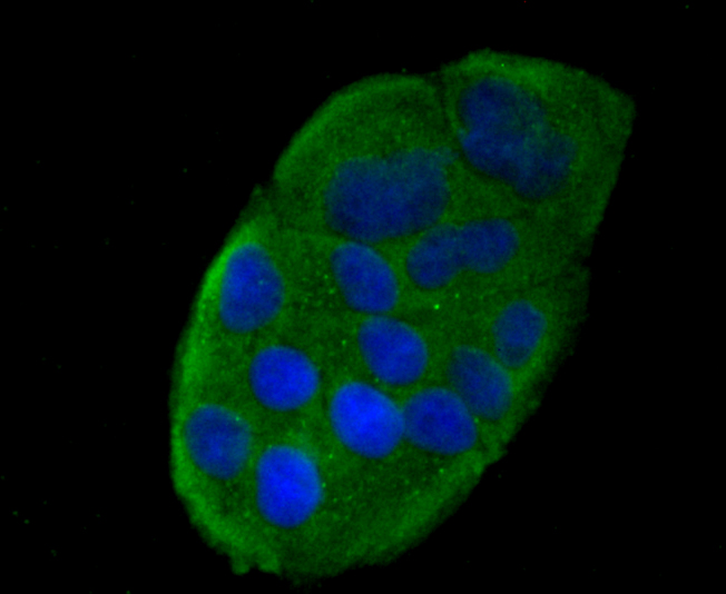
Fig2: ICC staining RANTES in Hela cells (green). The nuclear counter stain is DAPI (blue). Cells were fixed in paraformaldehyde, permeabilised with 0.25% Triton X100/PBS.Application
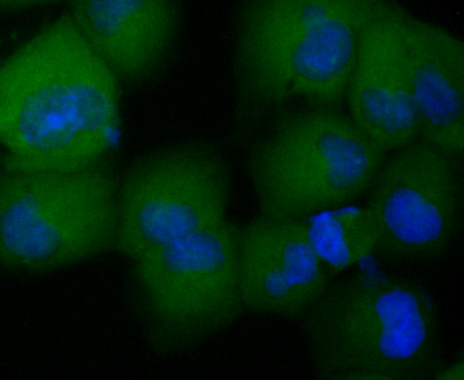
Fig3: ICC staining RANTES in HUVEC cells (green). The nuclear counter stain is DAPI (blue). Cells were fixed in paraformaldehyde, permeabilised with 0.25% Triton X100/PBS.Application
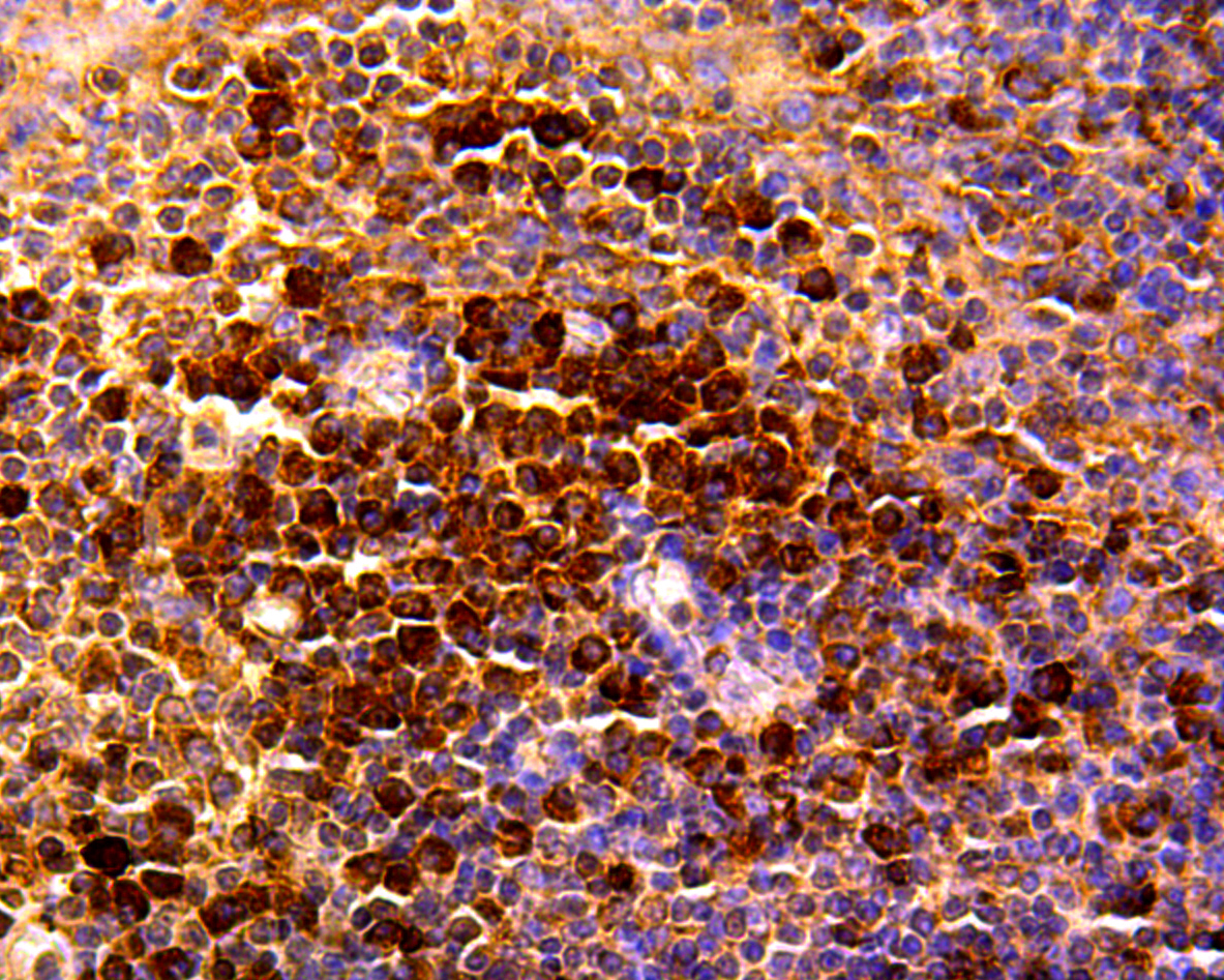
Fig4: Immunohistochemical analysis of paraffin-embedded human tonsil tissue using anti-RANTES antibody. Counter stained with hematoxylin.Application
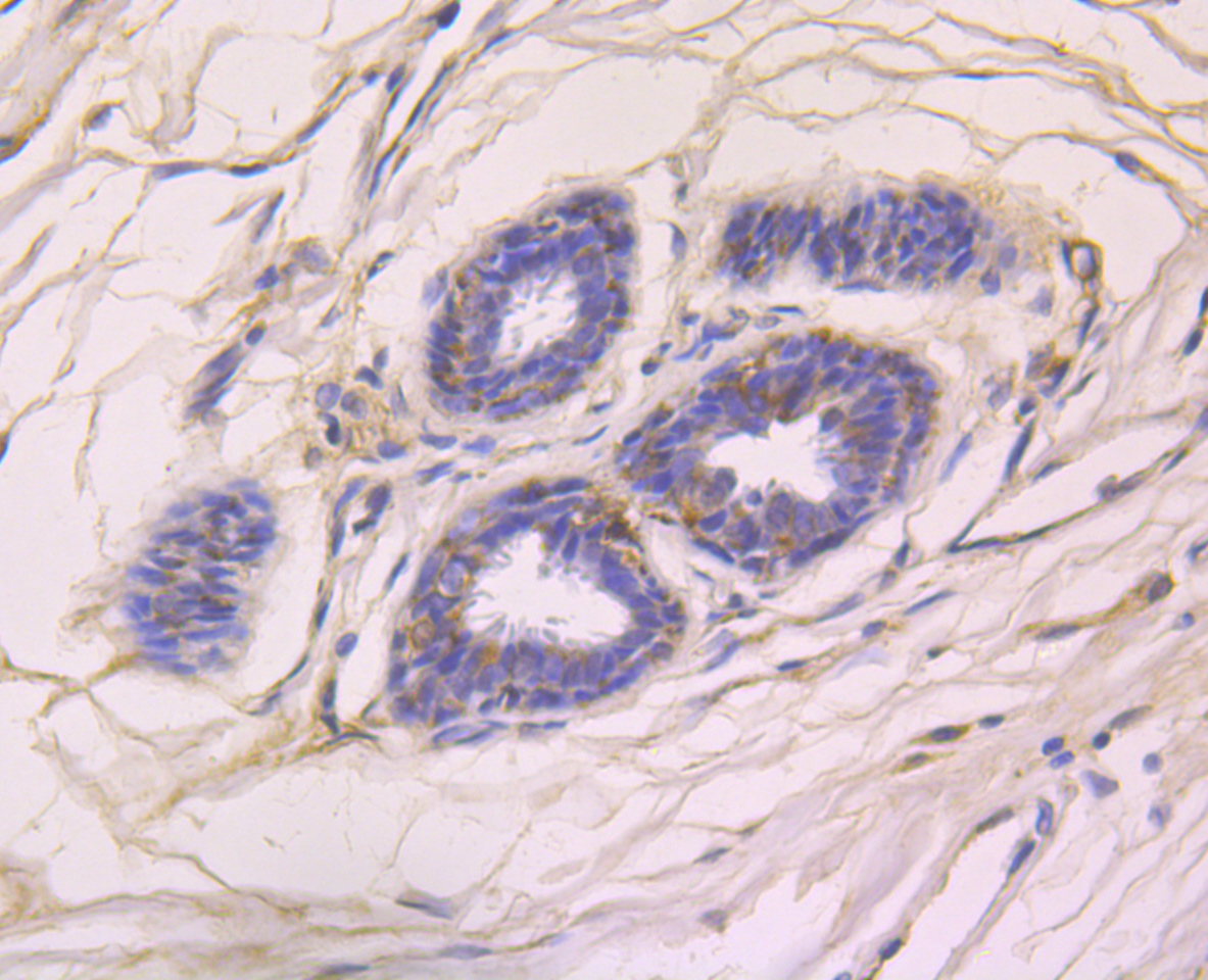
Fig5: Immunohistochemical analysis of paraffin-embedded human breast cancer tissue using anti-RANTES antibody. Counter stained with hematoxylin.Application
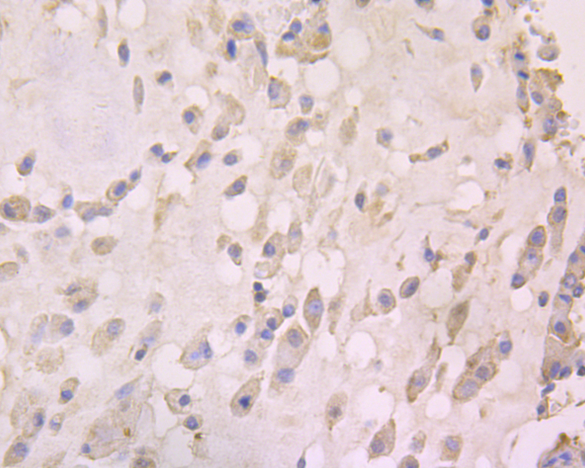
Fig6: Immunohistochemical analysis of paraffin-embedded human lung caner tissue using anti-RANTES antibody. Counter stained with hematoxylin.| Product Name | RANTES Recombinant Rabbit Monoclonal Antibody |
|---|---|
| Antibody Type | Primary Antibodies |
| Product description | Chemoattractant for blood monocytes, memory T-helper cells and eosinophils. Causes the release of histamine from basophils and activates eosinophils. Binds to CCR1, CCR3, CCR4 and CCR5. One of the major HIV-suppressive factors produced by CD8+ T-cells. Recombinant RANTES protein induces a dose-dependent inhibition of different strains of HIV-1, HIV-2, and simian immunodeficiency virus (SIV). The processed form RANTES(3-68) acts as a natural chemotaxis inhibitor and is a more potent inhibitor of HIV-1-infection. The second processed form RANTES(4-68) exhibits reduced chemotactic and HIV-suppressive activity compared with RANTES(1-68) and RANTES(3-68) and is generated by an unidentified enzyme associated with monocytes and neutrophils. |
| Immunogen | Recombinant protein within human RANTES aa 1-91. |
| Clonality | monoclonal |
|---|---|
| Isotype | IgG |
| Host Species | Recombinant rabbit |
| Tested Applications | ICCIFIHCWB |
| WB:1:500-1:1,000 ICC/IF:1:50-1:200 IHC:1:50-1:200 |
|
| Species Reactivity | HumanMouse |
| Concentration | 1mg/ml |
| Purification | Protein A |
| Alternative Names | Beta chemokine RANTES antibody Beta chemokine RANTES precursor antibody C C motif chemokine 5 antibody CCL 5 antibody CCL5 antibody CCL5_HUMAN antibody Chemokine (C C motif) ligand 5 antibody Chemokine CC Motif Ligand 5 antibody D17S136E antibody EoCP antibody Eosinophil chemotactic cytokine antibody MGC17164 antibody RANTES(4-68) antibody Regulated upon activation normally T expressed and presumably secreted antibody SCYA 5 antibody SCYA5 antibody SIS delta antibody SIS-delta antibody SISd antibody Small inducible cytokine A5 (RANTES) antibody Small inducible cytokine A5 antibody Small inducible cytokine subfamily A (Cys Cys) member 5 antibody Small-inducible cytokine A5 antibody T cell specific protein p288 antibody T cell specific RANTES protein antibody T cell-specific protein P228 antibody T-cell-specific protein RANTES antibody TCP 228 antibody TCP228 antibody |
|---|---|
| Molecular Weight(MW) | 10 kDa |
| Cellular Localization | Secreted. |
Application
Fig1: Western blot analysis of RANTES on mouse thymus tissue lysate using anti-RANTES antibody at 1/1,000 dilution.
Application
Fig2: ICC staining RANTES in Hela cells (green). The nuclear counter stain is DAPI (blue). Cells were fixed in paraformaldehyde, permeabilised with 0.25% Triton X100/PBS.
Application
Fig3: ICC staining RANTES in HUVEC cells (green). The nuclear counter stain is DAPI (blue). Cells were fixed in paraformaldehyde, permeabilised with 0.25% Triton X100/PBS.
Application
Fig4: Immunohistochemical analysis of paraffin-embedded human tonsil tissue using anti-RANTES antibody. Counter stained with hematoxylin.
Application
Fig5: Immunohistochemical analysis of paraffin-embedded human breast cancer tissue using anti-RANTES antibody. Counter stained with hematoxylin.
Application
Fig6: Immunohistochemical analysis of paraffin-embedded human lung caner tissue using anti-RANTES antibody. Counter stained with hematoxylin.| Positive Control | Mouse thymus tissue lysate, Hela, HUVEC, human tonsil tissue, human breast cancer tissue, human lung caner tissue. |
|---|---|
| Application Notes | WB:1:500-1:1,000 ICC/IF:1:50-1:200 IHC:1:50-1:200 |
| Form | Liquid |
|---|---|
| Storage Instructions | Store at +4℃ after thawing. Aliquot store at -20℃ or -80℃. Avoid repeated freeze / thaw cycles. |
| Storage Buffer | 1*TBS (pH7.4), 1%BSA, 40%Glycerol. Preservative: 0.05% Sodium Azide. |


