Application
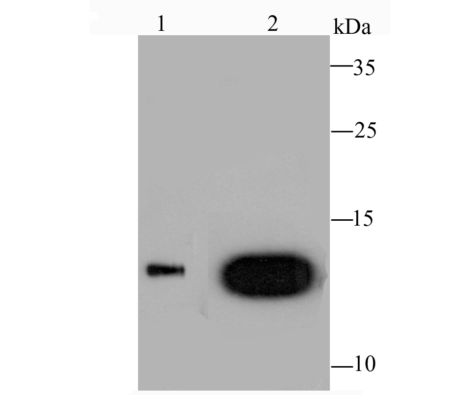
Fig1: Western blot analysis of Galectin 1 on HL-60 cell (1) and human skeletal muscle tissue (2) lysate using anti-Galectin 1 antibody at 1/1,000 dilution.Application
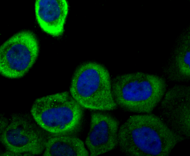
Fig2: ICC staining Galectin 1 in A431 cells (green). The nuclear counter stain is DAPI (blue). Cells were fixed in paraformaldehyde, permeabilised with 0.25% Triton X100/PBS.Application
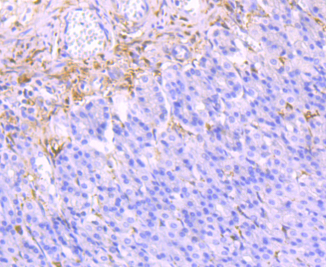
Fig3: Immunohistochemical analysis of paraffin-embedded rat stomach tissue using anti-Galectin 1 antibody. Counter stained with hematoxylin.Application
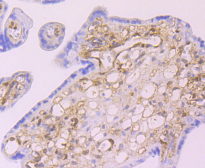
Fig4: Immunohistochemical analysis of paraffin-embedded human placenta tissue using anti-Galectin 1 antibody. Counter stained with hematoxylin.Application
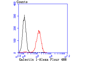
Fig5: Flow cytometric analysis of NIH-3T3 cells with Galectin 1 antibody at 1/100 dilution (red) compared with an unlabelled control (cells without incubation with primary antibody; black).| Product Name | Galectin 1 |
|---|---|
| Antibody Type | Primary Antibodies |
| Antigen Alias | 14 kDa laminin-binding protein antibody 14 kDa lectin antibody Beta galactoside binding lectin antibody Beta galactoside binding lectin L 14 I antibody beta galactoside binding protein antibody Beta-galactoside-binding lectin L-14-I antibody DKFZp686E23103 antibody Gal 1 antibody Gal-1 antibody GAL1 antibody Galaptin antibody Galbp antibody Galectin antibody Galectin-1 antibody Galectin1 antibody GBP antibody HBL antibody HLBP14 antibody HPL antibody L 14.5 antibody L-14.5 antibody L14 antibody Lactose binding lectin 1 antibody Lactose-binding lectin 1 antibody Lect14 antibody Lectin galactoside binding soluble 1 antibody Lectin galactoside-binding soluble 1 antibody LEG1_HUMAN antibody LGALS 1 antibody LGALS1 antibody Lgals1 lectin galactose binding soluble 1 antibody MAPK activating protein MP12 antibody Putative MAPK activating protein MP12 antibody Putative MAPK-activating protein PM12 antibody S Lac lectin 1 antibody S-Lac lectin 1 antibody |
| Product description | Galectins are a family of soluble b-galactoside-binding animal lectins that modulate cell-to-cell adhesion and cell-to-extracellular matrix (ECM) interactions and play a role in tumor progression, pre-mRNA splicing and apoptosis. Specifically, Galectin-1 is an autocrine regulator of cell-proliferation that plays a role in the maintenance of G0 and in the control of G2 traverse. Galectin-1, also known as LGALS1, is the protein product of a single gene linked to human chromosome 22q13.1. The Galectin-1 protein contains 135 amino acids, a single internal EcoRI site and a polyadenylation signal. Galectin-1 can localize to both intracellular and extracellular space. Galectin-1 is expressed in human placenta, human lung, HL-6, HepG2 and CEM cells. |
| Immunogen | Recombinant protein |
| Clonality | Monoclonal |
|---|---|
| Isotype | IgG |
| Host Species | Recombinant rabbit |
| Tested Applications | WBICC/IFIHCIPFC |
| WB:1:500-1:1,000 ICC/IF:1:50-1:200 IHC:1:400-1:800 FC:1:50-1:100 IP:1:10-1:50 |
|
| Species Reactivity | HumanRatMouse |
| Concentration | 1mg/ml |
| Alternative Names | 14 kDa laminin-binding protein antibody 14 kDa lectin antibody Beta galactoside binding lectin antibody Beta galactoside binding lectin L 14 I antibody beta galactoside binding protein antibody Beta-galactoside-binding lectin L-14-I antibody DKFZp686E23103 antibody Gal 1 antibody Gal-1 antibody GAL1 antibody Galaptin antibody Galbp antibody Galectin antibody Galectin-1 antibody Galectin1 antibody GBP antibody HBL antibody HLBP14 antibody HPL antibody L 14.5 antibody L-14.5 antibody L14 antibody Lactose binding lectin 1 antibody Lactose-binding lectin 1 antibody Lect14 antibody Lectin galactoside binding soluble 1 antibody Lectin galactoside-binding soluble 1 antibody LEG1_HUMAN antibody LGALS 1 antibody LGALS1 antibody Lgals1 lectin galactose binding soluble 1 antibody MAPK activating protein MP12 antibody Putative MAPK activating protein MP12 antibody Putative MAPK-activating protein PM12 antibody S Lac lectin 1 antibody S-Lac lectin 1 antibody |
|---|---|
| Cellular Localization | Secreted. Extracellular space. |
Application
Fig1: Western blot analysis of Galectin 1 on HL-60 cell (1) and human skeletal muscle tissue (2) lysate using anti-Galectin 1 antibody at 1/1,000 dilution.
Application
Fig2: ICC staining Galectin 1 in A431 cells (green). The nuclear counter stain is DAPI (blue). Cells were fixed in paraformaldehyde, permeabilised with 0.25% Triton X100/PBS.
Application
Fig3: Immunohistochemical analysis of paraffin-embedded rat stomach tissue using anti-Galectin 1 antibody. Counter stained with hematoxylin.
Application
Fig4: Immunohistochemical analysis of paraffin-embedded human placenta tissue using anti-Galectin 1 antibody. Counter stained with hematoxylin.
Application
Fig5: Flow cytometric analysis of NIH-3T3 cells with Galectin 1 antibody at 1/100 dilution (red) compared with an unlabelled control (cells without incubation with primary antibody; black).| Positive Control | HL-60, human skeletal muscle tissue lysate, A431, rat stomach tissue, human placenta tissue, NIH-3T3. |
|---|---|
| Application Notes | WB:1:500-1:1,000 ICC/IF:1:50-1:200 IHC:1:400-1:800 FC:1:50-1:100 IP:1:10-1:50 |
| Form | Liquid |
|---|---|
| Storage Instructions | Store at +4℃ after thawing. Aliquot store at -20℃ or -80℃. Avoid repeated freeze / thaw cycles. |
| Storage Buffer | 1*TBS (pH7.4), 1%BSA, 40%Glycerol. Preservative: 0.05% Sodium Azide. |


