Application
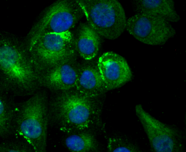
Fig1: ICC staining Estrogen Inducible Protein pS2 in A549 cells (green). The nuclear counter stain is DAPI (blue). Cells were fixed in paraformaldehyde, permeabilised with 0.25% Triton X100/PBS.Application
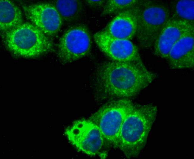
Fig2: ICC staining Estrogen Inducible Protein pS2 in MCF-7 cells (green). The nuclear counter stain is DAPI (blue). Cells were fixed in paraformaldehyde, permeabilised with 0.25% Triton X100/PBS.Application
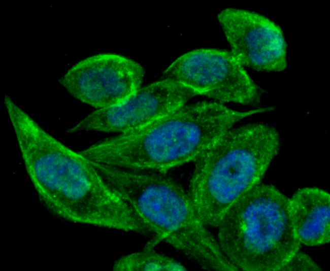
Fig3: ICC staining Estrogen Inducible Protein pS2 in PC-3M cells (green). The nuclear counter stain is DAPI (blue). Cells were fixed in paraformaldehyde, permeabilised with 0.25% Triton X100/PBS.Application
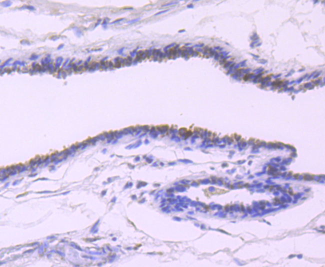
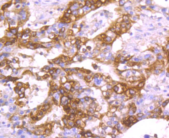
Fig4: Immunohistochemical analysis of paraffin-embedded human breast cancer tissue using anti-Estrogen Inducible Protein pS2 antibody. Counter stained with hematoxylin.Application
Fig5: Immunohistochemical analysis of paraffin-embedded human stomach cancer tissue using anti-Estrogen Inducible Protein pS2 antibody. Counter stained with hematoxylin.Application
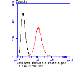
Fig6: Flow cytometric analysis of MCF-7 cells with Estrogen Inducible Protein pS2 antibody at 1/100 dilution (red) compared with an unlabelled control (cells without incubation with primary antibody; black).| Product Name | Estrogen Inducible Protein pS2 |
|---|---|
| Antibody Type | Primary Antibodies |
| Antigen Alias | BCEI antibody Breast cancer estrogen inducible protein antibody Breast cancer estrogen inducible sequence antibody Breast cancer estrogen-inducible protein antibody D21S21 antibody Gastrointestinal trefoil protein antibody Gastrointestinal trefoil protein pS2 antibody hP1.A antibody HP1A antibody HPS 2 antibody HPS2 antibody pNR 2 antibody PNR-2 antibody pNR2 antibody Polypeptide P1.A antibody Protein pS2 antibody PS 2 antibody pS2 antibody pS2 protein antibody TFF 1 antibody TFF1 antibody TFF1_HUMAN antibody Trefoil factor 1 antibody |
| Product description | Trefoil peptides are protease resistant molecules secreted throughout the gut that play a role in mucosal healing. These peptides contain three intrachain disulfide bonds, forming the trefoil motif, or P-domain. pS2, also designated Md2, BCEI, TFF1 and pNR-2, is a trefoil protein that is highly expressed in normal gastric mucosa. pS2 has also been detected in a number of carcinomas including cancers of the breast, pancreas and stomach. pS2 is not present in normal breast tissue but is inducible by estrogen in MCF7 cells. pS2 is known to form dimers and this dimerization is thought to play a role in its protective and healing properties. The gene encoding pS2 maps to human chromosome 21q22.3. |
| Immunogen | Recombinant protein |
| Clonality | Monoclonal |
|---|---|
| Isotype | IgG |
| Host Species | Recombinant rabbit |
| Tested Applications | WBICC/IFIHCIPFC |
| ICC/IF:1:50-1:200 IHC:1:50-1:200 FC:1:50-1:100 IP:1:10-1:50 WB:1:500 |
|
| Species Reactivity | Human |
| Concentration | 1mg/ml |
| Alternative Names | BCEI antibody Breast cancer estrogen inducible protein antibody Breast cancer estrogen inducible sequence antibody Breast cancer estrogen-inducible protein antibody D21S21 antibody Gastrointestinal trefoil protein antibody Gastrointestinal trefoil protein pS2 antibody hP1.A antibody HP1A antibody HPS 2 antibody HPS2 antibody pNR 2 antibody PNR-2 antibody pNR2 antibody Polypeptide P1.A antibody Protein pS2 antibody PS 2 antibody pS2 antibody pS2 protein antibody TFF 1 antibody TFF1 antibody TFF1_HUMAN antibody Trefoil factor 1 antibody |
|---|---|
| Cellular Localization | Secreted. |
| SwissProt ID | P04155 |
|---|
Application
Fig1: ICC staining Estrogen Inducible Protein pS2 in A549 cells (green). The nuclear counter stain is DAPI (blue). Cells were fixed in paraformaldehyde, permeabilised with 0.25% Triton X100/PBS.
Application
Fig2: ICC staining Estrogen Inducible Protein pS2 in MCF-7 cells (green). The nuclear counter stain is DAPI (blue). Cells were fixed in paraformaldehyde, permeabilised with 0.25% Triton X100/PBS.
Application
Fig3: ICC staining Estrogen Inducible Protein pS2 in PC-3M cells (green). The nuclear counter stain is DAPI (blue). Cells were fixed in paraformaldehyde, permeabilised with 0.25% Triton X100/PBS.
Application
Fig4: Immunohistochemical analysis of paraffin-embedded human breast cancer tissue using anti-Estrogen Inducible Protein pS2 antibody. Counter stained with hematoxylin.
Application
Fig5: Immunohistochemical analysis of paraffin-embedded human stomach cancer tissue using anti-Estrogen Inducible Protein pS2 antibody. Counter stained with hematoxylin.
Application
Fig6: Flow cytometric analysis of MCF-7 cells with Estrogen Inducible Protein pS2 antibody at 1/100 dilution (red) compared with an unlabelled control (cells without incubation with primary antibody; black).| Positive Control | A549, MCF-7, PC-3M, human breast cancer tissue, human stomach cancer tissue. |
|---|---|
| Application Notes | ICC/IF:1:50-1:200 IHC:1:50-1:200 FC:1:50-1:100 IP:1:10-1:50 WB:1:500 |
| Form | Liquid |
|---|---|
| Storage Instructions | Store at +4℃ after thawing. Aliquot store at -20℃ or -80℃. Avoid repeated freeze / thaw cycles. |
| Storage Buffer | 1*TBS (pH7.4), 1%BSA, 40%Glycerol. Preservative: 0.05% Sodium Azide. |


