Application
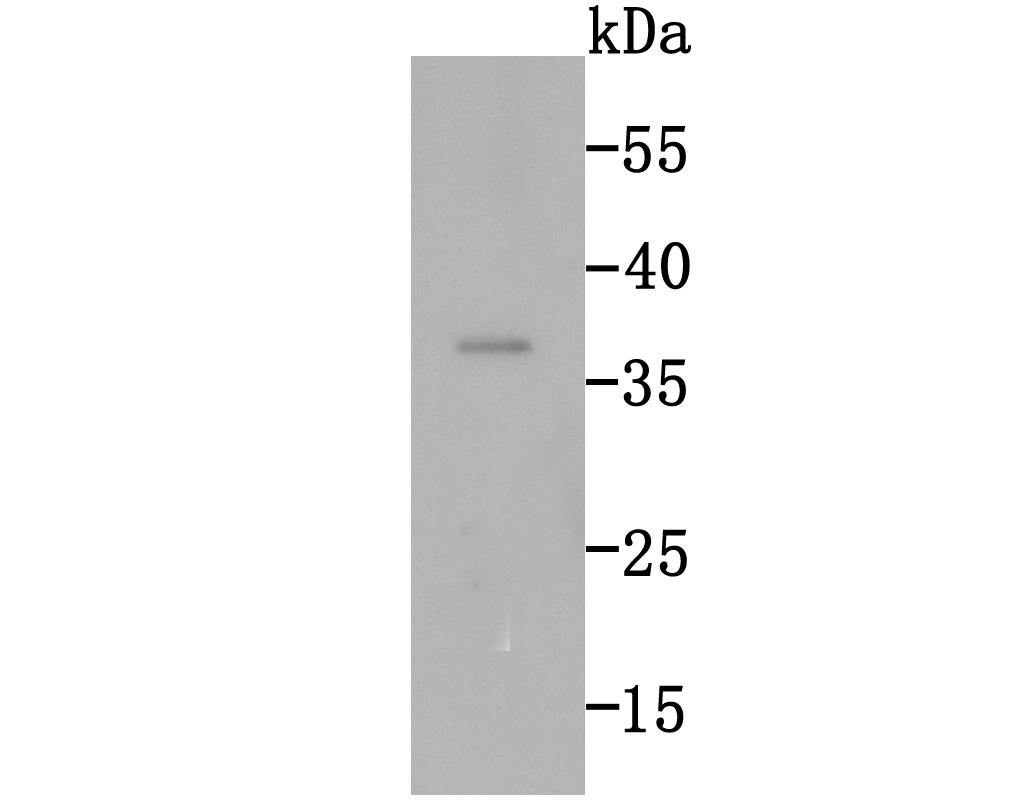
Fig1: Western blot analysis of Rad51 on mouse testie tissue lytase using anti-Rad51 antibody at 1/1,000 dilution.Application
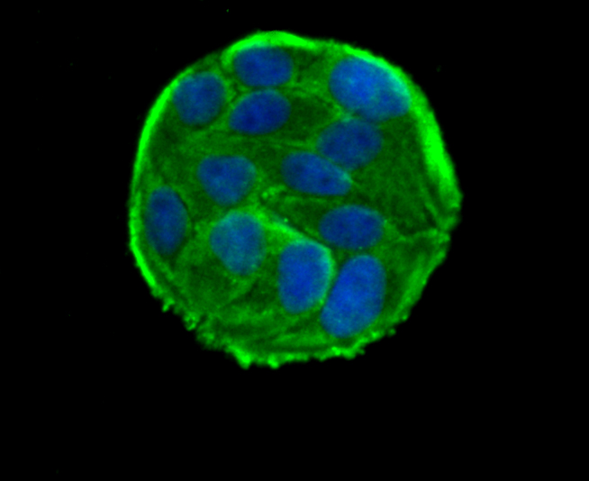
Fig2: ICC staining Rad51 in Hela cells (green). The nuclear counter stain is DAPI (blue). Cells were fixed in paraformaldehyde, permeabilised with 0.25% Triton X100/PBS.Application
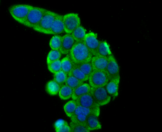
Fig3: ICC staining Rad51 in LOVO cells (green). The nuclear counter stain is DAPI (blue). Cells were fixed in paraformaldehyde, permeabilised with 0.25% Triton X100/PBS.Application
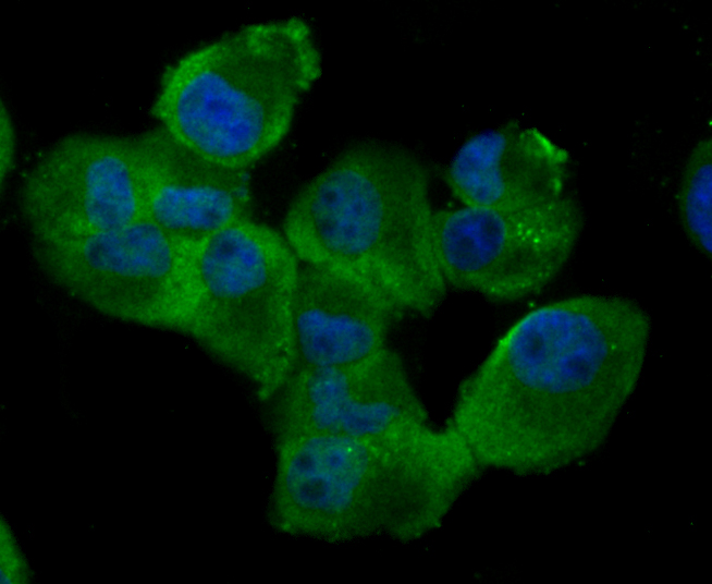
Fig4: ICC staining Rad51 in PANC-1 cells (green). The nuclear counter stain is DAPI (blue). Cells were fixed in paraformaldehyde, permeabilised with 0.25% Triton X100/PBS.Application
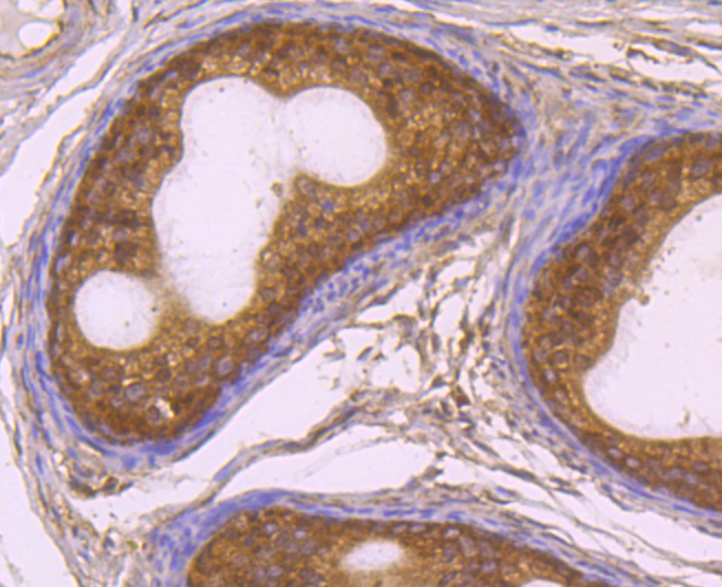
Fig5: Immunohistochemical analysis of paraffin-embedded rat epididymis tissue using anti-Rad51 antibody. Counter stained with hematoxylin.Application
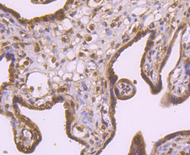
Fig6: Immunohistochemical analysis of paraffin-embedded human placenta tissue using anti-Rad51 antibody. Counter stained with hematoxylin.Application
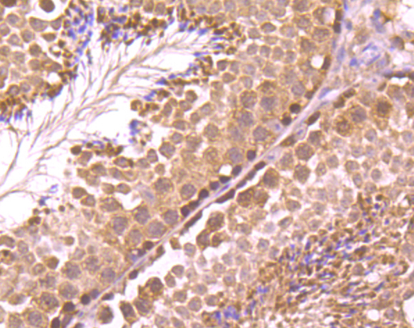
Fig7: Immunohistochemical analysis of paraffin-embedded mouse testis tissue using anti-Rad51 antibody. Counter stained with hematoxylin.Application
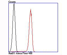
Fig8: Flow cytometric analysis of Jurkat cells with Rad51 antibody at 1/100 dilution (red) compared with an unlabelled control (cells without incubation with primary antibody; black).| Product Name | Rad51 [JM54-26] |
|---|---|
| Antibody Type | Primary Antibodies |
| Product description | d spleen, and mutations of this gene occur in primary lymphoma and colon cancer. MRE11 (meiotic recombination 11, ATLD, HNGS1) is a nuclear 3′-5′ exonuclease/endonuclease that associates with Rad50 and influences homologous recombination, telomere length maintenance, and DNA double-strand break repair. MRE11 is most abundant in proliferating tissues. |
| Immunogen | Recombinant protein |
| Clonality | Monoclonal |
|---|---|
| Isotype | IgG |
| Host Species | Recombinant rabbit |
| Tested Applications | WB,ICC/IF,IHC,IP,FC |
| WB:1:500 ICC/IF:1:50-1:200 IHC:1:50-1:200 IP:1:10-1:50 FC:1:50-1:100 |
|
| Species Reactivity | Human,Mouse,Rat |
| Concentration | 1mg/ml |
| Purification | Unpurified |
| Alternative Names | BRCA1/BRCA2 containing complex subunit 5 antibody BRCC 5 antibody BRCC5 antibody DNA repair protein RAD51 homolog 1 antibody DNA repair protein rhp51 antibody FANCR antibody hRAD51 antibody HsRAD51 antibody HsT16930 antibody MRMV2 antibody Rad 51 antibody RAD51 antibody RAD51 homolog (RecA homolog E. coli) (S. cerevisiae) antibody RAD51 homolog A antibody RAD51 homolog antibody RAD51 recombinase antibody RAD51 S. cerevisiae homolog of antibody RAD51_HUMAN antibody RAD51A antibody RECA antibody RecA like protein antibody RecA E. coli homolog of antibody Recombination protein A antibody |
|---|---|
| Molecular Weight(MW) | 37 kDa |
| Cellular Localization | Nucleus. Cytoplasm. |
Application
Fig1: Western blot analysis of Rad51 on mouse testie tissue lytase using anti-Rad51 antibody at 1/1,000 dilution.
Application
Fig2: ICC staining Rad51 in Hela cells (green). The nuclear counter stain is DAPI (blue). Cells were fixed in paraformaldehyde, permeabilised with 0.25% Triton X100/PBS.
Application
Fig3: ICC staining Rad51 in LOVO cells (green). The nuclear counter stain is DAPI (blue). Cells were fixed in paraformaldehyde, permeabilised with 0.25% Triton X100/PBS.
Application
Fig4: ICC staining Rad51 in PANC-1 cells (green). The nuclear counter stain is DAPI (blue). Cells were fixed in paraformaldehyde, permeabilised with 0.25% Triton X100/PBS.
Application
Fig5: Immunohistochemical analysis of paraffin-embedded rat epididymis tissue using anti-Rad51 antibody. Counter stained with hematoxylin.
Application
Fig6: Immunohistochemical analysis of paraffin-embedded human placenta tissue using anti-Rad51 antibody. Counter stained with hematoxylin.
Application
Fig7: Immunohistochemical analysis of paraffin-embedded mouse testis tissue using anti-Rad51 antibody. Counter stained with hematoxylin.
Application
Fig8: Flow cytometric analysis of Jurkat cells with Rad51 antibody at 1/100 dilution (red) compared with an unlabelled control (cells without incubation with primary antibody; black).| Positive Control | Mouse testie tissue lytase, LOVO, PANC-1, rat epididymis tissue, human placenta tissue, mouse testis tissue, Jurkat. |
|---|---|
| Application Notes | WB:1:500 ICC/IF:1:50-1:200 IHC:1:50-1:200 IP:1:10-1:50 FC:1:50-1:100 |
| Form | Liquid |
|---|---|
| Storage Instructions | Store at +4℃ after thawing. Aliquot store at -20℃ or -80℃. Avoid repeated freeze / thaw cycles. |
| Storage Buffer | 1*TBS (pH7.4), 1%BSA, 40%Glycerol. Preservative: 0.05% Sodium Azide. |


