WB
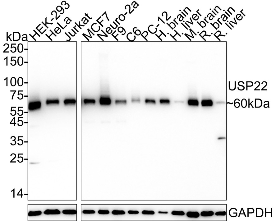
Western blot analysis of USP22 on different lysates with Rabbit anti-USP22 antibody at 1/1,000 dilution. Lane 1: HEK-293 cell lysate (15 µg/Lane), Lane 2: HeLa cell lysate (15 µg/Lane), Lane 3: Jurkat cell lysate (15 µg/Lane), Lane 4: MCF7 cell lysate (15 µg/Lane), Lane 5: Neuro-2a cell lysate (15 µg/Lane), Lane 6: F9 cell lysate (15 µg/Lane), Lane 7: C6 cell lysate (15 µg/Lane), Lane 8: PC-12 cell lysate (15 µg/Lane), Lane 9: Human brain tissue lysate (30 µg/Lane), Lane 10: Human liver tissue lysate (30 µg/Lane), Lane 11: Mouse brain tissue lysate (30 µg/Lane), Lane 12: Rat brain tissue lysate (30 µg/Lane), Lane 13: Rat liver tissue lysate (30 µg/Lane), Exposure time: Lane 1-3: 6 seconds; Lane 4-13: 21 seconds; 4-20% SDS-PAGE gel. Proteins were transferred to a PVDF membrane and blocked with 5% NFDM/TBST for 1 hour at room temperature. The primary antibody at 1/1,000 dilution was used in 5% NFDM/TBST at 4℃ overnight. Goat Anti-Rabbit IgG - HRP Secondary Antibody at 1/50,000 dilution was used for 1 hour at room temperature.IHC
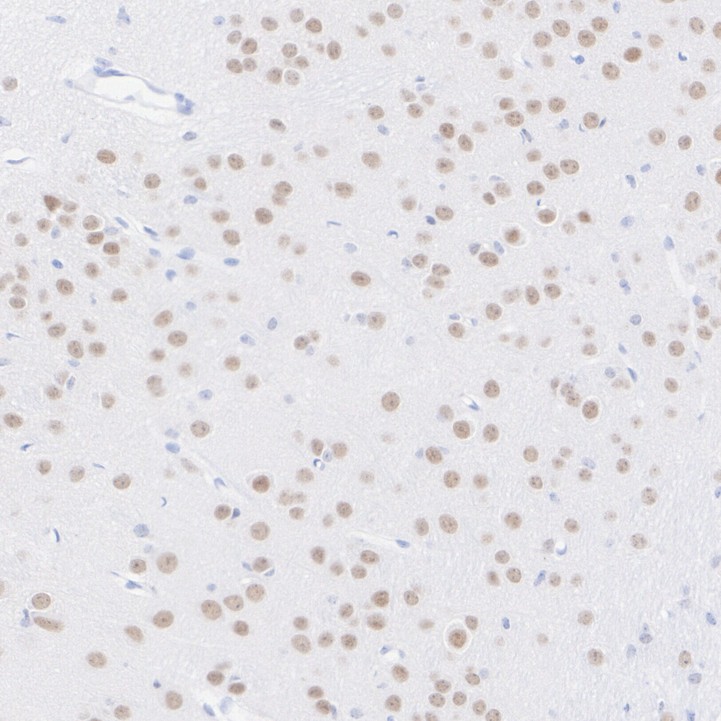
Immunohistochemical analysis of paraffin-embedded mouse brain tissue with Rabbit anti-USP22 antibody at 1/200 dilution. The section was pre-treated using heat mediated antigen retrieval with sodium citrate buffer (pH 6.0) (high pressure) for 2 minutes. The tissues were blocked in 1% BSA for 20 minutes at room temperature, washed with ddH2O and PBS, and then probed with the primary antibody at 1/200 dilution for 1 hour at room temperature. The detection was performed using an HRP conjugated compact polymer system. DAB was used as the chromogen. Tissues were counterstained with hematoxylin and mounted with DPX.IHC
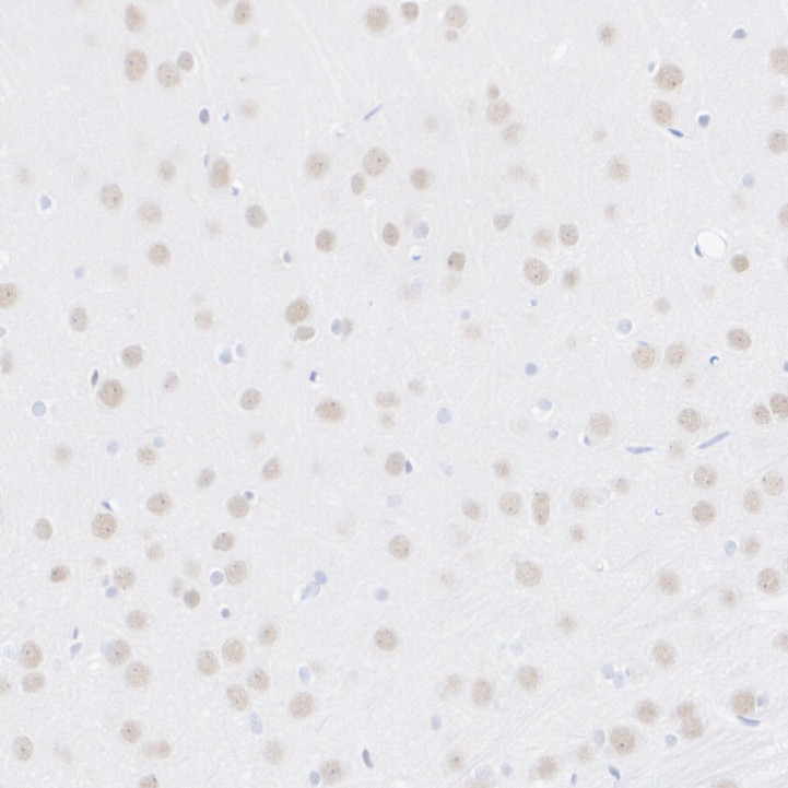
Immunohistochemical analysis of paraffin-embedded rat brain tissue with Rabbit anti-USP22 antibody at 1/200 dilution. The section was pre-treated using heat mediated antigen retrieval with sodium citrate buffer (pH 6.0) (high pressure) for 2 minutes. The tissues were blocked in 1% BSA for 20 minutes at room temperature, washed with ddH2O and PBS, and then probed with the primary antibody at 1/200 dilution for 1 hour at room temperature. The detection was performed using an HRP conjugated compact polymer system. DAB was used as the chromogen. Tissues were counterstained with hematoxylin and mounted with DPX.IF-P
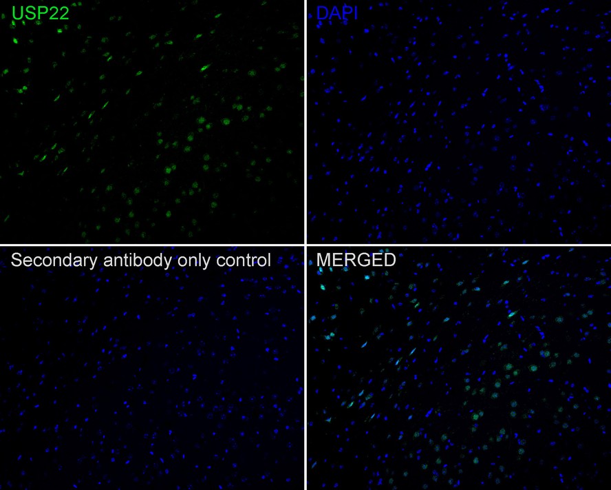
Immunofluorescence analysis of paraffin-embedded mouse brain tissue labeling USP22 with Rabbit anti-USP22 antibody at 1/50 dilution. The section was pre-treated using heat mediated antigen retrieval with sodium citrate buffer (pH 6.0) (high pressure) for 2 minutes. The tissues were blocked in 10% negative goat serum for 1 hour at room temperature, washed with PBS, and then probed with the primary antibody (green) at 1/50 dilution overnight at 4 ℃, washed with PBS. Goat Anti-Rabbit IgG H&L (488) was used as the secondary antibody at 1/1,000 dilution. Nuclei were counterstained with DAPI (blue).IP
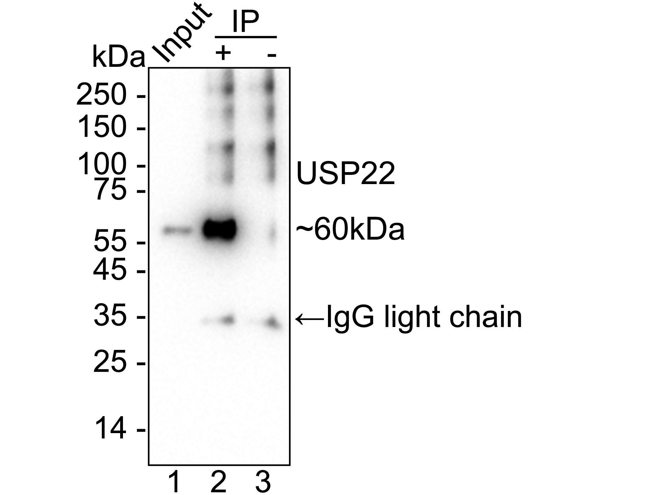
USP22 was immunoprecipitated from 0.2 mg HeLa cell lysate with Rabbit anti-USP22 antibody at 2 µg/10 µl beads. Western blot was performed from the immunoprecipitate using Rabbit anti-USP22 antibody at 1/2,000 dilution. Anti-Rabbit IgG for IP Nano-secondary antibody at 1/5,000 dilution was used for 1 hour at room temperature. Lane 1: HeLa cell lysate (input), Lane 2: Rabbit anti-USP22 antibody IP in HeLa cell lysate, Lane 3: Rabbit IgG instead of Rabbit anti-USP22 antibody in HeLa cell lysate, Blocking/Dilution buffer: 5% NFDM/TBST, Exposure time: 14 seconds.| Product Name | USP22 Recombinant Rabbit Monoclonal Antibody |
|---|---|
| Antibody Type | Primary Antibodies |
| Product description | The ubiquitin (Ub) pathway involves three sequential enzymatic steps that facilitate the conjugation of Ub and Ub-like molecules to specific protein substrates. Through the use of a wide range of enzymes that can add or remove ubiquitin, the Ub pathway controls many intracellular processes such as signal transduction, transcriptional activation and cell cycle progression. USP22 (ubiquitin specific peptidase 22), also known as USP3L, is a 525 amino acid protein that contains one UBP-type zinc finger and functions to catalyze the conversion of a ubiquitin C-terminal thioester to free ubiquitin and thiol, a reaction that may influence several cellular processes. Via its catalytic activity, USP22 is thought to play an important role in cell cycle progression and may also serve as a cancer stem cell marker. The gene encoding USP22 maps to human chromosome 17, which comprises over 2.5% of the human genome and encodes over 1,200 genes. |
| Immunogen | Synthetic peptide within Human USP22 aa 321-370 / 525. |
| Clonality | Monoclonal |
|---|---|
| Isotype | IgG |
| Host Species | Recombinant rabbit |
| Tested Applications | IF-PIHCIPWB |
| WB:1:1000-1:5000 IHC:1:200 IF-P:1:50 IP:1-2μg/sample |
|
| Species Reactivity | HumanMouseRat |
| Concentration | 1mg/ml |
| Purification | Protein A |
| Alternative Names | Deubiquitinating enzyme 22 antibody KIAA1063 antibody Ubiquitin carboxyl terminal hydrolase 22 antibody Ubiquitin carboxyl-terminal hydrolase 22 antibody Ubiquitin specific peptidase 22 antibody Ubiquitin specific peptidase 3 like antibody Ubiquitin specific processing protease 22 antibody Ubiquitin specific protease 22 antibody Ubiquitin thioesterase 22 antibody Ubiquitin thiolesterase 22 antibody Ubiquitin-specific-processing protease 22 antibody UBP22_HUMAN antibody USP 22 antibody Usp22 antibody USP3L antibody |
|---|---|
| Molecular Weight(MW) | 60 kDa |
| Cellular Localization | Nucleus. |
| SwissProt ID | Q9UPT9 |
|---|
WB
Western blot analysis of USP22 on different lysates with Rabbit anti-USP22 antibody at 1/1,000 dilution. Lane 1: HEK-293 cell lysate (15 µg/Lane), Lane 2: HeLa cell lysate (15 µg/Lane), Lane 3: Jurkat cell lysate (15 µg/Lane), Lane 4: MCF7 cell lysate (15 µg/Lane), Lane 5: Neuro-2a cell lysate (15 µg/Lane), Lane 6: F9 cell lysate (15 µg/Lane), Lane 7: C6 cell lysate (15 µg/Lane), Lane 8: PC-12 cell lysate (15 µg/Lane), Lane 9: Human brain tissue lysate (30 µg/Lane), Lane 10: Human liver tissue lysate (30 µg/Lane), Lane 11: Mouse brain tissue lysate (30 µg/Lane), Lane 12: Rat brain tissue lysate (30 µg/Lane), Lane 13: Rat liver tissue lysate (30 µg/Lane), Exposure time: Lane 1-3: 6 seconds; Lane 4-13: 21 seconds; 4-20% SDS-PAGE gel. Proteins were transferred to a PVDF membrane and blocked with 5% NFDM/TBST for 1 hour at room temperature. The primary antibody at 1/1,000 dilution was used in 5% NFDM/TBST at 4℃ overnight. Goat Anti-Rabbit IgG - HRP Secondary Antibody at 1/50,000 dilution was used for 1 hour at room temperature.
IHC
Immunohistochemical analysis of paraffin-embedded mouse brain tissue with Rabbit anti-USP22 antibody at 1/200 dilution. The section was pre-treated using heat mediated antigen retrieval with sodium citrate buffer (pH 6.0) (high pressure) for 2 minutes. The tissues were blocked in 1% BSA for 20 minutes at room temperature, washed with ddH2O and PBS, and then probed with the primary antibody at 1/200 dilution for 1 hour at room temperature. The detection was performed using an HRP conjugated compact polymer system. DAB was used as the chromogen. Tissues were counterstained with hematoxylin and mounted with DPX.
IHC
Immunohistochemical analysis of paraffin-embedded rat brain tissue with Rabbit anti-USP22 antibody at 1/200 dilution. The section was pre-treated using heat mediated antigen retrieval with sodium citrate buffer (pH 6.0) (high pressure) for 2 minutes. The tissues were blocked in 1% BSA for 20 minutes at room temperature, washed with ddH2O and PBS, and then probed with the primary antibody at 1/200 dilution for 1 hour at room temperature. The detection was performed using an HRP conjugated compact polymer system. DAB was used as the chromogen. Tissues were counterstained with hematoxylin and mounted with DPX.
IF-P
Immunofluorescence analysis of paraffin-embedded mouse brain tissue labeling USP22 with Rabbit anti-USP22 antibody at 1/50 dilution. The section was pre-treated using heat mediated antigen retrieval with sodium citrate buffer (pH 6.0) (high pressure) for 2 minutes. The tissues were blocked in 10% negative goat serum for 1 hour at room temperature, washed with PBS, and then probed with the primary antibody (green) at 1/50 dilution overnight at 4 ℃, washed with PBS. Goat Anti-Rabbit IgG H&L (488) was used as the secondary antibody at 1/1,000 dilution. Nuclei were counterstained with DAPI (blue).
IP
USP22 was immunoprecipitated from 0.2 mg HeLa cell lysate with Rabbit anti-USP22 antibody at 2 µg/10 µl beads. Western blot was performed from the immunoprecipitate using Rabbit anti-USP22 antibody at 1/2,000 dilution. Anti-Rabbit IgG for IP Nano-secondary antibody at 1/5,000 dilution was used for 1 hour at room temperature. Lane 1: HeLa cell lysate (input), Lane 2: Rabbit anti-USP22 antibody IP in HeLa cell lysate, Lane 3: Rabbit IgG instead of Rabbit anti-USP22 antibody in HeLa cell lysate, Blocking/Dilution buffer: 5% NFDM/TBST, Exposure time: 14 seconds.| Application Notes | WB:1:1000-1:5000 IHC:1:200 IF-P:1:50 IP:1-2μg/sample |
|---|
| Form | Liquid |
|---|---|
| Storage Instructions | Store at +4℃ after thawing. Aliquot store at -20℃ or -80℃. Avoid repeated freeze / thaw cycles. |
| Storage Buffer | 1*TBS (pH7.4), 1%BSA, 40%Glycerol. Preservative: 0.05% Sodium Azide. |


