Application
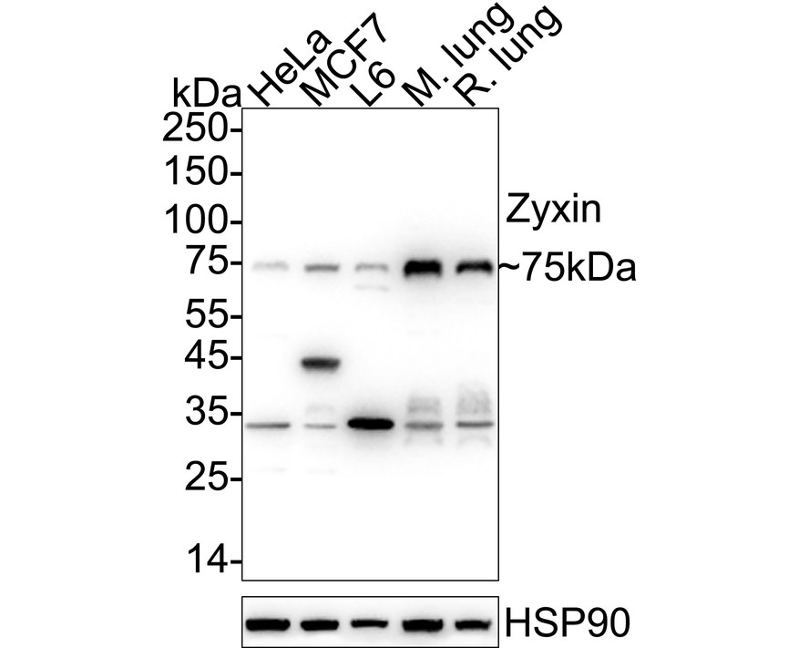
Fig1: Western blot analysis of Zyxin on different lysates with Rabbit anti-Zyxin antibody at 1/1,000 dilution. Lane 1: HeLa cell lysate (20 µg/Lane), Lane 2: MCF7 cell lysate (20 µg/Lane), Lane 3: L6 cell lysate (20 µg/Lane), Lane 4: Mouse lung tissue lysate (40 µg/Lane), Lane 5: Rat lung tissue lysate (40 µg/Lane), Exposure time: 6 seconds; 4-20% SDS-PAGE gel. Proteins were transferred to a PVDF membrane and blocked with 5% NFDM/TBST for 1 hour at room temperature. The primary antibody at 1/1,000 dilution was used in 5% NFDM/TBST at 4℃ overnight. Goat Anti-Rabbit IgG - HRP Secondary Antibody at 1/50,000 dilution was used for 1 hour at room temperature.Application
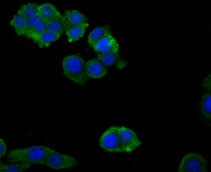
Fig2: ICC staining Zyxin in LOVO cells (green). The nuclear counter stain is DAPI (blue). Cells were fixed in paraformaldehyde, permeabilised with 0.25% Triton X100/PBS.Application
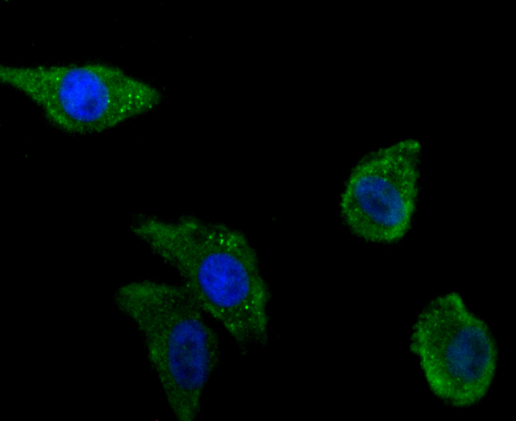
Fig3: ICC staining Zyxin in PC-3M cells (green). The nuclear counter stain is DAPI (blue). Cells were fixed in paraformaldehyde, permeabilised with 0.25% Triton X100/PBS.Application
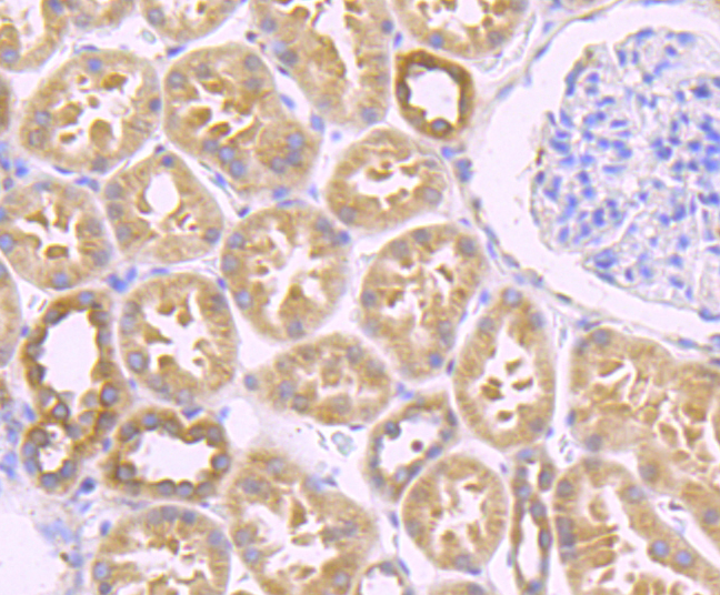
Fig4: Immunohistochemical analysis of paraffin-embedded human kidney tissue using anti-Zyxin antibody. Counter stained with hematoxylin.Application
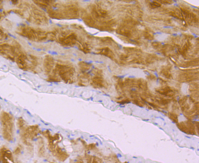
Fig5: Immunohistochemical analysis of paraffin-embedded mouse smooth muscle tissue using anti-Zyxin antibody. Counter stained with hematoxylin.Application
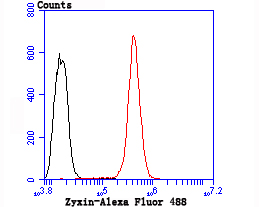
Fig6: Flow cytometric analysis of Hela cells with Zyxin antibody at 1/100 dilution (red) compared with an unlabelled control (cells without incubation with primary antibody; black). Alexa Fluor 488-conjugated goat anti rabbit IgG was used as the secondary antibody.| Product Name | Zyxin Recombinant Rabbit Monoclonal Antibody |
|---|---|
| Antibody Type | Primary Antibodies |
| Product description | Zyxin is a low abundance phosphoprotein localized to focal adhesion plaques and is thought to perform regulatory functions at these regions. The protein contains a number of proline-rich sequences as well as three LIM domains, zinc finger domains involved in protein binding. Zyxin interacts with several other proteins at cell adhesion sites, including members of the CRP (cysteine-rich protein) LIM domain containing protein family. The proline-rich domain of Zyxin associates with an SH3 domain of p95 Vav, but not with similar SH3 domains containing proteins such as GRB2 or PLCg. Zyxin has also been shown to interact with the focal adhesion protein VASP and may assist in the targeting of VASP to focal adhesions, microfilaments and membrane regions of high dynamic activity. |
| Immunogen | Synthetic peptide within Human Zyxin aa 284-343 / 572. |
| Clonality | Monoclonal |
|---|---|
| Isotype | IgG |
| Host Species | Recombinant rabbit |
| Tested Applications | FCICC/IFIHCIPWB |
| WB:1:500-1:1,000 ICC:1:50-1:200 IHC:1:50-1:200 FC:1:50-1:100 |
|
| Species Reactivity | HumanMouseRat |
| Concentration | 1mg/ml |
| Purification | Protein A |
| Alternative Names | ESP 2 antibody ESP2 antibody HED 2 antibody HED2 antibody ZYX antibody ZYX protein antibody Zyxin 2 antibody Zyxin2 antibody |
|---|---|
| Molecular Weight(MW) | 61kDa(Observed band size: 75kDa) |
| Cellular Localization | Cytoplasm, cytoskeleton and Nucleus. |
Application
Fig1: Western blot analysis of Zyxin on different lysates with Rabbit anti-Zyxin antibody at 1/1,000 dilution. Lane 1: HeLa cell lysate (20 µg/Lane), Lane 2: MCF7 cell lysate (20 µg/Lane), Lane 3: L6 cell lysate (20 µg/Lane), Lane 4: Mouse lung tissue lysate (40 µg/Lane), Lane 5: Rat lung tissue lysate (40 µg/Lane), Exposure time: 6 seconds; 4-20% SDS-PAGE gel. Proteins were transferred to a PVDF membrane and blocked with 5% NFDM/TBST for 1 hour at room temperature. The primary antibody at 1/1,000 dilution was used in 5% NFDM/TBST at 4℃ overnight. Goat Anti-Rabbit IgG - HRP Secondary Antibody at 1/50,000 dilution was used for 1 hour at room temperature.
Application
Fig2: ICC staining Zyxin in LOVO cells (green). The nuclear counter stain is DAPI (blue). Cells were fixed in paraformaldehyde, permeabilised with 0.25% Triton X100/PBS.
Application
Fig3: ICC staining Zyxin in PC-3M cells (green). The nuclear counter stain is DAPI (blue). Cells were fixed in paraformaldehyde, permeabilised with 0.25% Triton X100/PBS.
Application
Fig4: Immunohistochemical analysis of paraffin-embedded human kidney tissue using anti-Zyxin antibody. Counter stained with hematoxylin.
Application
Fig5: Immunohistochemical analysis of paraffin-embedded mouse smooth muscle tissue using anti-Zyxin antibody. Counter stained with hematoxylin.
Application
Fig6: Flow cytometric analysis of Hela cells with Zyxin antibody at 1/100 dilution (red) compared with an unlabelled control (cells without incubation with primary antibody; black). Alexa Fluor 488-conjugated goat anti rabbit IgG was used as the secondary antibody.| Application Notes | WB:1:500-1:1,000 ICC:1:50-1:200 IHC:1:50-1:200 FC:1:50-1:100 |
|---|
| Form | Liquid |
|---|---|
| Storage Instructions | Store at +4℃ after thawing. Aliquot store at -20℃ or -80℃. Avoid repeated freeze / thaw cycles. |
| Storage Buffer | 1*TBS (pH7.4), 1%BSA, 40%Glycerol. Preservative: 0.05% Sodium Azide. |


