Application
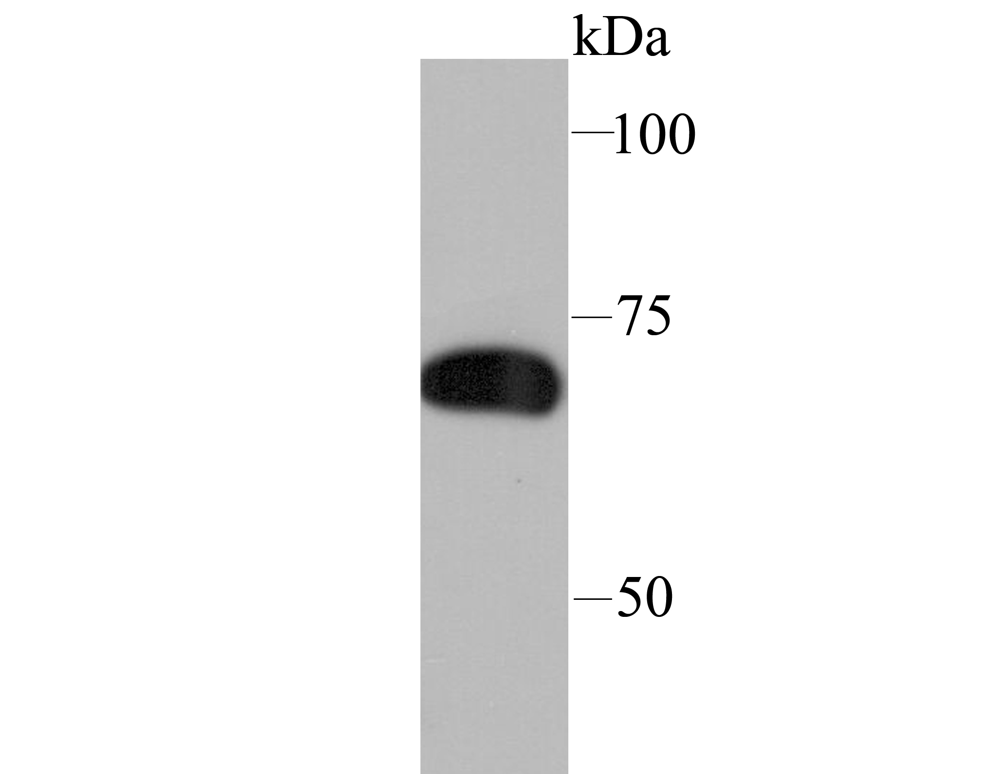
Fig1: Western blot analysis of SAMHD1 on THP-1 cell lysate using anti-SAMHD1 antibody at 1/1,000 dilution.Application
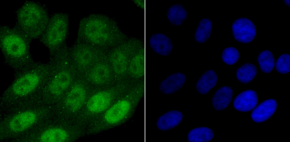
Fig2: ICC staining SAMHD1 in HepG2 cells (green). The nuclear counter stain is DAPI (blue). Cells were fixed in paraformaldehyde, permeabilised with 0.25% Triton X100/PBS.Application
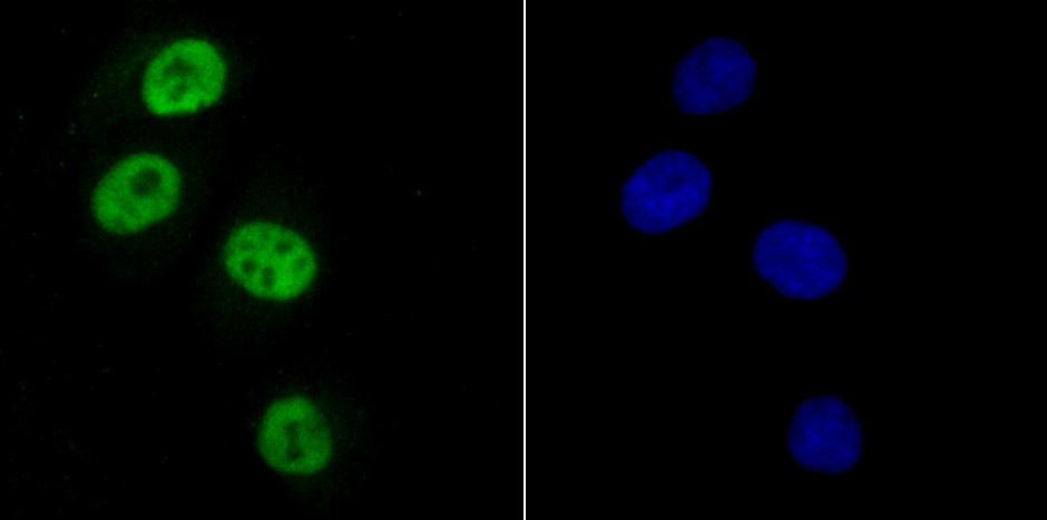
Fig3: ICC staining SAMHD1 in HUVEC cells (green). The nuclear counter stain is DAPI (blue). Cells were fixed in paraformaldehyde, permeabilised with 0.25% Triton X100/PBS.Application
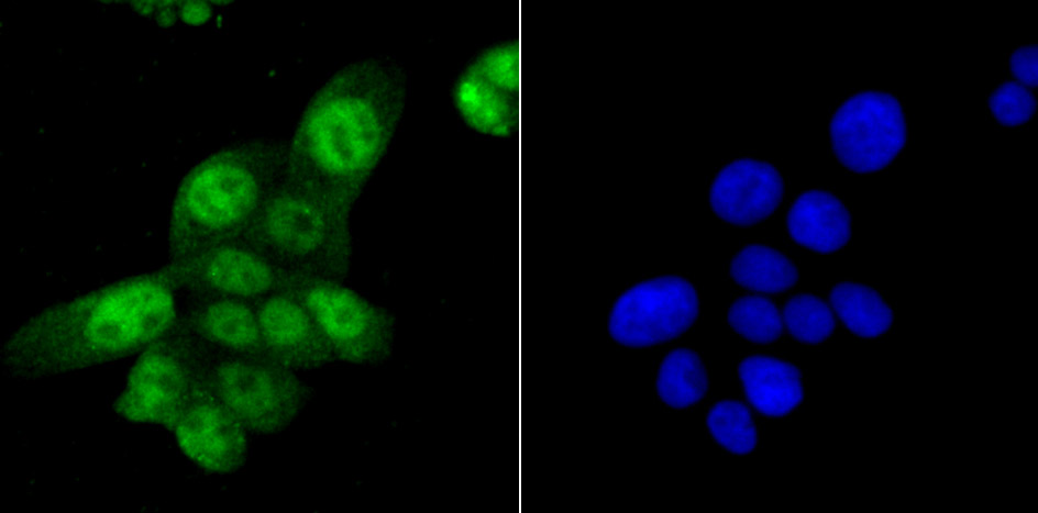
Fig4: ICC staining SAMHD1 in LOVO cells (green). The nuclear counter stain is DAPI (blue). Cells were fixed in paraformaldehyde, permeabilised with 0.25% Triton X100/PBS.Application
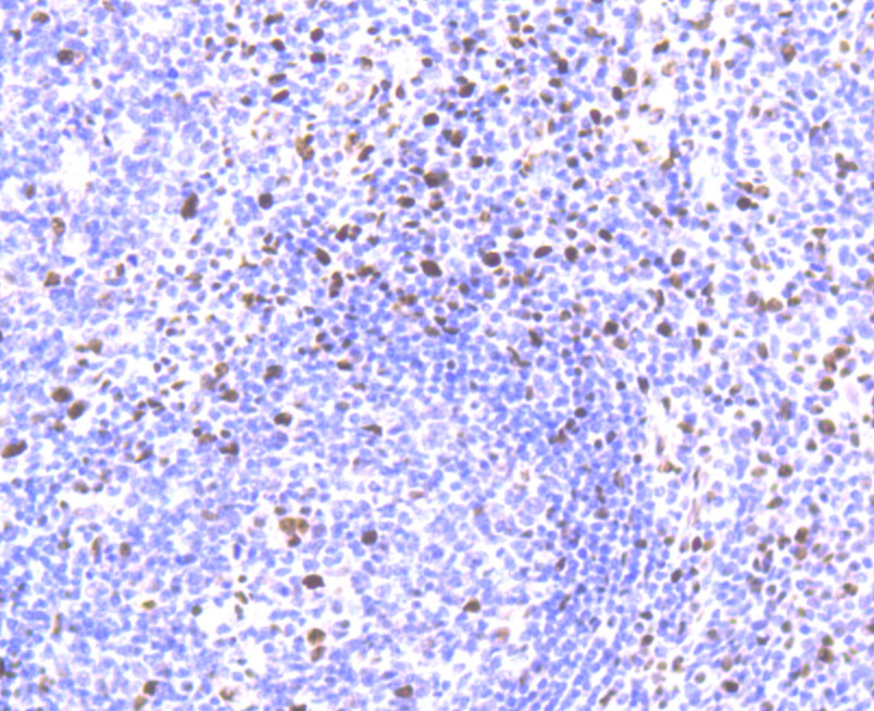
Fig5: Immunohistochemical analysis of paraffin-embedded human tonsil tissue using anti-SAMHD1 antibody. Counter stained with hematoxylin.Application
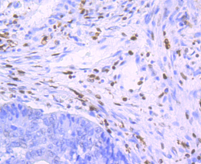
Fig6: Immunohistochemical analysis of paraffin-embedded human colon cancer tissue using anti-SAMHD1 antibody. Counter stained with hematoxylin.Application
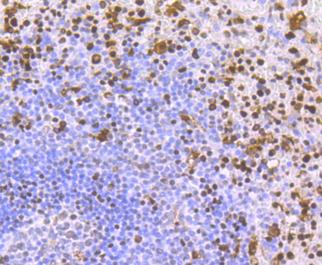
Fig7: Immunohistochemical analysis of paraffin-embedded human spleen tissue using anti-SAMHD1 antibody. Counter stained with hematoxylin.Application
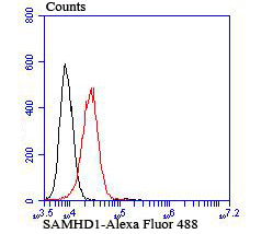
Fig8: Flow cytometric analysis of K562 cells with SAMHD1 antibody at 1/100 dilution (red) compared with an unlabelled control (cells without incubation with primary antibody; black).| Product Name | SAMHD1 [JU56-04] |
|---|---|
| Antibody Type | Primary Antibodies |
| Antigen Alias | CHBL2 antibody DCIP antibody Dendritic cell derived IFNG induced protein antibody Dendritic cell-derived IFNG-induced protein antibody Deoxynucleoside triphosphate triphosphohydrolase SAMHD1 antibody dNTPase antibody HD domain containing 1 antibody HDDC1 antibody Mg11 antibody Monocyte protein 5 antibody MOP 5 antibody MOP-5 antibody MOP5 antibody OTTHUMP00000030889 antibody SAM domain and HD domain 1 antibody SAM domain and HD domain containing protein 1 antibody SAM domain and HD domain-containing protein 1 antibody SAMH1_HUMAN antibody Samhd1 antibody SBBI88 antibody |
| Product description | SAM domain and HD domain-containing protein 1 is a protein that in humans is encoded by the SAMHD1 gene. SAMHD1 is a cellular enzyme, responsible for blocking replication of HIV in dendritic cells, macrophages and monocytes. It is an enzyme that exhibits phosphohydrolase activity, converting deoxynucleoside triphosphates (dNTPs) to inorganic phosphate (iPPP) and a 2'-deoxynucleoside (i.e. deoxynucleosides without a phosphate group). In doing so, SAMHD1 depletes the pool of dNTPs available to a reverse transcriptase for viral cDNA synthesis and thus prevents viral replication. SAMHD1 has also shown nuclease activity. Although a ribonuclease activity was described to be required for HIV-1 restriction, recent data confirmed that SAMHD1-mediated HIV-1 restriction in cells does not involve ribonuclease activity. |
| Immunogen | Recombinant protein |
| Clonality | Monoclonal |
|---|---|
| Isotype | IgG |
| Host Species | Recombinant rabbit |
| Tested Applications | WBICCIHCFC |
| WB:1:500-1:2,000 ICC:1:100-1:500 IHC:1:50-1:200 FC:1:50-1:100 |
|
| Species Reactivity | Human |
| Concentration | 1mg/ml |
| Alternative Names | CHBL2 antibody DCIP antibody Dendritic cell derived IFNG induced protein antibody Dendritic cell-derived IFNG-induced protein antibody Deoxynucleoside triphosphate triphosphohydrolase SAMHD1 antibody dNTPase antibody HD domain containing 1 antibody HDDC1 antibody Mg11 antibody Monocyte protein 5 antibody MOP 5 antibody MOP-5 antibody MOP5 antibody OTTHUMP00000030889 antibody SAM domain and HD domain 1 antibody SAM domain and HD domain containing protein 1 antibody SAM domain and HD domain-containing protein 1 antibody SAMH1_HUMAN antibody Samhd1 antibody SBBI88 antibody |
|---|---|
| Molecular Weight(MW) | 72 kDa |
| Cellular Localization | Nucleus. |
| SwissProt ID | Q9Y3Z3 |
|---|
Application
Fig1: Western blot analysis of SAMHD1 on THP-1 cell lysate using anti-SAMHD1 antibody at 1/1,000 dilution.
Application
Fig2: ICC staining SAMHD1 in HepG2 cells (green). The nuclear counter stain is DAPI (blue). Cells were fixed in paraformaldehyde, permeabilised with 0.25% Triton X100/PBS.
Application
Fig3: ICC staining SAMHD1 in HUVEC cells (green). The nuclear counter stain is DAPI (blue). Cells were fixed in paraformaldehyde, permeabilised with 0.25% Triton X100/PBS.
Application
Fig4: ICC staining SAMHD1 in LOVO cells (green). The nuclear counter stain is DAPI (blue). Cells were fixed in paraformaldehyde, permeabilised with 0.25% Triton X100/PBS.
Application
Fig5: Immunohistochemical analysis of paraffin-embedded human tonsil tissue using anti-SAMHD1 antibody. Counter stained with hematoxylin.
Application
Fig6: Immunohistochemical analysis of paraffin-embedded human colon cancer tissue using anti-SAMHD1 antibody. Counter stained with hematoxylin.
Application
Fig7: Immunohistochemical analysis of paraffin-embedded human spleen tissue using anti-SAMHD1 antibody. Counter stained with hematoxylin.
Application
Fig8: Flow cytometric analysis of K562 cells with SAMHD1 antibody at 1/100 dilution (red) compared with an unlabelled control (cells without incubation with primary antibody; black).| Positive Control | THP-1, HepG2, HUVEC, LOVO, human tonsil tissue, human colon cancer tissue, human spleen tissue, K562. |
|---|---|
| Application Notes | WB:1:500-1:2,000 ICC:1:100-1:500 IHC:1:50-1:200 FC:1:50-1:100 |
| Form | Liquid |
|---|---|
| Storage Instructions | Store at +4℃ after thawing. Aliquot store at -20℃ or -80℃. Avoid repeated freeze / thaw cycles. |
| Storage Buffer | 1*TBS (pH7.4), 1%BSA, 40%Glycerol. Preservative: 0.05% Sodium Azide. |


