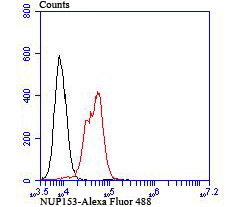
Application
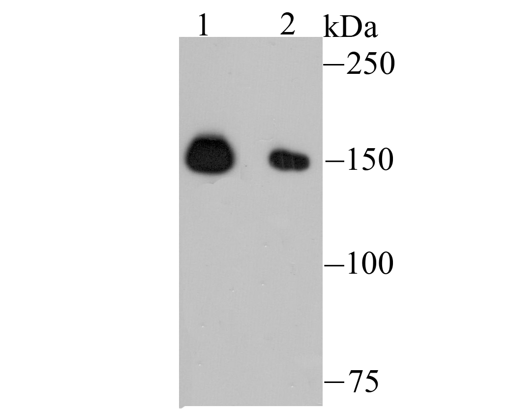
Fig1: Western blot analysis of NUP153 on K562 (1) and HepG2 (2) cell lysate using anti-NUP153 antibody at 1/500 dilution.Application
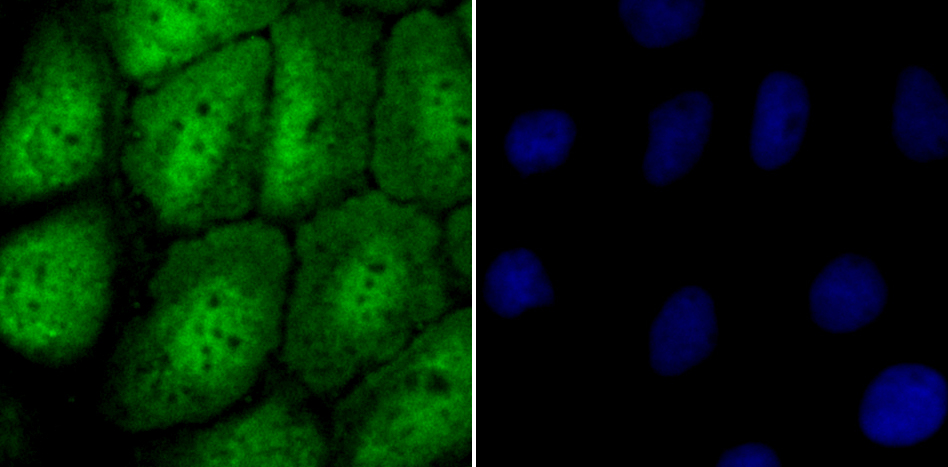
Fig2: ICC staining NUP153 in A431 cells (green). The nuclear counter stain is DAPI (blue). Cells were fixed in paraformaldehyde, permeabilised with 0.25% Triton X100/PBS.Application
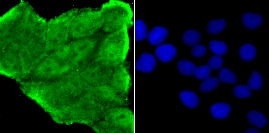
Fig3: ICC staining NUP153 in Hela cells (green). The nuclear counter stain is DAPI (blue). Cells were fixed in paraformaldehyde, permeabilised with 0.25% Triton X100/PBS.Application
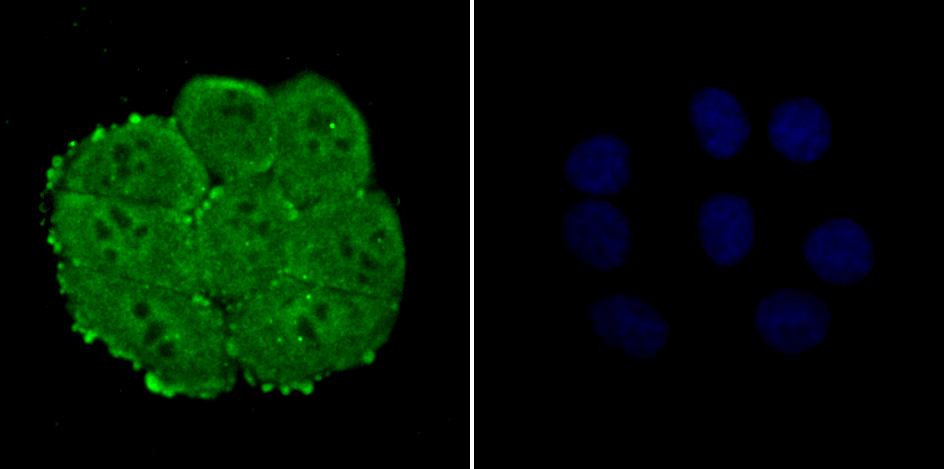
Fig4: ICC staining NUP153 in HepG2 cells (green). The nuclear counter stain is DAPI (blue). Cells were fixed in paraformaldehyde, permeabilised with 0.25% Triton X100/PBS.Application
Application| Product Name | NUP153 [JU40-41] |
|---|---|
| Antibody Type | Primary Antibodies |
| Antigen Alias | 153 kDa nucleoporin antibody HNUP153 antibody N153 antibody NU153_HUMAN antibody Nuclear pore complex protein hnup153 antibody Nuclear pore complex protein Nup153 antibody Nucleoporin 153kDa antibody Nucleoporin Nup153 antibody Nup 153 antibody Nup153 antibody |
| Product description | Nuclear pore complexes (NPCs) are the channels for the bi-directional movement of macromolecules between the nucleus and cytoplasm, and contain more than 100 different subunits. Many of them belong to a family called nucleoporins, which are characterized by the presence of O-linked N-acetylglucosamine moieties and a distinctive pentapeptide repeat (XFXFG). Nup153 is a peripheral NPC component that is implicated in protein and RNP transport and in the interaction of NPCs with the nuclear lamina. Nup153 contains a unique N-terminal region, a central domain consisting of four to five zinc fingers and a C-terminal region containing about 30 irregularly spaced FXFG repeats. Nup153 is cleaved by caspases during apoptosis. Nup153 interacts wtih TAP, which is essential for mRNA export, and associates with chromatin towards the end of anaphase, in parallel with the inner nuclear membrane protein LAP2. Nup153 is involved in NPC assembly, in anchoring NPCs within the nuclear envelope and in mediating specific nuclear import events. |
| Immunogen | Recombinant protein |
| Clonality | Monoclonal |
|---|---|
| Isotype | IgG |
| Host Species | Recombinant rabbit |
| Tested Applications | WBICC/IFFC |
| WB:1:500-1:2,000 ICC:1:100-1:500 FC:1:50-1:100 |
|
| Species Reactivity | Human |
| Concentration | 1mg/ml |
| Alternative Names | 153 kDa nucleoporin antibody HNUP153 antibody N153 antibody NU153_HUMAN antibody Nuclear pore complex protein hnup153 antibody Nuclear pore complex protein Nup153 antibody Nucleoporin 153kDa antibody Nucleoporin Nup153 antibody Nup 153 antibody Nup153 antibody |
|---|---|
| Molecular Weight(MW) | 154 kDa |
| Cellular Localization | Nucleus, nuclear pore complex. |
| SwissProt ID | P49790 |
|---|
Application
Fig1: Western blot analysis of NUP153 on K562 (1) and HepG2 (2) cell lysate using anti-NUP153 antibody at 1/500 dilution.
Application
Fig2: ICC staining NUP153 in A431 cells (green). The nuclear counter stain is DAPI (blue). Cells were fixed in paraformaldehyde, permeabilised with 0.25% Triton X100/PBS.
Application
Fig3: ICC staining NUP153 in Hela cells (green). The nuclear counter stain is DAPI (blue). Cells were fixed in paraformaldehyde, permeabilised with 0.25% Triton X100/PBS.
Application
Fig4: ICC staining NUP153 in HepG2 cells (green). The nuclear counter stain is DAPI (blue). Cells were fixed in paraformaldehyde, permeabilised with 0.25% Triton X100/PBS.
Application
Application| Positive Control | K562, HepG2, A431, Hela. |
|---|---|
| Application Notes | WB:1:500-1:2,000 ICC:1:100-1:500 FC:1:50-1:100 |
| Form | Liquid |
|---|---|
| Storage Instructions | Store at +4℃ after thawing. Aliquot store at -20℃ or -80℃. Avoid repeated freeze / thaw cycles. |
| Storage Buffer | 1*TBS (pH7.4), 1%BSA, 40%Glycerol. Preservative: 0.05% Sodium Azide. |


