WB
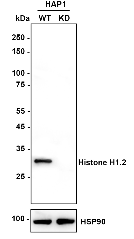
Western blot analysis of Histone H1.2 on different lysates with Rabbit anti-Histone H1.2 antibody at 1/1,000 dilution. Lane 1: HAP1-parental cell lysate, Lane 2: HAP1-Histone H1.2 KD cell lysate, Lysates/proteins at 10 µg/Lane. Exposure time: 15 seconds; 4-20% SDS-PAGE gel. Proteins were transferred to a PVDF membrane and blocked with 5% NFDM/TBST for 1 hour at room temperature. The primary antibody at 1/1,000 dilution was used in K1803 at 4℃ overnight. Goat Anti-Rabbit IgG - HRP Secondary Antibody at 1/50,000 dilution was used for 1 hour at room temperature.WB
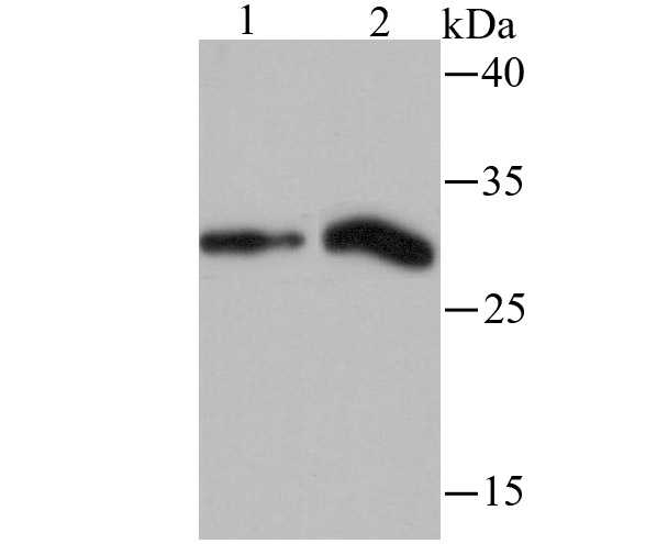
Western blot analysis of Histone H1.2 on Rat liver and Mouse lung tissue lysates using anti-Histone H1.2 antibody at 1/500 dilution.ICC/IF
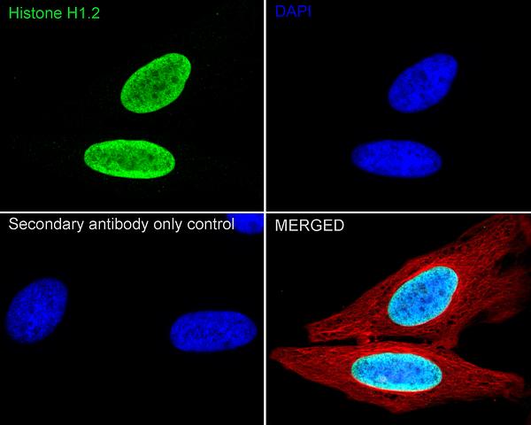
Immunocytochemistry analysis of HeLa cells labeling Histone H1.2 with Rabbit anti-Histone H1.2 antibody at 1/500 dilution. Cells were fixed in 4% paraformaldehyde for 20 minutes at room temperature, permeabilized with 0.1% Triton X-100 in PBS for 5 minutes at room temperature, then blocked with 1% BSA in 10% negative goat serum for 1 hour at room temperature. Cells were then incubated with Rabbit anti-Histone H1.2 antibody at 1/100 dilution in 1% BSA in PBST overnight at 4 ℃. Goat Anti-Rabbit IgG H&L (488) was used as the secondary antibody at 1/1,000 dilution. PBS instead of the primary antibody was used as the secondary antibody only control. Nuclear DNA was labelled in blue with DAPI. Beta tubulin (red) was stained at 1/100 dilution overnight at +4℃. Goat Anti-Mouse IgG H&L (594) was used as the secondary antibody at 1/1,000 dilution.ICC/IF
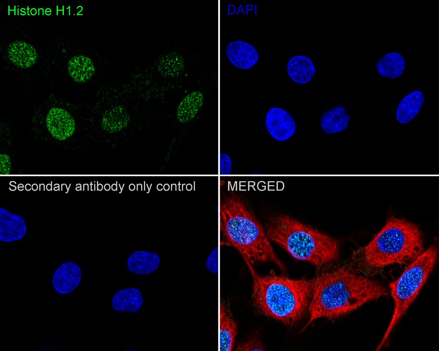
Immunocytochemistry analysis of C6 cells labeling Histone H1.2 with Rabbit anti-Histone H1.2 antibody at 1/100 dilution. Cells were fixed in 4% paraformaldehyde for 20 minutes at room temperature, permeabilized with 0.1% Triton X-100 in PBS for 5 minutes at room temperature, then blocked with 1% BSA in 10% negative goat serum for 1 hour at room temperature. Cells were then incubated with Rabbit anti-Histone H1.2 antibody at 1/100 dilution in 1% BSA in PBST overnight at 4 ℃. Goat Anti-Rabbit IgG H&L (488) was used as the secondary antibody at 1/1,000 dilution. PBS instead of the primary antibody was used as the secondary antibody only control. Nuclear DNA was labelled in blue with DAPI. Beta tubulin (red) was stained at 1/100 dilution overnight at +4℃. Goat Anti-Mouse IgG H&L (594) was used as the secondary antibody at 1/1,000 dilution.IHC
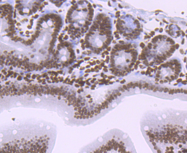
Immunohistochemical analysis of paraffin-embedded mouse colon tissue using anti-Histone H1.2 antibody. The section was pre-treated using heat mediated antigen retrieval with sodium citrate buffer (pH6) for 20 mins. Counter stained with hematoxylin.IHC
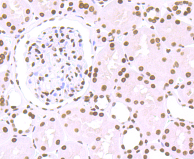
Immunohistochemical analysis of paraffin-embedded human kidney tissue using anti-Histone H1.2 antibody. The section was pre-treated using heat mediated antigen retrieval with sodium citrate buffer (pH6) for 20 mins. Counter stained with hematoxylin.IHC
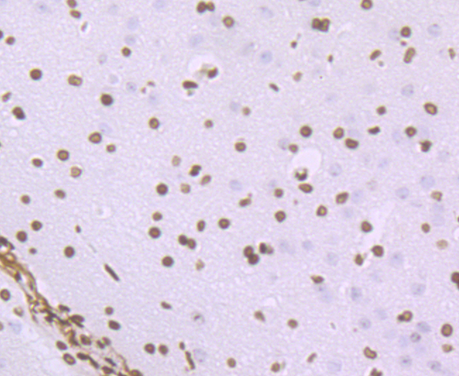
Immunohistochemical analysis of paraffin-embedded mouse colon tissue using anti-Histone H1.2 antibody. The section was pre-treated using heat mediated antigen retrieval with sodium citrate buffer (pH6) for 20 mins. Counter stained with hematoxylin.ChIP
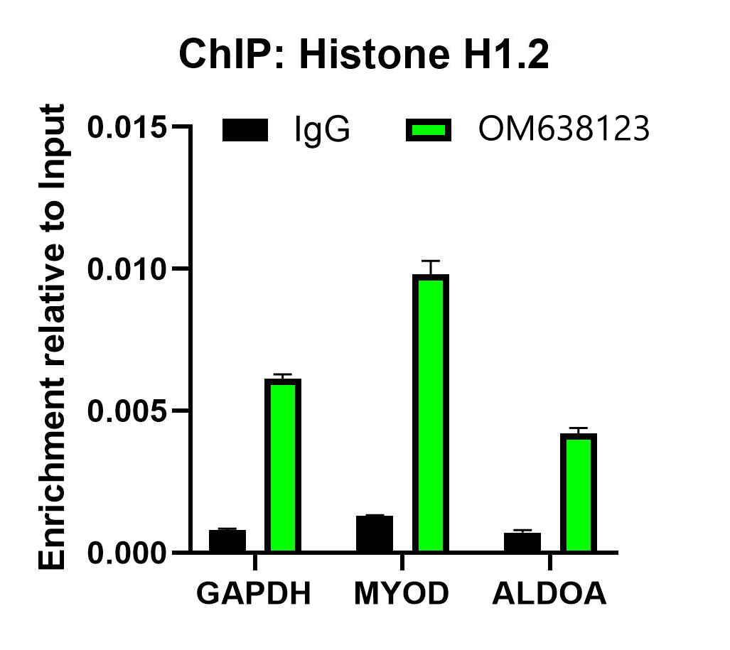
Chromatin immunoprecipitations were performed with cross-linked chromatin from HeLa cells with Histone H1.2 or Normal Rabbit IgG according to the ChIP protocol. The enriched DNA was quantified by real-time PCR using indicated primers. The amount of immunoprecipitated DNA in each sample is represented as signal relative to the total amount of input chromatin, which is equivalent to one.| Product Name | Histone H1.2 Recombinant Rabbit Monoclonal Antibody |
|---|---|
| Antibody Type | Primary Antibodies |
| Product description | Histone H1 protein binds to linker DNA between nucleosomes forming the macromolecular structure known as the chromatin fiber. Histones H1 are necessary for the condensation of nucleosome chains into higher-order structured fibers. Acts also as a regulator of individual gene transcription through chromatin remodeling, nucleosome spacing and DNA methylation. |
| Immunogen | Synthetic peptide within Human Histone H12 aa 1-50 / 213. |
| Clonality | Monoclonal |
|---|---|
| Isotype | IgG |
| Host Species | Recombinant rabbit |
| Tested Applications | ChIPICC/IFIHCWB |
| WB:1:1000-1:2000 IHC:1:50-1:200 ICC/IF:1:100-1:500 ChIP:Use 0.5~2 μg for 25 μg of chromatin. |
|
| Species Reactivity | HumanMouseRat |
| Concentration | 1mg/ml |
| Purification | Protein A |
| Alternative Names | H1 histone family member 2 antibody H1.a antibody H12_HUMAN antibody H1F2 antibody H1s-1 antibody HIST1H1C antibody Histone 1 H1c antibody Histone cluster 1 H1c antibody Histone H1.2 antibody Histone H1c antibody Histone H1d antibody Histone H1s-1 antibody MGC3992 antibody |
|---|---|
| Molecular Weight(MW) | 21kDa(Observed band size: 30kDa) |
| Cellular Localization | Nucleus. Chromosome. |
WB
Western blot analysis of Histone H1.2 on different lysates with Rabbit anti-Histone H1.2 antibody at 1/1,000 dilution. Lane 1: HAP1-parental cell lysate, Lane 2: HAP1-Histone H1.2 KD cell lysate, Lysates/proteins at 10 µg/Lane. Exposure time: 15 seconds; 4-20% SDS-PAGE gel. Proteins were transferred to a PVDF membrane and blocked with 5% NFDM/TBST for 1 hour at room temperature. The primary antibody at 1/1,000 dilution was used in K1803 at 4℃ overnight. Goat Anti-Rabbit IgG - HRP Secondary Antibody at 1/50,000 dilution was used for 1 hour at room temperature.
WB
Western blot analysis of Histone H1.2 on Rat liver and Mouse lung tissue lysates using anti-Histone H1.2 antibody at 1/500 dilution.
ICC/IF
Immunocytochemistry analysis of HeLa cells labeling Histone H1.2 with Rabbit anti-Histone H1.2 antibody at 1/500 dilution. Cells were fixed in 4% paraformaldehyde for 20 minutes at room temperature, permeabilized with 0.1% Triton X-100 in PBS for 5 minutes at room temperature, then blocked with 1% BSA in 10% negative goat serum for 1 hour at room temperature. Cells were then incubated with Rabbit anti-Histone H1.2 antibody at 1/100 dilution in 1% BSA in PBST overnight at 4 ℃. Goat Anti-Rabbit IgG H&L (488) was used as the secondary antibody at 1/1,000 dilution. PBS instead of the primary antibody was used as the secondary antibody only control. Nuclear DNA was labelled in blue with DAPI. Beta tubulin (red) was stained at 1/100 dilution overnight at +4℃. Goat Anti-Mouse IgG H&L (594) was used as the secondary antibody at 1/1,000 dilution.
ICC/IF
Immunocytochemistry analysis of C6 cells labeling Histone H1.2 with Rabbit anti-Histone H1.2 antibody at 1/100 dilution. Cells were fixed in 4% paraformaldehyde for 20 minutes at room temperature, permeabilized with 0.1% Triton X-100 in PBS for 5 minutes at room temperature, then blocked with 1% BSA in 10% negative goat serum for 1 hour at room temperature. Cells were then incubated with Rabbit anti-Histone H1.2 antibody at 1/100 dilution in 1% BSA in PBST overnight at 4 ℃. Goat Anti-Rabbit IgG H&L (488) was used as the secondary antibody at 1/1,000 dilution. PBS instead of the primary antibody was used as the secondary antibody only control. Nuclear DNA was labelled in blue with DAPI. Beta tubulin (red) was stained at 1/100 dilution overnight at +4℃. Goat Anti-Mouse IgG H&L (594) was used as the secondary antibody at 1/1,000 dilution.
IHC
Immunohistochemical analysis of paraffin-embedded mouse colon tissue using anti-Histone H1.2 antibody. The section was pre-treated using heat mediated antigen retrieval with sodium citrate buffer (pH6) for 20 mins. Counter stained with hematoxylin.
IHC
Immunohistochemical analysis of paraffin-embedded human kidney tissue using anti-Histone H1.2 antibody. The section was pre-treated using heat mediated antigen retrieval with sodium citrate buffer (pH6) for 20 mins. Counter stained with hematoxylin.
IHC
Immunohistochemical analysis of paraffin-embedded mouse colon tissue using anti-Histone H1.2 antibody. The section was pre-treated using heat mediated antigen retrieval with sodium citrate buffer (pH6) for 20 mins. Counter stained with hematoxylin.
ChIP
Chromatin immunoprecipitations were performed with cross-linked chromatin from HeLa cells with Histone H1.2 or Normal Rabbit IgG according to the ChIP protocol. The enriched DNA was quantified by real-time PCR using indicated primers. The amount of immunoprecipitated DNA in each sample is represented as signal relative to the total amount of input chromatin, which is equivalent to one.| Application Notes | WB:1:1000-1:2000 IHC:1:50-1:200 ICC/IF:1:100-1:500 ChIP:Use 0.5~2 μg for 25 μg of chromatin. |
|---|
| Form | Liquid |
|---|---|
| Storage Instructions | Store at +4℃ after thawing. Aliquot store at -20℃ or -80℃. Avoid repeated freeze / thaw cycles. |
| Storage Buffer | 1*TBS (pH7.4), 1%BSA, 40%Glycerol. Preservative: 0.05% Sodium Azide. |


