Application
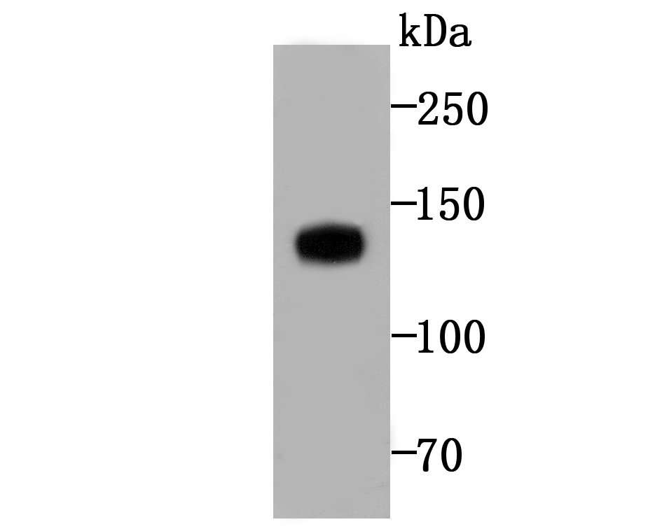
Fig1: Western blot analysis of Involucrin on human skin tissue lysate using Involucrin antibody at 1/500 dilution.Application
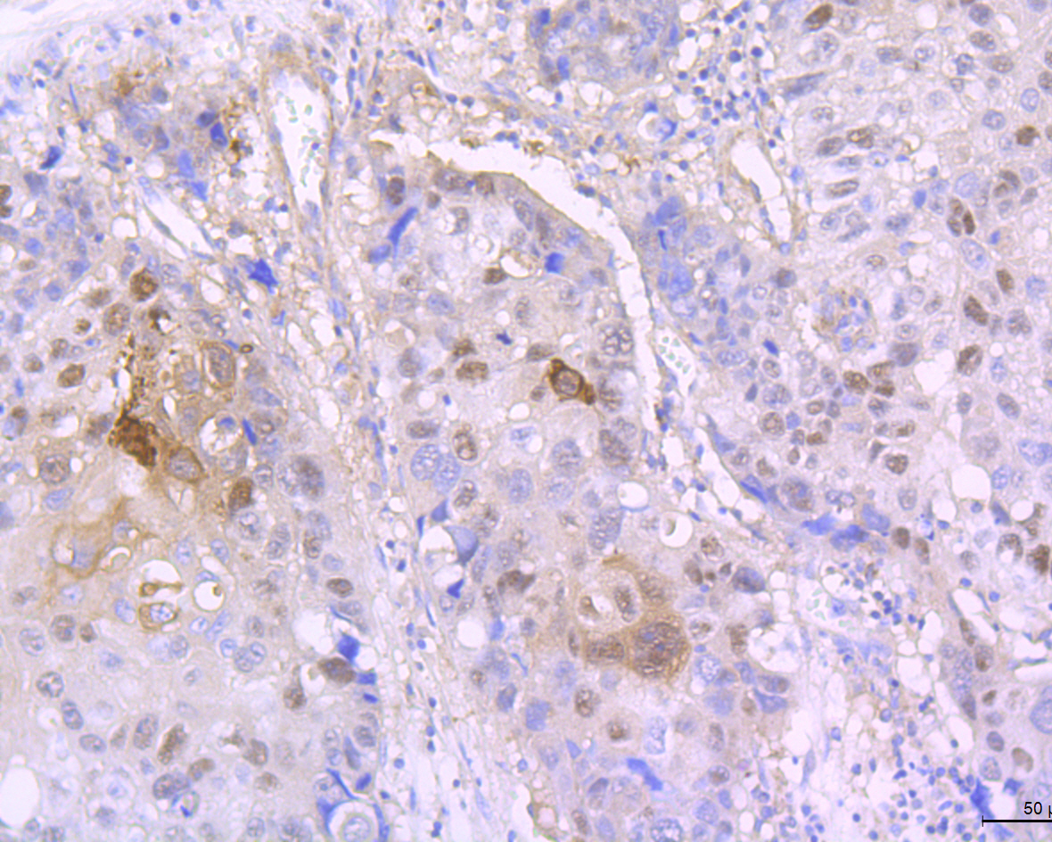
Fig2: Immunohistochemical analysis of paraffin-embedded human lung cancer tissue using anti-Involucrin antibody. Counter stained with hematoxylin.Application
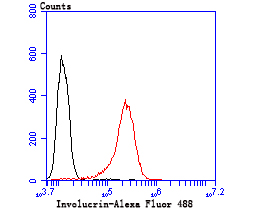
Fig3: Flow cytometric analysis of A431 cells with Involucrin antibody at 1/50 dilution (red) compared with an unlabelled control (cells without incubation with primary antibody; black). Alexa Fluor 488-conjugated goat anti-rabbit IgG was used as the secondary antibody.| Product Name | Involucrin [JU73-02] |
|---|---|
| Antibody Type | Primary Antibodies |
| Antigen Alias | INVO_HUMAN antibody Involucrin antibody IVL antibody |
| Product description | Involucrin is a precursor protein of the keratinocyte cornified envelope, which is formed beneath the inner surface of the cell membrane during terminal differentiation. During keratinocyte terminal differentiation, glutamine residues of involucrin become covalently cross-linked to other envelope precursors via covalent epsilon-(gamma-glutamyl) lysine bonds. Moreover, its large size allows involucrin to cross-link molecules that are separated by substantial distances in the cornified envelope. These properties allow a single involucrin molecule to form multiple cross-links, in multiple spatial planes, with other envelope precursors. |
| Immunogen | Recombinant protein |
| Clonality | Monoclonal |
|---|---|
| Isotype | IgG |
| Host Species | Recombinant rabbit |
| Tested Applications | WBIHCFC |
| WB:1:500-1:2,000 IHC:1:50-1:200 FC:1:50-1:200 |
|
| Species Reactivity | HumanRat |
| Concentration | 1mg/ml |
| Alternative Names | INVO_HUMAN antibody Involucrin antibody IVL antibody |
|---|---|
| Molecular Weight(MW) | 120 kDa |
| Cellular Localization | Cytoplasm. |
Application
Fig1: Western blot analysis of Involucrin on human skin tissue lysate using Involucrin antibody at 1/500 dilution.
Application
Fig2: Immunohistochemical analysis of paraffin-embedded human lung cancer tissue using anti-Involucrin antibody. Counter stained with hematoxylin.
Application
Fig3: Flow cytometric analysis of A431 cells with Involucrin antibody at 1/50 dilution (red) compared with an unlabelled control (cells without incubation with primary antibody; black). Alexa Fluor 488-conjugated goat anti-rabbit IgG was used as the secondary antibody.| Positive Control | Human skin tissue lysate, human lung cancer tissue, A431 cell. |
|---|---|
| Application Notes | WB:1:500-1:2,000 IHC:1:50-1:200 FC:1:50-1:200 |
| Form | Liquid |
|---|---|
| Storage Instructions | Store at +4℃ after thawing. Aliquot store at -20℃ or -80℃. Avoid repeated freeze / thaw cycles. |
| Storage Buffer | 1*TBS (pH7.4), 1%BSA, 40%Glycerol. Preservative: 0.05% Sodium Azide. |


