Application
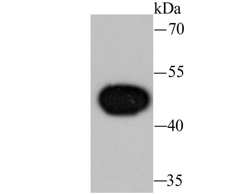
Fig1: Western blot analysis of Wnt5a on Hela cell lysate using anti-Wnt5a antibody at 1/1,000 dilution.Application
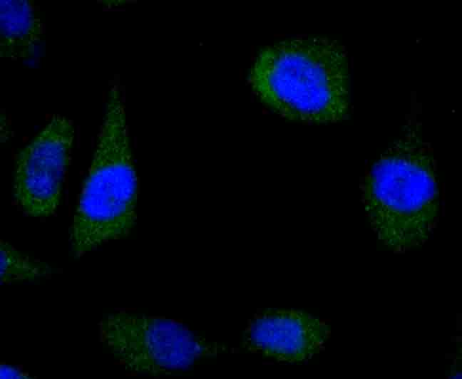
Fig2: ICC staining Wnt5a in PC-3M cells (green). The nuclear counter stain is DAPI (blue). Cells were fixed in paraformaldehyde, permeabilised with 0.25% Triton X100/PBS.Application
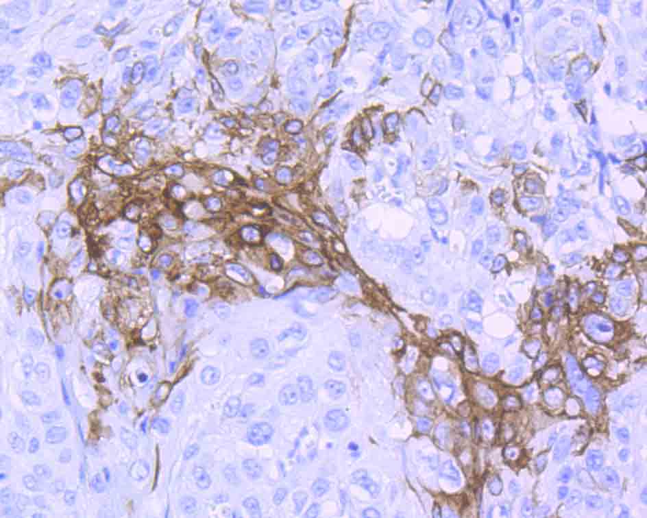
Fig3: Immunohistochemical analysis of paraffin-embedded human lung cancer tissue using anti-Wnt5a antibody. Counter stained with hematoxylin.Application
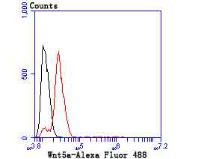
Fig4: Flow cytometric analysis of SH-SY5Y cells with Wnt5a antibody at 1/50 dilution (red) compared with an unlabelled control (cells without incubation with primary antibody; black). Alexa Fluor 488-conjugated goat anti rabbit IgG was used as the secondary antibody.| Product Name | Wnt5a [JU30-30] |
|---|---|
| Antibody Type | Primary Antibodies |
| Antigen Alias | hWNT 5A antibody hWNT5A antibody Protein Wnt 5a antibody Protein Wnt-5a antibody Protein Wnt5a antibody Wingless type MMTV integration site family member 5A antibody Wnt 5a antibody WNT 5A protein antibody WNT 5A protein precursor antibody WNT5A antibody WNT5A protein precursor antibody WNT5A_HUMAN antibody |
| Product description | The Wnt genes belong to a family of protooncogenes with at least 13 known members that are expressed in species ranging from Drosophila to man. The name Wnt denotes the relationship of this family to the Drosophila segment polarity gene "wingless" and to its vertebrate ortholog, Int1, a mouse protooncogene. Transcription of Wnt family genes appears to be developmentally regulated in a precise temporal and spatial manner. The Wnt genes encode cysteine-rich putative glycoproteins, which have features typical of secreted growth factors. Northern blot analysis detects expression of Wnt-5a in brain, lung, and heart. At least five distinct Wnt-5a transcripts are observed, which are due to transcript variability 5-prime to the initiation methionine. In situ hybridization detects a complex spatial and temporal pattern of Wnt-5a in the mouse, including expression in the developing central nervous system, limbs, facial processes, and the posterior region of the fetus. Human frizzled-5 is the receptor for the Wnt-5a ligand. It is suggested that Wnt-5a augments primitive hematopoietic development in vivo and represents an in vivo regulator of hematopoietic stem cell function in the human. |
| Immunogen | Recombinant protein |
| Clonality | Monoclonal |
|---|---|
| Isotype | IgG |
| Host Species | Recombinant rabbit |
| Tested Applications | WBICCIHCFC |
| WB:1:500-1:2,000 IHC:1:50-1:200 ICC:1:50 FC:1:50-1:100: |
|
| Species Reactivity | Human |
| Concentration | 1mg/ml |
| Alternative Names | hWNT 5A antibody hWNT5A antibody Protein Wnt 5a antibody Protein Wnt-5a antibody Protein Wnt5a antibody Wingless type MMTV integration site family member 5A antibody Wnt 5a antibody WNT 5A protein antibody WNT 5A protein precursor antibody WNT5A antibody WNT5A protein precursor antibody WNT5A_HUMAN antibody |
|---|---|
| Molecular Weight(MW) | 42 kDa |
| Cellular Localization | Secreted, extracellular space, extracellular matrix. |
| SwissProt ID | P41221 |
|---|
Application
Fig1: Western blot analysis of Wnt5a on Hela cell lysate using anti-Wnt5a antibody at 1/1,000 dilution.
Application
Fig2: ICC staining Wnt5a in PC-3M cells (green). The nuclear counter stain is DAPI (blue). Cells were fixed in paraformaldehyde, permeabilised with 0.25% Triton X100/PBS.
Application
Fig3: Immunohistochemical analysis of paraffin-embedded human lung cancer tissue using anti-Wnt5a antibody. Counter stained with hematoxylin.
Application
Fig4: Flow cytometric analysis of SH-SY5Y cells with Wnt5a antibody at 1/50 dilution (red) compared with an unlabelled control (cells without incubation with primary antibody; black). Alexa Fluor 488-conjugated goat anti rabbit IgG was used as the secondary antibody.| Positive Control | Hela, PC-3M, human lung cancer tissue, SH-SY5Y. |
|---|---|
| Application Notes | WB:1:500-1:2,000 IHC:1:50-1:200 ICC:1:50 FC:1:50-1:100: |
| Form | Liquid |
|---|---|
| Storage Instructions | Store at +4℃ after thawing. Aliquot store at -20℃ or -80℃. Avoid repeated freeze / thaw cycles. |
| Storage Buffer | 1*TBS (pH7.4), 1%BSA, 40%Glycerol. Preservative: 0.05% Sodium Azide. |


