Application
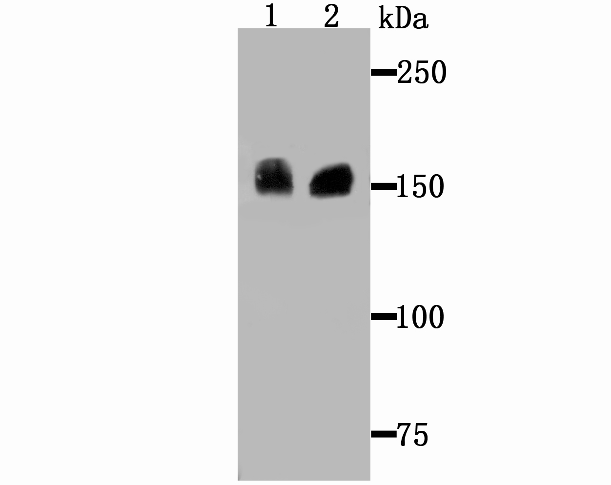
Fig1: Western blot analysis of Drosha on Hela cell and SiHa cell lysates using anti-Drosha antibody at 1/500 dilution.Application
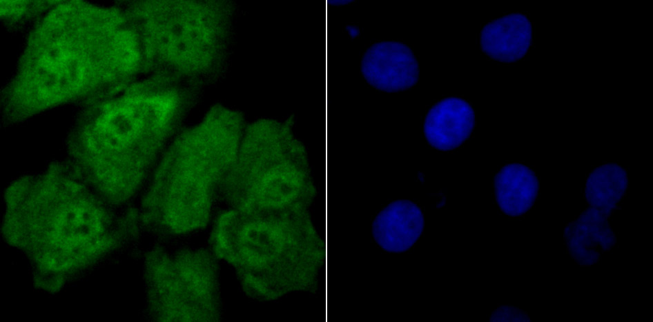
Fig2: ICC staining Drosha in A549 cells (green). The nuclear counter stain is DAPI (blue). Cells were fixed in paraformaldehyde, permeabilised with 0.25% Triton X100/PBS.Application
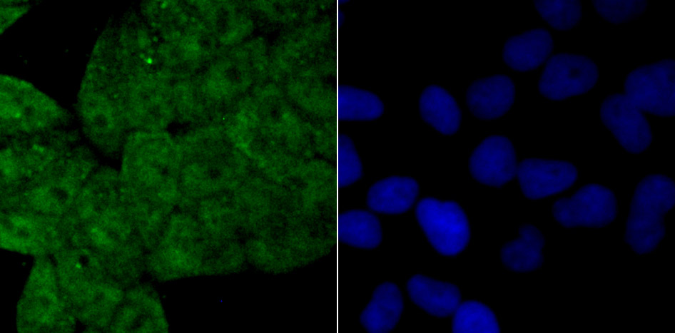
Fig3: ICC staining Drosha in Hela cells (green). The nuclear counter stain is DAPI (blue). Cells were fixed in paraformaldehyde, permeabilised with 0.25% Triton X100/PBS.Application
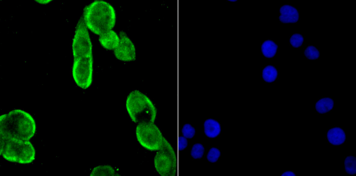
Fig4: ICC staining Drosha in LOVO cells (green). The nuclear counter stain is DAPI (blue). Cells were fixed in paraformaldehyde, permeabilised with 0.25% Triton X100/PBS.Application
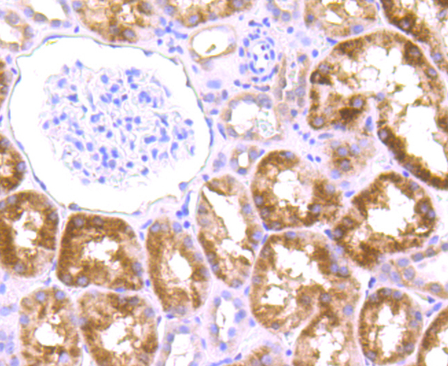
Fig5: Immunohistochemical analysis of paraffin-embedded human kidney tissue using anti-Drosha antibody. Counter stained with hematoxylin.Application
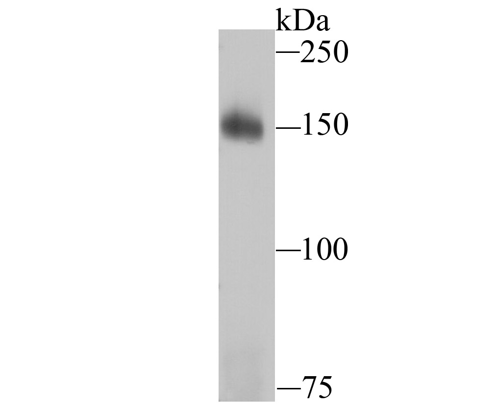
Fig6: Western blot analysis of Drosha on K562 cell lysates using anti-Drosha antibody at 1/1,000 dilution.Application
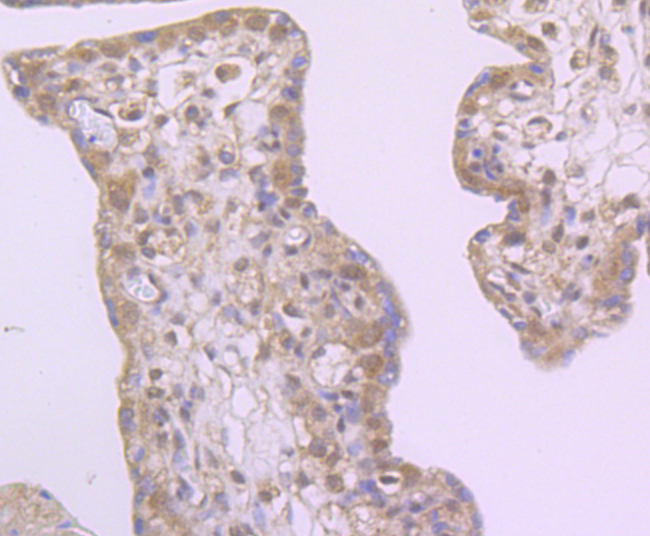
Fig7: Immunohistochemical analysis of paraffin-embedded human placenta tissue using anti-Drosha antibody. Counter stained with hematoxylin.Application
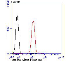
Fig8: Flow cytometric analysis of Jurkat cells with Drosha antibody at 1/100 dilution (red) compared with an unlabelled control (cells without incubation with primary antibody; black).| Product Name | Drosha [JU33-01] |
|---|---|
| Antibody Type | Primary Antibodies |
| Antigen Alias | DROSHA antibody Drosha double stranded RNA specific endoribonuclease antibody Drosha ribonuclease type III antibody Etohi2 antibody HSA242976 antibody Nuclear RNase III Drosha antibody p241 antibody Protein Drosha antibody Putative protein p241 which interacts with transcription factor Sp1 antibody Putative ribonuclease III antibody RANSE3L antibody Ribonuclease 3 antibody Ribonuclease III antibody Ribonuclease III nuclear antibody Ribonuclease type III nuclear antibody RibonucleaseIII antibody RN 3 antibody RN3 antibody RNase 3 antibody RNase III antibody RNase3 antibody RNASE3L antibody RNaseIII antibody RNASEN antibody RNC_HUMAN antibody |
| Product description | The ribonuclease III superfamily represents a structurally distinct group of double-strand-specific endonucleases with essential roles in RNA maturation, RNA decay, and gene silencing. Initial cleavage of microRNAs is catalysed by Drosha, a nuclease of the RNase III family, which acts on primary miRNA transcripts (pri-miRNAs) in the nucleus. Human Drosha is a component of two multi-protein complexes. The larger complex contains multiple classes of RNA-associated proteins including RNA helicases, proteins that bind double-stranded RNA, novel heterogeneous nuclear ribonucleoproteins and the Ewing's sarcoma family of proteins. The smaller complex is composed of Drosha and the double-stranded-RNA-binding protein, DGCR8. |
| Immunogen | Recombinant protein |
| Clonality | Monoclonal |
|---|---|
| Isotype | IgG |
| Host Species | Recombinant rabbit |
| Tested Applications | WBICCIFIHCFC |
| WB:1:500-1:1,000 ICC:1:50-1:200 IHC:1:50-1:200 FC:1:50-1:100 |
|
| Species Reactivity | Human |
| Concentration | 1mg/ml |
| Alternative Names | DROSHA antibody Drosha double stranded RNA specific endoribonuclease antibody Drosha ribonuclease type III antibody Etohi2 antibody HSA242976 antibody Nuclear RNase III Drosha antibody p241 antibody Protein Drosha antibody Putative protein p241 which interacts with transcription factor Sp1 antibody Putative ribonuclease III antibody RANSE3L antibody Ribonuclease 3 antibody Ribonuclease III antibody Ribonuclease III nuclear antibody Ribonuclease type III nuclear antibody RibonucleaseIII antibody RN 3 antibody RN3 antibody RNase 3 antibody RNase III antibody RNase3 antibody RNASE3L antibody RNaseIII antibody RNASEN antibody RNC_HUMAN antibody |
|---|---|
| Molecular Weight(MW) | 159 kDa |
| Cellular Localization | Nucleus. |
| SwissProt ID | Q9NRR4 |
|---|
Application
Fig1: Western blot analysis of Drosha on Hela cell and SiHa cell lysates using anti-Drosha antibody at 1/500 dilution.
Application
Fig2: ICC staining Drosha in A549 cells (green). The nuclear counter stain is DAPI (blue). Cells were fixed in paraformaldehyde, permeabilised with 0.25% Triton X100/PBS.
Application
Fig3: ICC staining Drosha in Hela cells (green). The nuclear counter stain is DAPI (blue). Cells were fixed in paraformaldehyde, permeabilised with 0.25% Triton X100/PBS.
Application
Fig4: ICC staining Drosha in LOVO cells (green). The nuclear counter stain is DAPI (blue). Cells were fixed in paraformaldehyde, permeabilised with 0.25% Triton X100/PBS.
Application
Fig5: Immunohistochemical analysis of paraffin-embedded human kidney tissue using anti-Drosha antibody. Counter stained with hematoxylin.
Application
Fig6: Western blot analysis of Drosha on K562 cell lysates using anti-Drosha antibody at 1/1,000 dilution.
Application
Fig7: Immunohistochemical analysis of paraffin-embedded human placenta tissue using anti-Drosha antibody. Counter stained with hematoxylin.
Application
Fig8: Flow cytometric analysis of Jurkat cells with Drosha antibody at 1/100 dilution (red) compared with an unlabelled control (cells without incubation with primary antibody; black).| Positive Control | Hela, SiHa, K562, A549, LOVO, Jurkat, human kidney tissue, human placenta tissue. |
|---|---|
| Application Notes | WB:1:500-1:1,000 ICC:1:50-1:200 IHC:1:50-1:200 FC:1:50-1:100 |
| Form | Liquid |
|---|---|
| Storage Instructions | Store at +4℃ after thawing. Aliquot store at -20℃ or -80℃. Avoid repeated freeze / thaw cycles. |
| Storage Buffer | 1*TBS (pH7.4), 1%BSA, 40%Glycerol. Preservative: 0.05% Sodium Azide. |


