Application
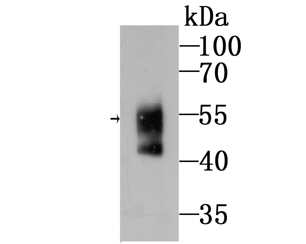
Fig1: Western blot analysis of WASL on mouse Human serum lysates using anti-Ubiquitin antibody at 1/500 dilution.Application
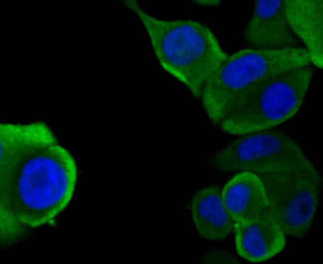
Fig2: ICC staining WASL in SK-Br-3 cells (green). The nuclear counter stain is DAPI (blue). Cells were fixed in paraformaldehyde, permeabilised with 0.25% Triton X100/PBS.Application
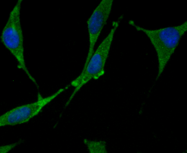
Fig3: ICC staining WASL in SH-SY5Y cells (green). The nuclear counter stain is DAPI (blue). Cells were fixed in paraformaldehyde, permeabilised with 0.25% Triton X100/PBS.Application
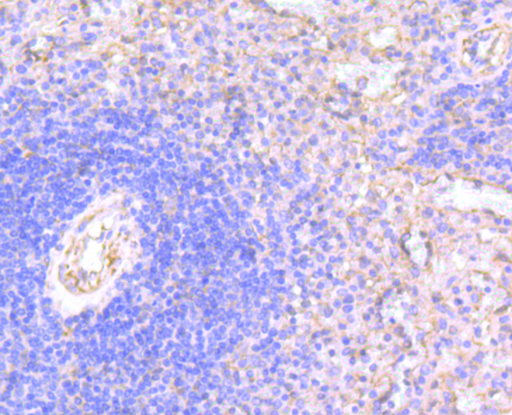
Fig4: Immunohistochemical analysis of paraffin-embedded human spleen tissue using anti-WASL antibody. Counter stained with hematoxylin.Application
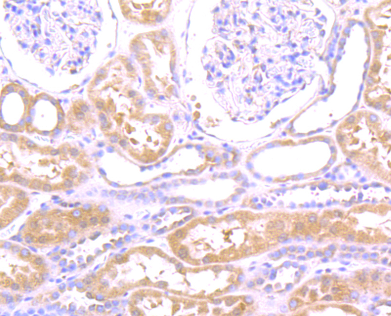
Fig5: Immunohistochemical analysis of paraffin-embedded human kidney tissue using anti-WASL antibody. Counter stained with hematoxylin.Application
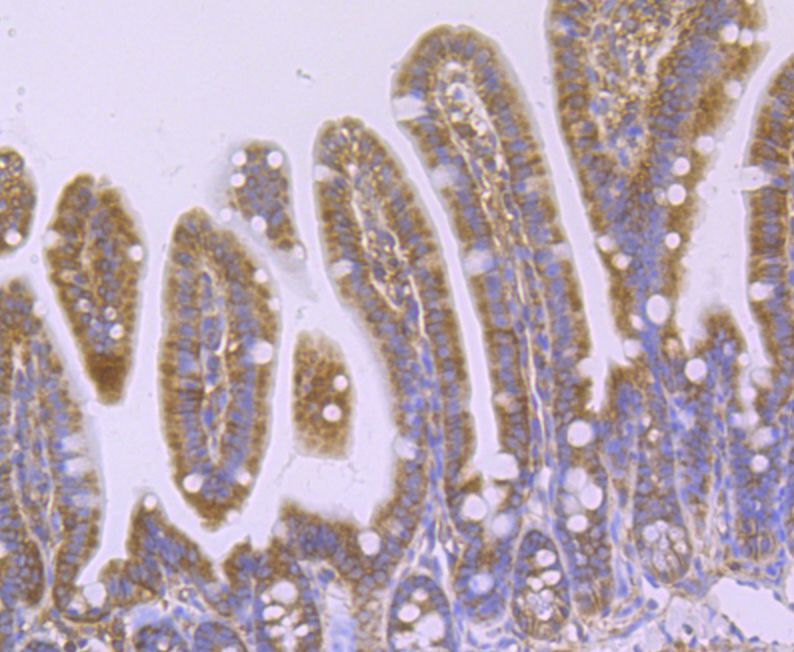
Fig6: Immunohistochemical analysis of paraffin-embedded mouse colon tissue using anti-WASL antibody. Counter stained with hematoxylin.Application
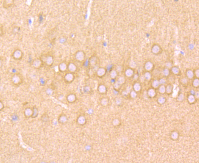
Fig7: Immunohistochemical analysis of paraffin-embedded mouse brain tissue using anti-WASL antibody. Counter stained with hematoxylin.Application
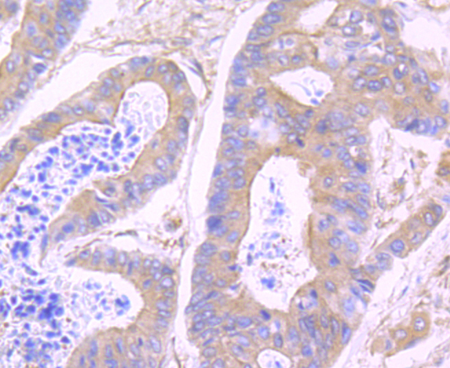
Fig8: Immunohistochemical analysis of paraffin-embedded human colon cancer tissue using anti-WASL antibody. Counter stained with hematoxylin.Application
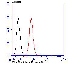
Fig9: Flow cytometric analysis of K562 cells with WASL antibody at 1/100 dilution (red) compared with an unlabelled control (cells without incubation with primary antibody; black).| Product Name | WASL [JU07-31] |
|---|---|
| Antibody Type | Primary Antibodies |
| Antigen Alias | WASP family, verprolin homology domain-containing protein 1 antibody WASP family member 1 antibody FLJ31482 antibody homology of dictyostelium scar 1 antibody KIAA0269 antibody Protein WAVE-1 antibodyv Protein WAVE1 antibody scar, Dictyostelium, homology of, 1 antibody SCAR1 antibody Similar to a plant extensin like protein antibody Verprolin homology domain containing protein 1 antibody Verprolin homology domain-containing protein 1 antibody WAS protein family, member 1 antibody Wasf1 antibody WASF1_HUMAN antibody WASL antibody WASP family 1 antibody WASP family protein member 1 antibody WASP family, verprolin homology domain-containing protein antibody WAVE antibody WAVE1 antibody Wiskott Aldrich syndrome protein family member 1 antibody Wiskott-Aldrich syndrome protein family member 1 antibody |
| Product description | This gene encodes a member of the Wiskott-Aldrich syndrome (WAS) protein family. Wiskott-Aldrich syndrome proteins share similar domain structure, and associate with a variety of signaling molecules to alter the actin cytoskeleton. The encoded protein is highly expressed in neural tissues, and interacts with several proteins involved in cytoskeletal organization, including cell division control protein 42 (CDC42) and the actin-related protein-2/3 (ARP2/3) complex. The encoded protein may be involved in the formation of long actin microspikes, and in neurite extension. Regulates actin polymerization by stimulating the actin-nucleating activity of the Arp2/3 complex. Involved in mitosis and cytokinesis, via its role in the regulation of actin polymerization. Binds to HSF1/HSTF1 and forms a complex on heat shock promoter elements (HSE) that negatively regulates HSP90 expression.. |
| Immunogen | Recombinant protein |
| Clonality | Monoclonal |
|---|---|
| Isotype | IgG |
| Host Species | Recombinant rabbit |
| Tested Applications | WBICCIHCFC |
| WB:1:500-1:2,000 ICC:1:50-1:200 IHC:1:50-1:200 FC:1:50-1:100 |
|
| Species Reactivity | HumanMouse |
| Concentration | 1mg/ml |
| Alternative Names | WASP family verprolin homology domain-containing protein 1 antibody WASP family member 1 antibody FLJ31482 antibody homology of dictyostelium scar 1 antibody KIAA0269 antibody Protein WAVE-1 antibodyv Protein WAVE1 antibody scar Dictyostelium homology of 1 antibody SCAR1 antibody Similar to a plant extensin like protein antibody Verprolin homology domain containing protein 1 antibody Verprolin homology domain-containing protein 1 antibody WAS protein family member 1 antibody Wasf1 antibody WASF1_HUMAN antibody WASL antibody WASP family 1 antibody WASP family protein member 1 antibody WASP family verprolin homology domain-containing protein antibody WAVE antibody WAVE1 antibody Wiskott Aldrich syndrome protein family member 1 antibody Wiskott-Aldrich syndrome protein family member 1 antibody |
|---|---|
| Molecular Weight(MW) | 55 kDa |
| Cellular Localization | Cytoplasm, cytoskeleton. Nucleus. |
Application
Fig1: Western blot analysis of WASL on mouse Human serum lysates using anti-Ubiquitin antibody at 1/500 dilution.
Application
Fig2: ICC staining WASL in SK-Br-3 cells (green). The nuclear counter stain is DAPI (blue). Cells were fixed in paraformaldehyde, permeabilised with 0.25% Triton X100/PBS.
Application
Fig3: ICC staining WASL in SH-SY5Y cells (green). The nuclear counter stain is DAPI (blue). Cells were fixed in paraformaldehyde, permeabilised with 0.25% Triton X100/PBS.
Application
Fig4: Immunohistochemical analysis of paraffin-embedded human spleen tissue using anti-WASL antibody. Counter stained with hematoxylin.
Application
Fig5: Immunohistochemical analysis of paraffin-embedded human kidney tissue using anti-WASL antibody. Counter stained with hematoxylin.
Application
Fig6: Immunohistochemical analysis of paraffin-embedded mouse colon tissue using anti-WASL antibody. Counter stained with hematoxylin.
Application
Fig7: Immunohistochemical analysis of paraffin-embedded mouse brain tissue using anti-WASL antibody. Counter stained with hematoxylin.
Application
Fig8: Immunohistochemical analysis of paraffin-embedded human colon cancer tissue using anti-WASL antibody. Counter stained with hematoxylin.
Application
Fig9: Flow cytometric analysis of K562 cells with WASL antibody at 1/100 dilution (red) compared with an unlabelled control (cells without incubation with primary antibody; black).| Positive Control | Human serum, SH-SY5Y, SK-Br-3, human colon cancer tissue, human kidney tissue, human spleen tissue, mouse colon tissue, mouse brain tissue, K562. |
|---|---|
| Application Notes | WB:1:500-1:2,000 ICC:1:50-1:200 IHC:1:50-1:200 FC:1:50-1:100 |
| Form | Liquid |
|---|---|
| Storage Instructions | Store at +4℃ after thawing. Aliquot store at -20℃ or -80℃. Avoid repeated freeze / thaw cycles. |
| Storage Buffer | 1*TBS (pH7.4), 1%BSA, 40%Glycerol. Preservative: 0.05% Sodium Azide. |


