Application
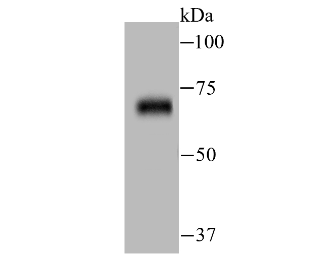
Fig1: Western blot analysis of ZAP70 on human thymus tissue lysate using anti-ZAP70 antibody at 1/500 dilution.Application
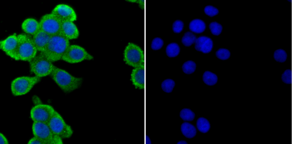
Fig2: ICC staining ZAP70 in LOVO cells (green). The nuclear counter stain is DAPI (blue). Cells were fixed in paraformaldehyde, permeabilised with 0.25% Triton X100/PBS.Application
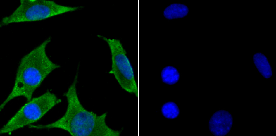
Fig3: ICC staining ZAP70 in SH-SY5Y cells (green). The nuclear counter stain is DAPI (blue). Cells were fixed in paraformaldehyde, permeabilised with 0.25% Triton X100/PBS.Application
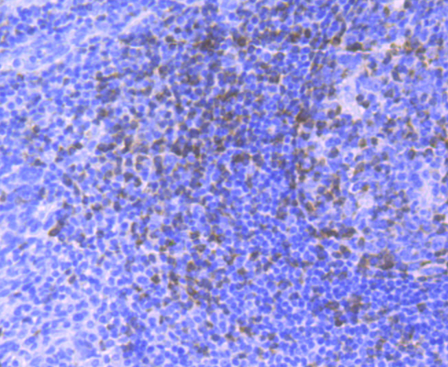
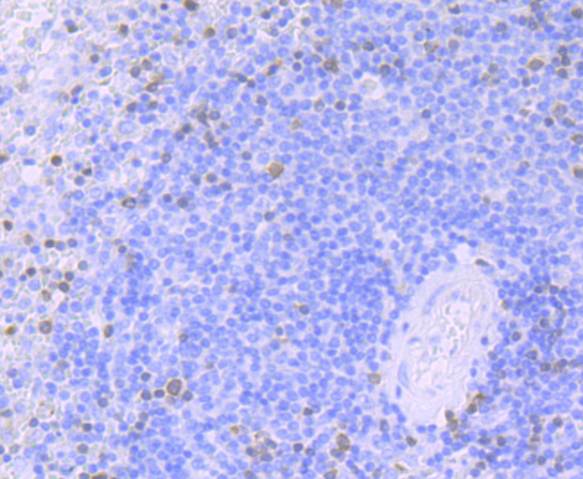
Fig4: Immunohistochemical analysis of paraffin-embedded human tonsil tissue using anti-ZAP70 antibody. Counter stained with hematoxylin.Application
Fig5: Immunohistochemical analysis of paraffin-embedded human spleen tissue using anti-ZAP70 antibody. Counter stained with hematoxylin.Application
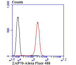
Fig6: Flow cytometric analysis of Jurkat cells with ZAP70 antibody at 1/100 dilution (red) compared with an unlabelled control (cells without incubation with primary antibody; black). Alexa Fluor® 488-conjugated goat anti-rabbit IgG was used as the secondary antibody.| Product Name | ZAP70 [JU08-39] |
|---|---|
| Antibody Type | Primary Antibodies |
| Antigen Alias | 70 kDa zeta associated protein antibody 70 kDa zeta-associated protein antibody EC 2.7.10.2 antibody FLJ17670 antibody FLJ17679 antibody Selective T cell defect antibody SRK antibody STD antibody Syk related tyrosine kinase antibody Syk-related tyrosine kinase antibody Truncated ZAP kinase antibody Tyrosine protein kinase ZAP70 antibody Tyrosine-protein kinase ZAP-70 antibody TZK antibody ZAP 70 antibody ZAP70 antibody ZAP70_HUMAN antibody Zeta chain associated protein kinase 70kD antibody Zeta chain associated protein kinase 70kDa antibody Zeta chain associated protein kinase 70kDa isoform 1 antibody Zeta chain associated protein kinase 70kDa isoform 2 antibody Zeta chain of T cell receptor associated protein kinase 70 antibody Zeta chain TCR associated protein kinase 70kD antibody Zeta chain TCR associated protein kinase 70kDa antibody |
| Product description | The activation of T lymphocytes by antigens is mediated by the T cell receptor (TCR) which is a multisubunit complex assembled from at least six different genes. The TCR subunits include the Ti α and β chains, the CD3 γ, δ and e chains and a ζ-containing homodimer or heterodimer. The disulfide-linked Ti α-β heterodimer is responsible for antigen recognition, but the short 5 amino acid cytoplasmic domains of Ti α and β are unlikely to be sufficient to couple to intracellular signaling pathways. In contrast, the structured features of the CD3 and ζ subunits suggest a role in signal transduction. Of these, the ζ chain, which is expressed as either a homodimer or heterodimer, has a short extracellular domain of only 9 amino acids, but a larger 113 amino acid cytoplasmic domain. A tyrosine phosphoprotein, ZAP-70, has been identified that associates with z and undergoes tyrosine phosphorylation following TCR stimulation. |
| Immunogen | Recombinant protein |
| Clonality | Monoclonal |
|---|---|
| Isotype | IgG |
| Host Species | Recombinant rabbit |
| Tested Applications | WBICCIHCFCIP |
| WB:1:500-1:1,000 ICC:1:50-1:200 IHC:1:50-1:200 FC:1:50-1:100 IP:1:10-1:50 |
|
| Species Reactivity | Human |
| Concentration | 1mg/ml |
| Alternative Names | 70 kDa zeta associated protein antibody 70 kDa zeta-associated protein antibody EC 2.7.10.2 antibody FLJ17670 antibody FLJ17679 antibody Selective T cell defect antibody SRK antibody STD antibody Syk related tyrosine kinase antibody Syk-related tyrosine kinase antibody Truncated ZAP kinase antibody Tyrosine protein kinase ZAP70 antibody Tyrosine-protein kinase ZAP-70 antibody TZK antibody ZAP 70 antibody ZAP70 antibody ZAP70_HUMAN antibody Zeta chain associated protein kinase 70kD antibody Zeta chain associated protein kinase 70kDa antibody Zeta chain associated protein kinase 70kDa isoform 1 antibody Zeta chain associated protein kinase 70kDa isoform 2 antibody Zeta chain of T cell receptor associated protein kinase 70 antibody Zeta chain TCR associated protein kinase 70kD antibody Zeta chain TCR associated protein kinase 70kDa antibody |
|---|---|
| Molecular Weight(MW) | 70 kDa |
| Cellular Localization | Cytoplasm. Cell membrane. |
| SwissProt ID | P43403 |
|---|
Application
Fig1: Western blot analysis of ZAP70 on human thymus tissue lysate using anti-ZAP70 antibody at 1/500 dilution.
Application
Fig2: ICC staining ZAP70 in LOVO cells (green). The nuclear counter stain is DAPI (blue). Cells were fixed in paraformaldehyde, permeabilised with 0.25% Triton X100/PBS.
Application
Fig3: ICC staining ZAP70 in SH-SY5Y cells (green). The nuclear counter stain is DAPI (blue). Cells were fixed in paraformaldehyde, permeabilised with 0.25% Triton X100/PBS.
Application
Fig4: Immunohistochemical analysis of paraffin-embedded human tonsil tissue using anti-ZAP70 antibody. Counter stained with hematoxylin.
Application
Fig5: Immunohistochemical analysis of paraffin-embedded human spleen tissue using anti-ZAP70 antibody. Counter stained with hematoxylin.
Application
Fig6: Flow cytometric analysis of Jurkat cells with ZAP70 antibody at 1/100 dilution (red) compared with an unlabelled control (cells without incubation with primary antibody; black). Alexa Fluor® 488-conjugated goat anti-rabbit IgG was used as the secondary antibody.| Positive Control | Human thymus tissue lysate, LOVO, SH-SY5Y, human tonsil tissue, human spleen tissue, Jurkat. |
|---|---|
| Application Notes | WB:1:500-1:1,000 ICC:1:50-1:200 IHC:1:50-1:200 FC:1:50-1:100 IP:1:10-1:50 |
| Form | Liquid |
|---|---|
| Storage Instructions | Store at +4℃ after thawing. Aliquot store at -20℃ or -80℃. Avoid repeated freeze / thaw cycles. |
| Storage Buffer | 1*TBS (pH7.4), 1%BSA, 40%Glycerol. Preservative: 0.05% Sodium Azide. |


