Application
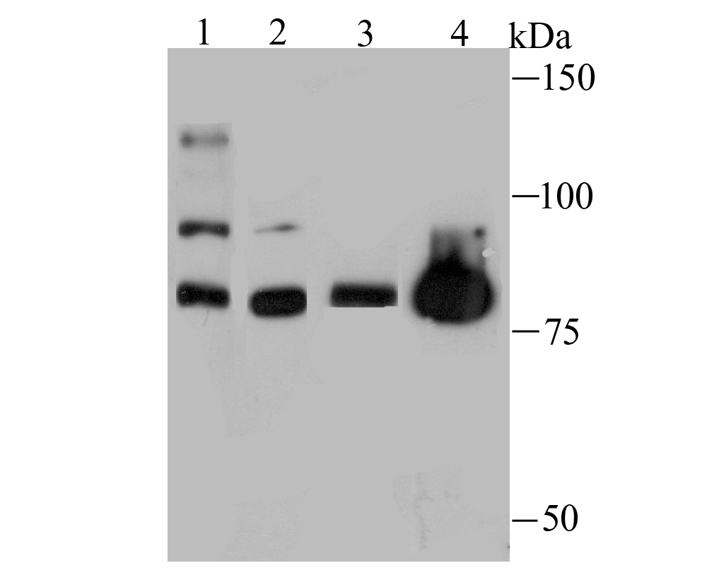
Fig1: Western blot analysis of Nuclear Matrix Protein p84 on different lysates using anti-Nuclear Matrix Protein p84 antibody at 1/500 dilution. Positive control: Lane 1: Hela Lane 2: A431 Lane 3: Mouse skeletal muscle Lane 4: PC-12Application
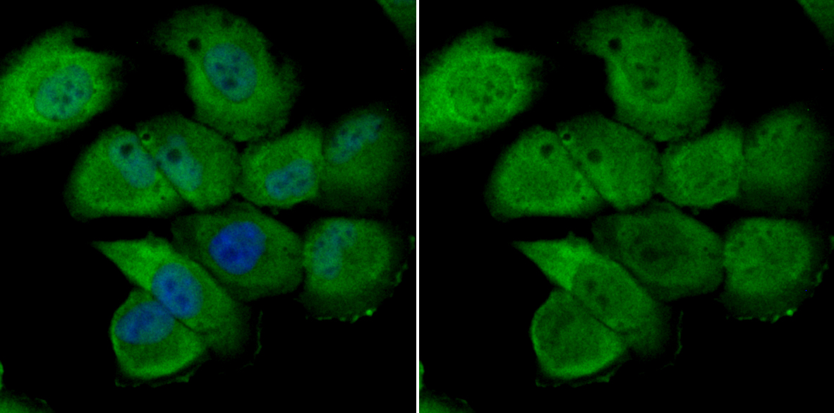
Fig2: ICC staining Nuclear Matrix Protein p84 in HUVEC cells (green). The nuclear counter stain is DAPI (blue). Cells were fixed in paraformaldehyde, permeabilised with 0.25% Triton X100/PBS.Application
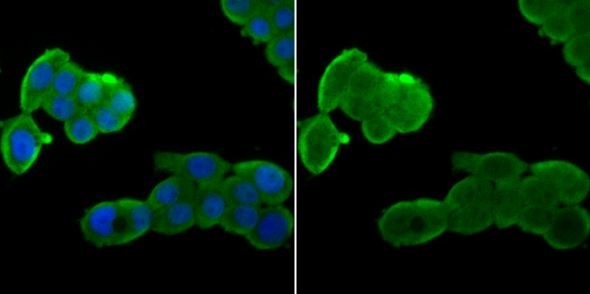
Fig3: ICC staining Nuclear Matrix Protein p84 in LOVO cells (green). The nuclear counter stain is DAPI (blue). Cells were fixed in paraformaldehyde, permeabilised with 0.25% Triton X100/PBS.Application
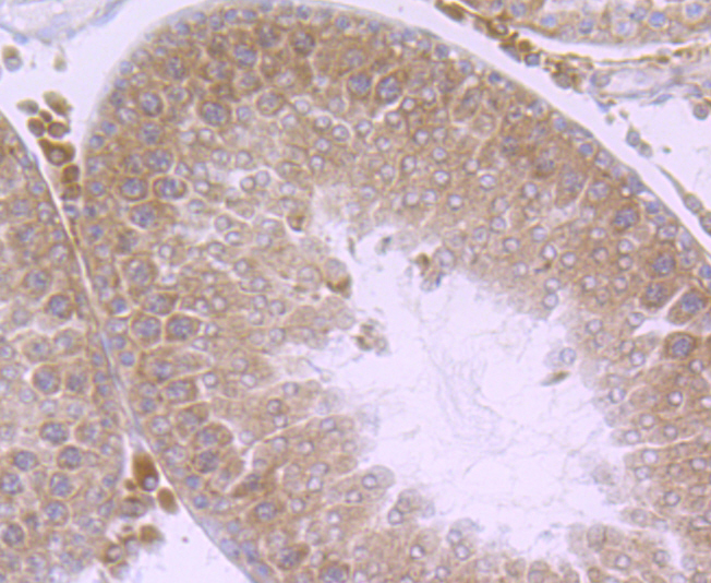
Fig4: Immunohistochemical analysis of paraffin-embedded rat testis tissue using anti-Nuclear Matrix Protein p84 antibody. Counter stained with hematoxylin.Application
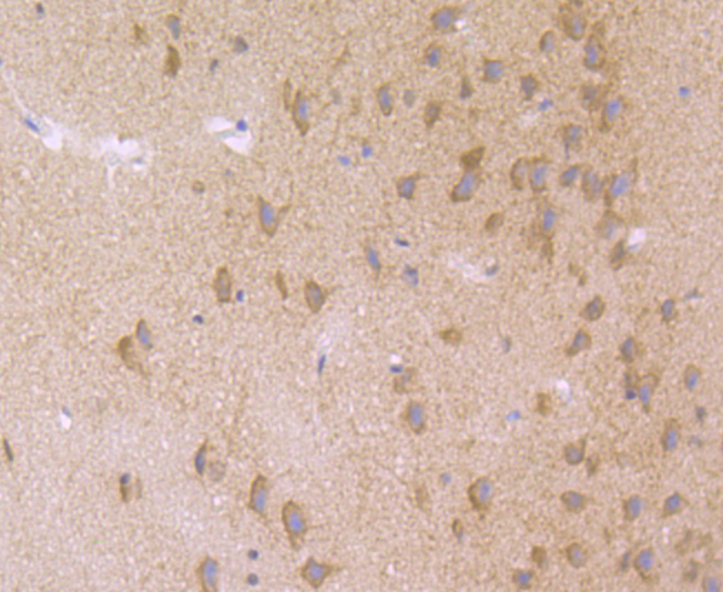
Fig5: Immunohistochemical analysis of paraffin-embedded rat brain tissue using anti-Nuclear Matrix Protein p84 antibody. Counter stained with hematoxylin.Application
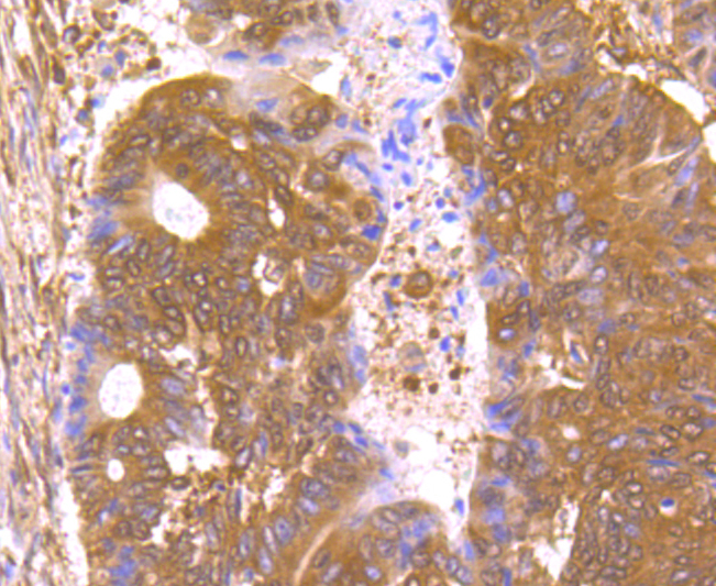
Fig6: Immunohistochemical analysis of paraffin-embedded human colon cancer tissue using anti-Nuclear Matrix Protein p84 antibody. Counter stained with hematoxylin.Application
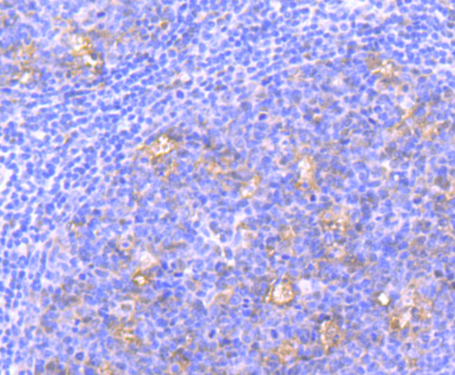
Fig7: Immunohistochemical analysis of paraffin-embedded human tonsil tissue using anti-Nuclear Matrix Protein p84 antibody. Counter stained with hematoxylin.Application
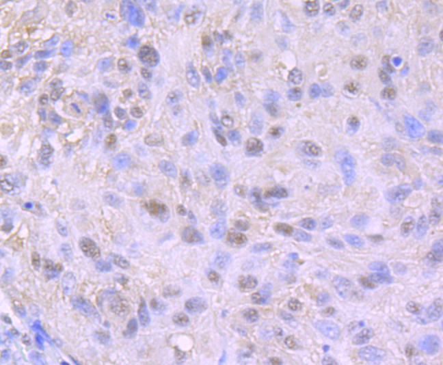
Fig8: Immunohistochemical analysis of paraffin-embedded human lung cancer tissue using anti-Nuclear Matrix Protein p84 antibody. Counter stained with hematoxylin.| Product Name | Nuclear Matrix Protein p84 [JU53-18] |
|---|---|
| Antibody Type | Primary Antibodies |
| Antigen Alias | hTREX84 antibody Death domain containing protein p84N5 antibody HPR 1 antibody HPR1 antibody hTREX84 antibody Nuclear matrix protein p84 antibody P84 antibody p84N5 antibody Tho 1 antibody THO complex 1 antibody THO complex subunit 1 antibody Tho1 antibody THOC 1 antibody Thoc1 antibody THOC1_HUMAN antibody |
| Product description | THOC1 (THO complex subunit 1), also known as Tho1, P84, HPR1 or P84N5, is a 657 amino acid nuclear matrix protein and is evolutionarily conserved from yeast to humans. THOC1 contains one death domain and is a component of the heteromultimeric THO/TREX (transcription/export) complex along with THOC2, THOC3, BAT1 and ALY. The THO/TREX complex is recruited to transcribed genes and travels along with RNA polymerase II (Pol II) during elongation, coupling elongating Pol II with RNA splicing and export factors. THOC1 is expressed at high levels in breast cancer cells and at relatively low levels in normal epithelia. A reduction of THOC1 in cancer cell lines results in reduced cell proliferation. This suggests that cancer cells are dependent on the high levels of THOC1 expression and therefore THOC1 may be a good target for cancer therapy. |
| Immunogen | Recombinant protein |
| Clonality | Monoclonal |
|---|---|
| Isotype | IgG |
| Host Species | Recombinant rabbit |
| Tested Applications | WBICCIHC |
| WB:1:500-1:2,000 ICC:1:50-1:200 IHC:1:50-1:200 |
|
| Species Reactivity | HumanMouseRat |
| Concentration | 1mg/ml |
| Alternative Names | hTREX84 antibody Death domain containing protein p84N5 antibody HPR 1 antibody HPR1 antibody hTREX84 antibody Nuclear matrix protein p84 antibody P84 antibody p84N5 antibody Tho 1 antibody THO complex 1 antibody THO complex subunit 1 antibody Tho1 antibody THOC 1 antibody Thoc1 antibody THOC1_HUMAN antibody |
|---|---|
| Molecular Weight(MW) | 76 kDa |
| Cellular Localization | SwissProt: Q96FV9(Human) Q8R3N6(Mouse) P59924(Rat) |
Application
Fig1: Western blot analysis of Nuclear Matrix Protein p84 on different lysates using anti-Nuclear Matrix Protein p84 antibody at 1/500 dilution. Positive control: Lane 1: Hela Lane 2: A431 Lane 3: Mouse skeletal muscle Lane 4: PC-12
Application
Fig2: ICC staining Nuclear Matrix Protein p84 in HUVEC cells (green). The nuclear counter stain is DAPI (blue). Cells were fixed in paraformaldehyde, permeabilised with 0.25% Triton X100/PBS.
Application
Fig3: ICC staining Nuclear Matrix Protein p84 in LOVO cells (green). The nuclear counter stain is DAPI (blue). Cells were fixed in paraformaldehyde, permeabilised with 0.25% Triton X100/PBS.
Application
Fig4: Immunohistochemical analysis of paraffin-embedded rat testis tissue using anti-Nuclear Matrix Protein p84 antibody. Counter stained with hematoxylin.
Application
Fig5: Immunohistochemical analysis of paraffin-embedded rat brain tissue using anti-Nuclear Matrix Protein p84 antibody. Counter stained with hematoxylin.
Application
Fig6: Immunohistochemical analysis of paraffin-embedded human colon cancer tissue using anti-Nuclear Matrix Protein p84 antibody. Counter stained with hematoxylin.
Application
Fig7: Immunohistochemical analysis of paraffin-embedded human tonsil tissue using anti-Nuclear Matrix Protein p84 antibody. Counter stained with hematoxylin.
Application
Fig8: Immunohistochemical analysis of paraffin-embedded human lung cancer tissue using anti-Nuclear Matrix Protein p84 antibody. Counter stained with hematoxylin.| Positive Control | Hela, A431, mouse skeletal muscle tissue lysate, PC-12, HUVEC, LOVO, rat testis tissue, rat brain tissue, human tonsil tissue, human lung cancer tissue, human colon cancer tissue. |
|---|---|
| Application Notes | WB:1:500-1:2,000 ICC:1:50-1:200 IHC:1:50-1:200 |
| Form | Liquid |
|---|---|
| Storage Instructions | Store at +4℃ after thawing. Aliquot store at -20℃ or -80℃. Avoid repeated freeze / thaw cycles. |
| Storage Buffer | 1*TBS (pH7.4), 1%BSA, 40%Glycerol. Preservative: 0.05% Sodium Azide. |


