WB
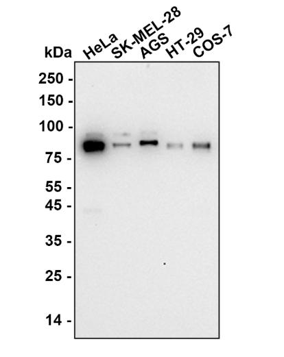
Western blot analysis of Cullin 4a on different lysates with Rabbit anti-Cullin 4a antibody at 1/2,000 dilution. Lane 1: HeLa (Human cervical adenocarcinoma cell), Lane 2: SK-MEL-28 (Human malignant melanoma cell), Lane 3: AGS (Human stomach adenocarcinoma cell), Lane 4: HT-29 (Human colorectal adenocarcinoma cell), Lane 5: COS-7 (African green monkey kidney fibroblast), Lysates/proteins at 10 µg/Lane. Exposure time: 3 minutes; Blocking: 5% NFDM/TBST, 1 hour at room temperature, Primary antibody:1/2,000 in primary antibody dilution buffer, overnight at 4 ℃ Secondary antibody: Goat anti-Rabbit IgG-HR, 1/50,000 in 5% NFDM/TBST, 1 hour at room temperatureIHC
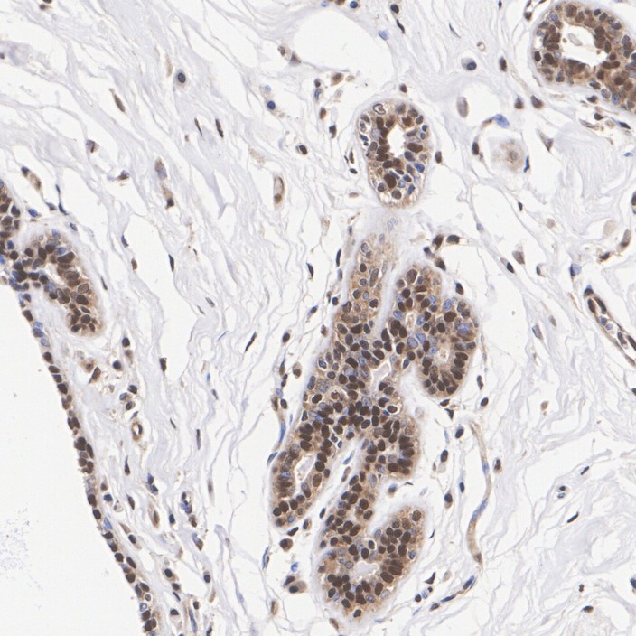
Immunohistochemical analysis of paraffin-embedded human breast cancer tissue with Rabbit anti-Cullin 4a antibody at 1/1,000 dilution. The section was pre-treated using heat mediated antigen retrieval with Tris-EDTA buffer (pH 9.0) for 20 minutes. The tissues were blocked in 1% BSA for 20 minutes at room temperature, washed with ddH2O and PBS, and then probed with the primary antibody at 1/1,000 dilution for 1 hour at room temperature. The detection was performed using an HRP conjugated compact polymer system. DAB was used as the chromogen. Tissues were counterstained with hematoxylin and mounted with DPX.IHC
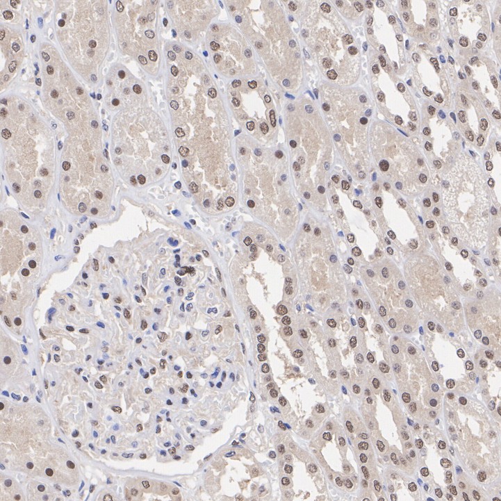
Immunohistochemical analysis of paraffin-embedded human kidney tissue with Rabbit anti-Cullin 4a antibody at 1/1,000 dilution. The section was pre-treated using heat mediated antigen retrieval with Tris-EDTA buffer (pH 9.0) for 20 minutes. The tissues were blocked in 1% BSA for 20 minutes at room temperature, washed with ddH2O and PBS, and then probed with the primary antibody at 1/1,000 dilution for 1 hour at room temperature. The detection was performed using an HRP conjugated compact polymer system. DAB was used as the chromogen. Tissues were counterstained with hematoxylin and mounted with DPX.IHC
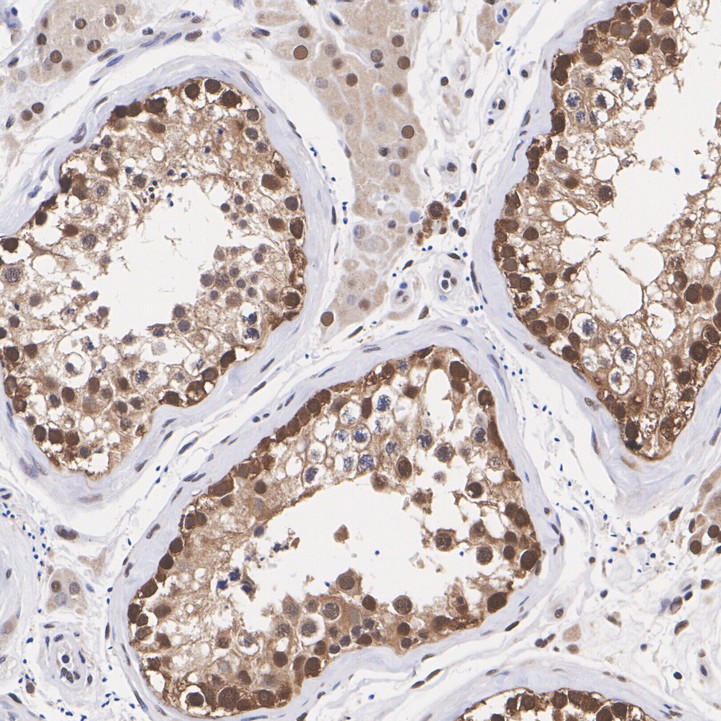
Immunohistochemical analysis of paraffin-embedded human testis tissue with Rabbit anti-Cullin 4a antibody at 1/1,000 dilution. The section was pre-treated using heat mediated antigen retrieval with Tris-EDTA buffer (pH 9.0) for 20 minutes. The tissues were blocked in 1% BSA for 20 minutes at room temperature, washed with ddH2O and PBS, and then probed with the primary antibody at 1/1,000 dilution for 1 hour at room temperature. The detection was performed using an HRP conjugated compact polymer system. DAB was used as the chromogen. Tissues were counterstained with hematoxylin and mounted with DPX.| Product Name | Cullin 4a Recombinant Rabbit Monoclonal Antibody |
|---|---|
| Antibody Type | Primary Antibodies |
| Product description | Core component of multiple cullin-RING-based E3 ubiquitin-protein ligase complexes which mediate the ubiquitination of target proteins. As a scaffold protein may contribute to catalysis through positioning of the substrate and the ubiquitin-conjugating enzyme. The E3 ubiquitin-protein ligase activity of the complex is dependent on the neddylation of the cullin subunit and is inhibited by the association of the deneddylated cullin subunit with TIP120A/CAND1. The functional specificity of the E3 ubiquitin-protein ligase complex depends on the variable substrate recognition component. DCX(DET1-COP1) directs ubiquitination of JUN. DCX(DDB2) directs ubiquitination of XPC. DCX(DDB2) ubiquitinates histones H3-H4 and is required for efficient histone deposition during replication-coupled (H3.1) and replication-independent (H3.3) nucleosome assembly, probably by facilitating the transfer of H3 from ASF1A/ASF1B to other chaperones involved in histone deposition. |
| Immunogen | Synthetic peptide within Human Cullin 4a aa 710-759 / 759. |
| Clonality | Monoclonal |
|---|---|
| Isotype | IgG |
| Host Species | Recombinant rabbit |
| Tested Applications | IHCWB |
| WB:1:2000 IHC:1:1000 |
|
| Species Reactivity | Human |
| Concentration | 1mg/ml |
| Purification | Protein A |
| Alternative Names | 2810470J21Rik antibody AW495282 antibody CUL 4A antibody CUL-4A antibody Cul4a antibody Cul4a protein antibody CUL4A_HUMAN antibody Cullin-4A antibody MGC36573 antibody MGC64071 antibody |
|---|---|
| Molecular Weight(MW) | 88 kDa(Observed band size:88,92 kDa) |
| Cellular Localization | Nucleus. |
| SwissProt ID | Q13619 |
|---|
WB
Western blot analysis of Cullin 4a on different lysates with Rabbit anti-Cullin 4a antibody at 1/2,000 dilution. Lane 1: HeLa (Human cervical adenocarcinoma cell), Lane 2: SK-MEL-28 (Human malignant melanoma cell), Lane 3: AGS (Human stomach adenocarcinoma cell), Lane 4: HT-29 (Human colorectal adenocarcinoma cell), Lane 5: COS-7 (African green monkey kidney fibroblast), Lysates/proteins at 10 µg/Lane. Exposure time: 3 minutes; Blocking: 5% NFDM/TBST, 1 hour at room temperature, Primary antibody:1/2,000 in primary antibody dilution buffer, overnight at 4 ℃ Secondary antibody: Goat anti-Rabbit IgG-HR, 1/50,000 in 5% NFDM/TBST, 1 hour at room temperature
IHC
Immunohistochemical analysis of paraffin-embedded human breast cancer tissue with Rabbit anti-Cullin 4a antibody at 1/1,000 dilution. The section was pre-treated using heat mediated antigen retrieval with Tris-EDTA buffer (pH 9.0) for 20 minutes. The tissues were blocked in 1% BSA for 20 minutes at room temperature, washed with ddH2O and PBS, and then probed with the primary antibody at 1/1,000 dilution for 1 hour at room temperature. The detection was performed using an HRP conjugated compact polymer system. DAB was used as the chromogen. Tissues were counterstained with hematoxylin and mounted with DPX.
IHC
Immunohistochemical analysis of paraffin-embedded human kidney tissue with Rabbit anti-Cullin 4a antibody at 1/1,000 dilution. The section was pre-treated using heat mediated antigen retrieval with Tris-EDTA buffer (pH 9.0) for 20 minutes. The tissues were blocked in 1% BSA for 20 minutes at room temperature, washed with ddH2O and PBS, and then probed with the primary antibody at 1/1,000 dilution for 1 hour at room temperature. The detection was performed using an HRP conjugated compact polymer system. DAB was used as the chromogen. Tissues were counterstained with hematoxylin and mounted with DPX.
IHC
Immunohistochemical analysis of paraffin-embedded human testis tissue with Rabbit anti-Cullin 4a antibody at 1/1,000 dilution. The section was pre-treated using heat mediated antigen retrieval with Tris-EDTA buffer (pH 9.0) for 20 minutes. The tissues were blocked in 1% BSA for 20 minutes at room temperature, washed with ddH2O and PBS, and then probed with the primary antibody at 1/1,000 dilution for 1 hour at room temperature. The detection was performed using an HRP conjugated compact polymer system. DAB was used as the chromogen. Tissues were counterstained with hematoxylin and mounted with DPX.| Application Notes | WB:1:2000 IHC:1:1000 |
|---|
| Form | Liquid |
|---|---|
| Storage Instructions | Store at +4℃ after thawing. Aliquot store at -20℃ or -80℃. Avoid repeated freeze / thaw cycles. |
| Storage Buffer | 1*TBS (pH7.4), 1%BSA, 40%Glycerol. Preservative: 0.05% Sodium Azide. |


