Application
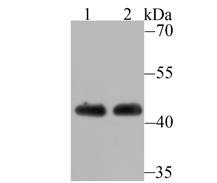
Fig1: Western blot analysis of ACTL6A on Jurkat cell(1) and mouse brain tissue(2) lysate using anti-ACTL6A antibody at 1/500 dilution.Application
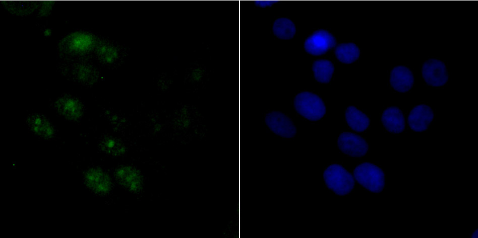
Fig2: ICC staining ACTL6A in LOVO cells (green). The nuclear counter stain is DAPI (blue). Cells were fixed in paraformaldehyde, permeabilised with 0.25% Triton X100/PBS.Application
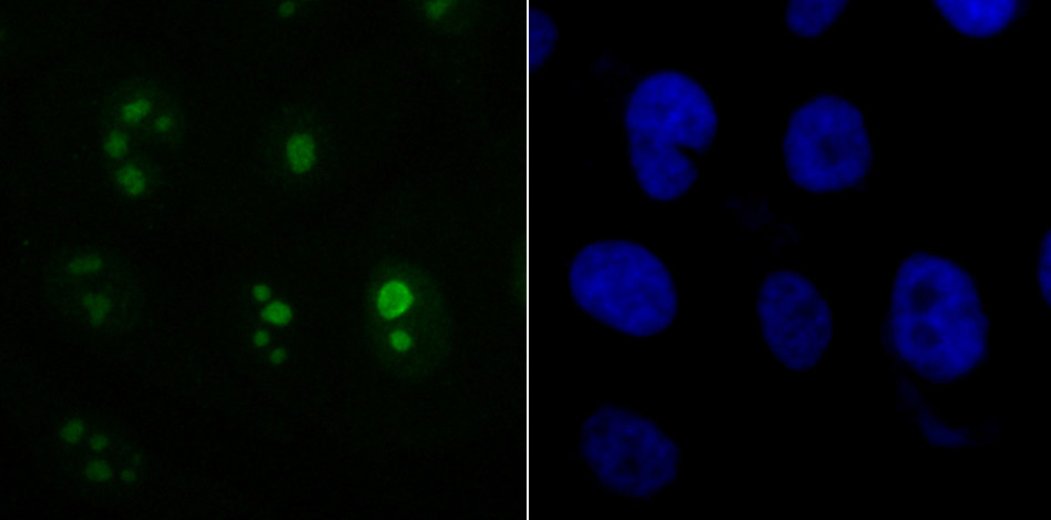
Fig3: ICC staining ACTL6A in PC-3M cells (green). The nuclear counter stain is DAPI (blue). Cells were fixed in paraformaldehyde, permeabilised with 0.25% Triton X100/PBS.Application
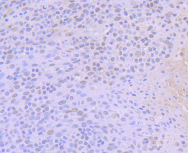
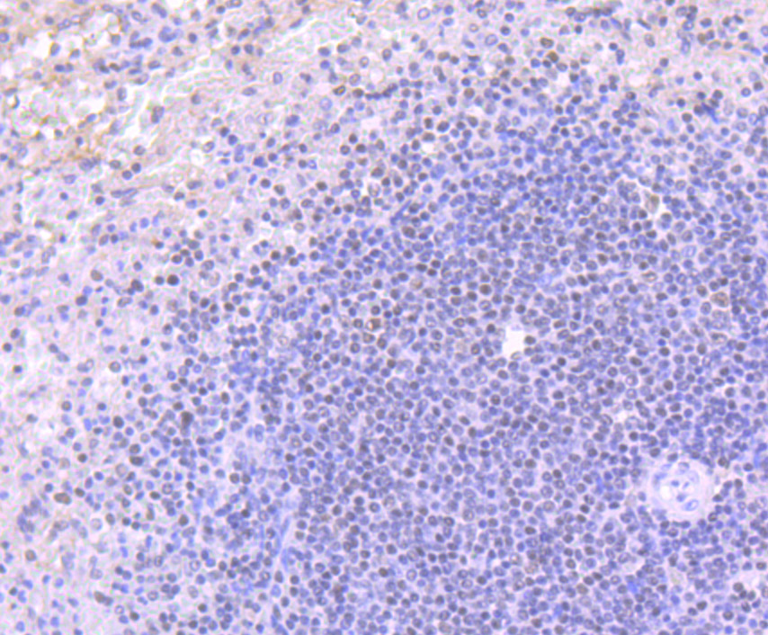
Fig4: Immunohistochemical analysis of paraffin-embedded human tonsil tissue using anti-ACTL6A antibody. Counter stained with hematoxylin.Application
Fig5: Immunohistochemical analysis of paraffin-embedded human spleen tissue using anti-ACTL6A antibody. Counter stained with hematoxylin.Application
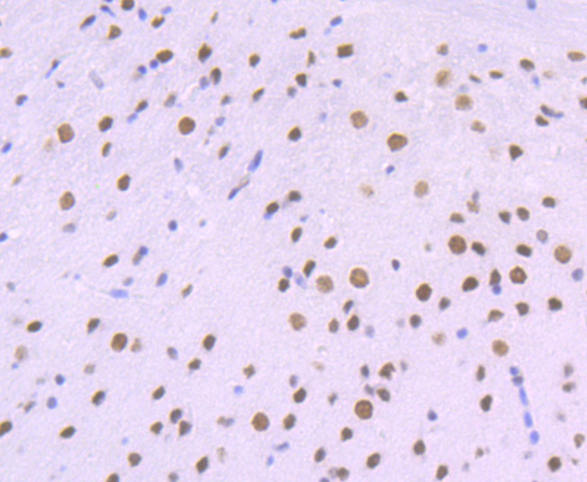
Fig6: Immunohistochemical analysis of paraffin-embedded mouse brain tissue using anti-ACTL6A antibody. Counter stained with hematoxylin.| Product Name | ACTL6A [JU40-32] |
|---|---|
| Antibody Type | Primary Antibodies |
| Antigen Alias | 53 kDa BRG1 associated factor A antibody 53 kDa BRG1-associated factor A antibody ACL6A_HUMAN antibody Actin like 6A antibody Actin like protein 6A antibody Actin related protein 4 antibody Actin related protein antibody Actin related protein Baf53a antibody Actin-like protein 6A antibody Actin-related protein Baf53a antibody ACTL 6 antibody ACTL 6A antibody ACTL6 antibody ACTL6A antibody Arp4 antibody ARPN BETA antibody ArpNbeta antibody BAF 53A antibody BAF complex 53 kDa subunit antibody BAF53 antibody BAF53A antibody BRG1 associated factor antibody BRG1-associated factor 53A antibody hArpN beta antibody INO80 complex subunit K antibody INO80K antibody MGC5382 antibody |
| Product description | ACTL6A also named BAF53. The SWI/SNF complex regulates gene expression via ATP-dependent chromatin remodeling. Brm (SNF2-a), Brg-1 (SNF2-b), Ini1 (integrase interactor 1, SNF5), BAF53 (ARPnβ), BAF57, BAF155 (SRG3), and BAF170 make up the functional core. BAF53 homologues from yeast to humans contain a conserved N-terminal motif, which contains residues at serine 2 and tyrosine 6, which play important roles in BAF53 activity. The BAF53 protein shuttles between the nucleus and cytoplasm. BAF53 also forms a complex with TIP49 and TIP48, which mediates c-Myc oncogenic activity. |
| Immunogen | Recombinant protein |
| Clonality | Monoclonal |
|---|---|
| Isotype | IgG |
| Host Species | Recombinant rabbit |
| Tested Applications | WBICCIFIHCIP |
| WB:1:500-1:2,000 ICC/IF:1:50-1:100 IHC:1:50-1:100 IP:1:10-1:50 |
|
| Species Reactivity | HumanMouse |
| Concentration | 1mg/ml |
| Alternative Names | 53 kDa BRG1 associated factor A antibody 53 kDa BRG1-associated factor A antibody ACL6A_HUMAN antibody Actin like 6A antibody Actin like protein 6A antibody Actin related protein 4 antibody Actin related protein antibody Actin related protein Baf53a antibody Actin-like protein 6A antibody Actin-related protein Baf53a antibody ACTL 6 antibody ACTL 6A antibody ACTL6 antibody ACTL6A antibody Arp4 antibody ARPN BETA antibody ArpNbeta antibody BAF 53A antibody BAF complex 53 kDa subunit antibody BAF53 antibody BAF53A antibody BRG1 associated factor antibody BRG1-associated factor 53A antibody hArpN beta antibody INO80 complex subunit K antibody INO80K antibody MGC5382 antibody |
|---|---|
| Molecular Weight(MW) | 47 kDa |
| Cellular Localization | Nucleus. |
Application
Fig1: Western blot analysis of ACTL6A on Jurkat cell(1) and mouse brain tissue(2) lysate using anti-ACTL6A antibody at 1/500 dilution.
Application
Fig2: ICC staining ACTL6A in LOVO cells (green). The nuclear counter stain is DAPI (blue). Cells were fixed in paraformaldehyde, permeabilised with 0.25% Triton X100/PBS.
Application
Fig3: ICC staining ACTL6A in PC-3M cells (green). The nuclear counter stain is DAPI (blue). Cells were fixed in paraformaldehyde, permeabilised with 0.25% Triton X100/PBS.
Application
Fig4: Immunohistochemical analysis of paraffin-embedded human tonsil tissue using anti-ACTL6A antibody. Counter stained with hematoxylin.
Application
Fig5: Immunohistochemical analysis of paraffin-embedded human spleen tissue using anti-ACTL6A antibody. Counter stained with hematoxylin.
Application
Fig6: Immunohistochemical analysis of paraffin-embedded mouse brain tissue using anti-ACTL6A antibody. Counter stained with hematoxylin.| Positive Control | Jurkat, mouse brain tissue, LOVO, PC-3M, human tonsil tissue, human spleen tissue. |
|---|---|
| Application Notes | WB:1:500-1:2,000 ICC/IF:1:50-1:100 IHC:1:50-1:100 IP:1:10-1:50 |
| Form | Liquid |
|---|---|
| Storage Instructions | Store at +4℃ after thawing. Aliquot store at -20℃ or -80℃. Avoid repeated freeze / thaw cycles. |
| Storage Buffer | 1*TBS (pH7.4), 1%BSA, 40%Glycerol. Preservative: 0.05% Sodium Azide. |


