Application
Fig1: Western blot analysis of IL13 Receptor alpha 1 on A549 cell lysates using anti-IL13 Receptor alpha 1 antibody at 1/500 dilution.Application
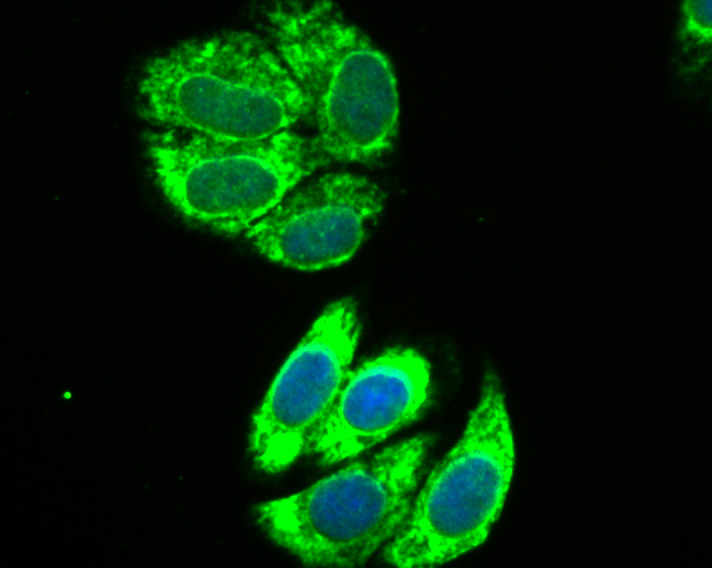
Fig2: ICC staining IL13 Receptor alpha 1 in HepG2 cells (green). The nuclear counter stain is DAPI (blue). Cells were fixed in paraformaldehyde, permeabilised with 0.25% Triton X100/PBS.Application
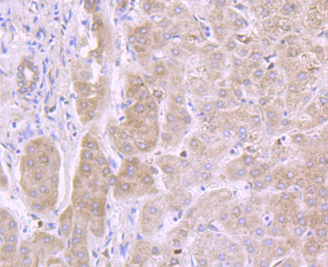
Fig3: Immunohistochemical analysis of paraffin-embedded human liver tissue using anti-IL13 Receptor alpha 1 antibody. Counter stained with hematoxylin.| Product Name | IL13 Receptor alpha 1 [JU38-89] |
|---|---|
| Antibody Type | Primary Antibodies |
| Antigen Alias | bB128O4.2.1 (interleukin 13 receptor, alpha 1) antibody Cancer/testis antigen 19 antibody CD213a1 antibody CD213A1 Antigen antibody CT 19 antibody CT19 antibody I13R1_HUMAN antibody IL 13 receptor subunit alpha 1 antibody IL 13R alpha 1 antibody IL 13R subunit alpha 1 antibody IL 13Ra antibody IL 13RA1 antibody IL-13 receptor subunit alpha-1 antibody IL-13R subunit alpha-1 antibody IL-13R-alpha-1 antibody IL-13RA1 antibody IL13 receptor alpha 1 chain antibody IL13R antibody IL13RA antibody Il13ra1 antibody Interleukin 13 receptor alpha 1 antibody Interleukin 13 receptor alpha 1 chain antibody interleukin 13 receptor subunit alpha 1 antibody Interleukin-13 receptor subunit alpha-1 antibody NR 4 antibody NR4 antibody |
| Product description | Interleukin 13 receptor, alpha 1, also known as IL13RA1 and CD213A1 (cluster of differentiation 213A1), is a human gene. Binds with low affinity to interleukin-13 (IL13). Together with IL4RA can form a functional receptor for IL13. Also serves as an alternate accessory protein to the common cytokine receptor gamma chain for interleukin-4 (IL4) signaling, but cannot replace the function of IL2RG in allowing enhanced interleukin-2 (IL2) binding activity. This protein has been shown to bind tyrosine kinase TYK2, and thus may mediate the signaling processes that lead to the activation of JAK1, STAT3 and STAT6 induced by IL13 and IL4. |
| Immunogen | Recombinant protein |
| Clonality | Monoclonal |
|---|---|
| Isotype | IgG |
| Host Species | Recombinant rabbit |
| Tested Applications | WBICCIHC |
| WB:1:500-2,000 ICC:1:100-1:500 IHC:1:50-1:200 |
|
| Species Reactivity | Human |
| Concentration | 1mg/ml |
| Alternative Names | bB128O4.2.1 (interleukin 13 receptor alpha 1) antibody Cancer/testis antigen 19 antibody CD213a1 antibody CD213A1 Antigen antibody CT 19 antibody CT19 antibody I13R1_HUMAN antibody IL 13 receptor subunit alpha 1 antibody IL 13R alpha 1 antibody IL 13R subunit alpha 1 antibody IL 13Ra antibody IL 13RA1 antibody IL-13 receptor subunit alpha-1 antibody IL-13R subunit alpha-1 antibody IL-13R-alpha-1 antibody IL-13RA1 antibody IL13 receptor alpha 1 chain antibody IL13R antibody IL13RA antibody Il13ra1 antibody Interleukin 13 receptor alpha 1 antibody Interleukin 13 receptor alpha 1 chain antibody interleukin 13 receptor subunit alpha 1 antibody Interleukin-13 receptor subunit alpha-1 antibody NR 4 antibody NR4 antibody |
|---|---|
| Molecular Weight(MW) | 50/40 kDa |
| Cellular Localization | Membrane. |
| SwissProt ID | P78552 |
|---|
Application
Fig1: Western blot analysis of IL13 Receptor alpha 1 on A549 cell lysates using anti-IL13 Receptor alpha 1 antibody at 1/500 dilution.
Application
Fig2: ICC staining IL13 Receptor alpha 1 in HepG2 cells (green). The nuclear counter stain is DAPI (blue). Cells were fixed in paraformaldehyde, permeabilised with 0.25% Triton X100/PBS.
Application
Fig3: Immunohistochemical analysis of paraffin-embedded human liver tissue using anti-IL13 Receptor alpha 1 antibody. Counter stained with hematoxylin.| Positive Control | A549, HepG2, human liver tissue. |
|---|---|
| Application Notes | WB:1:500-2,000 ICC:1:100-1:500 IHC:1:50-1:200 |
| Form | Liquid |
|---|---|
| Storage Instructions | Store at +4℃ after thawing. Aliquot store at -20℃ or -80℃. Avoid repeated freeze / thaw cycles. |
| Storage Buffer | 1*TBS (pH7.4), 1%BSA, 40%Glycerol. Preservative: 0.05% Sodium Azide. |


