Application
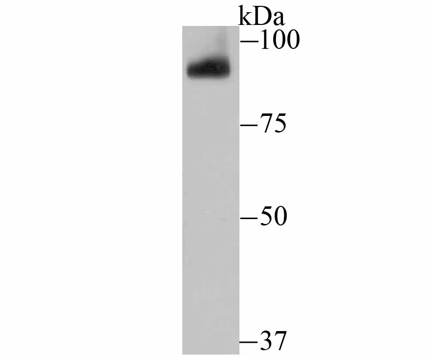
Fig1: Western blot analysis of PIWIL1 on mouse testis tissue lysate using anti-PIWIL1 antibody at 1/500 dilution.Application
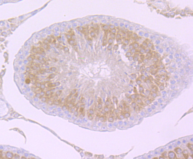
Fig2: Immunohistochemical analysis of paraffin-embedded rat testis tissue using anti-PIWIL1 antibody. Counter stained with hematoxylin.Application
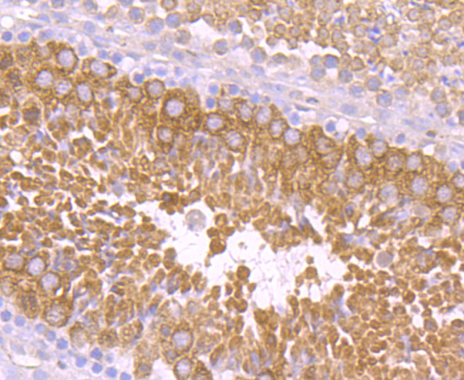
Fig3: Immunohistochemical analysis of paraffin-embedded mouse testis tissue using anti-PIWIL1 antibody. Counter stained with hematoxylin.| Product Name | PIWIL1 [JU35-12] |
|---|---|
| Antibody Type | Primary Antibodies |
| Antigen Alias | HIWI antibody MIWI antibody Piwi (Drosophila) like 1 antibody PIWI antibody Piwi homolog antibody Piwi like 1 (drosophila) antibody Piwi like 1 antibody Piwi-like protein 1 antibody PIWIL1 antibody PIWL1_HUMAN antibody |
| Product description | Endoribonuclease that plays a central role in postnatal germ cells by repressing transposable elements and preventing their mobilization, which is essential for the germline integrity. Acts via the piRNA metabolic process, which mediates the repression of transposable elements during meiosis by forming complexes composed of piRNAs and Piwi proteins and governs the methylation and subsequent repression of transposons. Directly binds methylated piRNAs, a class of 24 to 30 nucleotide RNAs that are generated by a Dicer-independent mechanism and are primarily derived from transposons and other repeated sequence elements. Strongly prefers a uridine in the first position of their guide (g1U preference, also named 1U-bias). Not involved in the piRNA amplification loop, also named ping-pong amplification cycle. Acts as an endoribonuclease that cleaves transposon messenger RNAs. Besides their function in transposable elements repression, piRNAs are probably involved in other processes during meiosis such as translation regulation. Probable component of some RISC complex, which mediates RNA cleavage and translational silencing. Also plays a role in the formation of chromatoid bodies and is required for some miRNAs stability. Required to sequester RNF8 in the cytoplasm until late spermatogenesis; RNF8 being released upon ubiquitination and degradation of PIWIL1. |
| Immunogen | Recombinant protein |
| Clonality | Monoclonal |
|---|---|
| Isotype | IgG |
| Host Species | Recombinant rabbit |
| Tested Applications | WBIHC |
| WB:1:500-1:1,000 IHC:1:50-1:200 |
|
| Species Reactivity | HumanMouseRat |
| Concentration | 1mg/ml |
| Alternative Names | HIWI antibody MIWI antibody Piwi (Drosophila) like 1 antibody PIWI antibody Piwi homolog antibody Piwi like 1 (drosophila) antibody Piwi like 1 antibody Piwi-like protein 1 antibody PIWIL1 antibody PIWL1_HUMAN antibody |
|---|---|
| Molecular Weight(MW) | 98 kDa |
| Cellular Localization | Cytoplasm. |
Application
Fig1: Western blot analysis of PIWIL1 on mouse testis tissue lysate using anti-PIWIL1 antibody at 1/500 dilution.
Application
Fig2: Immunohistochemical analysis of paraffin-embedded rat testis tissue using anti-PIWIL1 antibody. Counter stained with hematoxylin.
Application
Fig3: Immunohistochemical analysis of paraffin-embedded mouse testis tissue using anti-PIWIL1 antibody. Counter stained with hematoxylin.| Positive Control | Mouse testis tissue, rat testis tissue. |
|---|---|
| Application Notes | WB:1:500-1:1,000 IHC:1:50-1:200 |
| Form | Liquid |
|---|---|
| Storage Instructions | Store at +4℃ after thawing. Aliquot store at -20℃ or -80℃. Avoid repeated freeze / thaw cycles. |
| Storage Buffer | 1*TBS (pH7.4), 1%BSA, 40%Glycerol. Preservative: 0.05% Sodium Azide. |


