Application
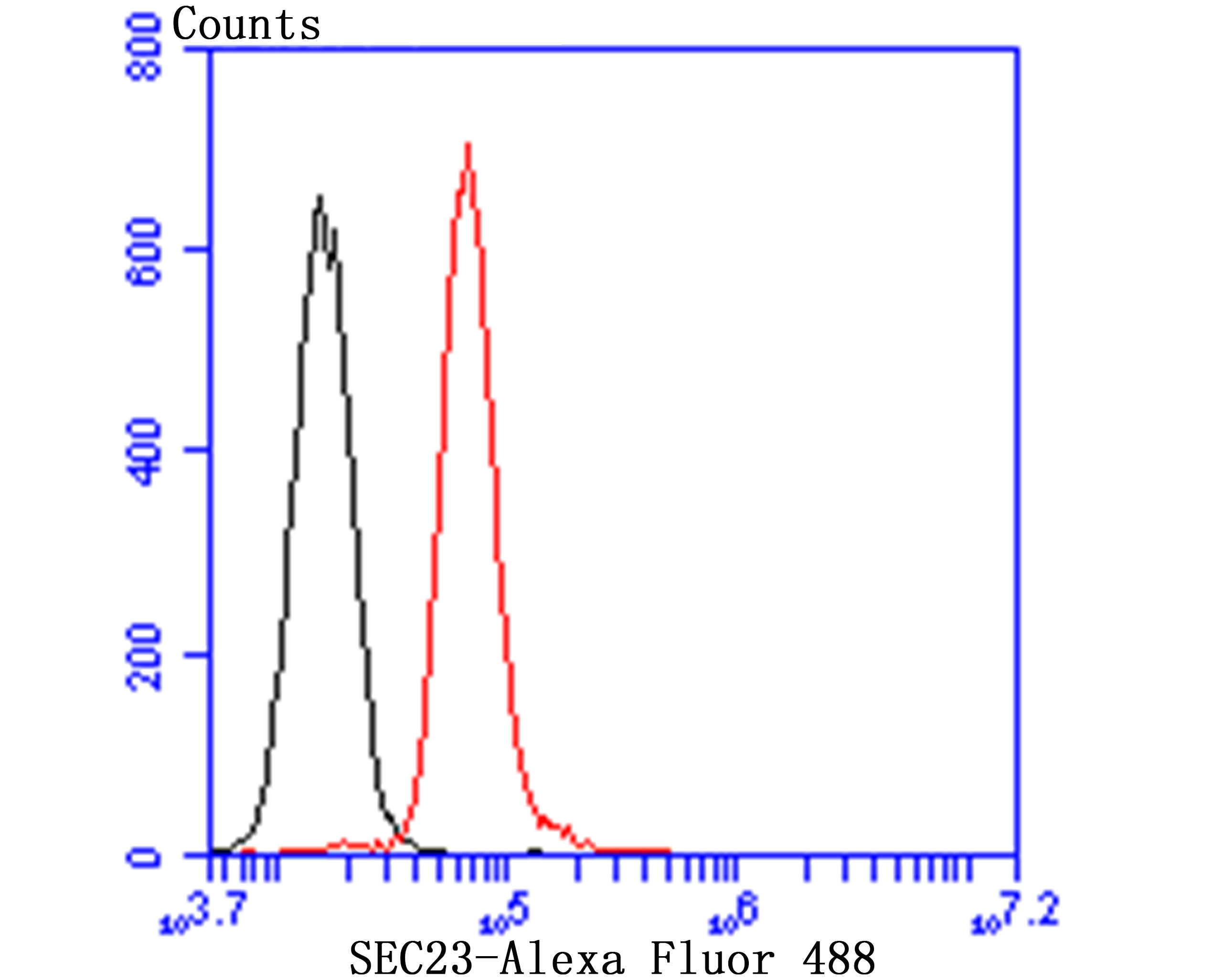
Fig6: Flow cytometric analysis of HepG2 cells with SEC23 antibody at 1/100 dilution (red) compared with an unlabelled control (cells without incubation with primary antibody; black). Goat anti rabbit IgG (FITC) was used as the secondary antibody.Application
Fig6: Flow cytometric analysis of HepG2 cells with SEC23 antibody at 1/100 dilution (red) compared with an unlabelled control (cells without incubation with primary antibody; black). Goat anti rabbit IgG (FITC) was used as the secondary antibody.Application
Fig6: Flow cytometric analysis of HepG2 cells with SEC23 antibody at 1/100 dilution (red) compared with an unlabelled control (cells without incubation with primary antibody; black). Goat anti rabbit IgG (FITC) was used as the secondary antibody.Application
Fig6: Flow cytometric analysis of HepG2 cells with SEC23 antibody at 1/100 dilution (red) compared with an unlabelled control (cells without incubation with primary antibody; black). Goat anti rabbit IgG (FITC) was used as the secondary antibody.Application
Fig6: Flow cytometric analysis of HepG2 cells with SEC23 antibody at 1/100 dilution (red) compared with an unlabelled control (cells without incubation with primary antibody; black). Goat anti rabbit IgG (FITC) was used as the secondary antibody.Application
Fig6: Flow cytometric analysis of HepG2 cells with SEC23 antibody at 1/100 dilution (red) compared with an unlabelled control (cells without incubation with primary antibody; black). Goat anti rabbit IgG (FITC) was used as the secondary antibody.| Product Name | SEC23 [JU37-48] |
|---|---|
| Antibody Type | Primary Antibodies |
| Antigen Alias | CLSD antibody Protein transport protein Sec23A antibody SC23A_HUMAN antibody Sec23 homolog A (S. cerevisiae) antibody SEC23-related protein A antibody sec23a antibody |
| Product description | Component of the coat protein complex II (COPII) which promotes the formation of transport vesicles from the endoplasmic reticulum (ER). The coat has two main functions, the physical deformation of the endoplasmic reticulum membrane into vesicles and the selection of cargo molecules for their transport to the Golgi complex. |
| Immunogen | Recombinant protein |
| Clonality | Monoclonal |
|---|---|
| Isotype | IgG |
| Host Species | Recombinant rabbit |
| Tested Applications | WBICCIHCFC |
| WB:1:500-1:1,000 ICC:1:50-1:200 IHC:1:50-1:200 FC:1:50-1:100 |
|
| Species Reactivity | HumanMouse |
| Concentration | 1mg/ml |
| Alternative Names | CLSD antibody Protein transport protein Sec23A antibody SC23A_HUMAN antibody Sec23 homolog A (S. cerevisiae) antibody SEC23-related protein A antibody sec23a antibody |
|---|---|
| Molecular Weight(MW) | 86/63 kDa |
| Cellular Localization | Cytosol. Endoplasmic reticulum. |
Application
Fig6: Flow cytometric analysis of HepG2 cells with SEC23 antibody at 1/100 dilution (red) compared with an unlabelled control (cells without incubation with primary antibody; black). Goat anti rabbit IgG (FITC) was used as the secondary antibody.
Application
Fig6: Flow cytometric analysis of HepG2 cells with SEC23 antibody at 1/100 dilution (red) compared with an unlabelled control (cells without incubation with primary antibody; black). Goat anti rabbit IgG (FITC) was used as the secondary antibody.
Application
Fig6: Flow cytometric analysis of HepG2 cells with SEC23 antibody at 1/100 dilution (red) compared with an unlabelled control (cells without incubation with primary antibody; black). Goat anti rabbit IgG (FITC) was used as the secondary antibody.
Application
Fig6: Flow cytometric analysis of HepG2 cells with SEC23 antibody at 1/100 dilution (red) compared with an unlabelled control (cells without incubation with primary antibody; black). Goat anti rabbit IgG (FITC) was used as the secondary antibody.
Application
Fig6: Flow cytometric analysis of HepG2 cells with SEC23 antibody at 1/100 dilution (red) compared with an unlabelled control (cells without incubation with primary antibody; black). Goat anti rabbit IgG (FITC) was used as the secondary antibody.
Application
Fig6: Flow cytometric analysis of HepG2 cells with SEC23 antibody at 1/100 dilution (red) compared with an unlabelled control (cells without incubation with primary antibody; black). Goat anti rabbit IgG (FITC) was used as the secondary antibody.| Positive Control | NIH-3T3, HepG2, HUVEC, human tonsil tissue, human fetal skeletal muscle tissue, HepG2. |
|---|---|
| Application Notes | WB:1:500-1:1,000 ICC:1:50-1:200 IHC:1:50-1:200 FC:1:50-1:100 |
| Form | Liquid |
|---|---|
| Storage Instructions | Store at +4℃ after thawing. Aliquot store at -20℃ or -80℃. Avoid repeated freeze / thaw cycles. |
| Storage Buffer | 1*TBS (pH7.4), 1%BSA, 40%Glycerol. Preservative: 0.05% Sodium Azide. |


