Application
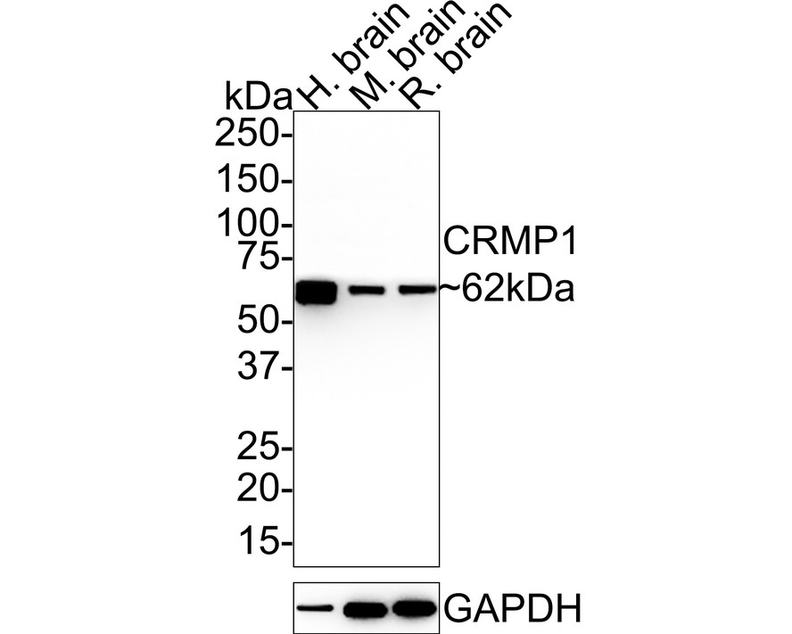
Fig1: Western blot analysis of CRMP1 on different lysates with Rabbit anti-CRMP1 antibody at 1/5,000 dilution. Lane 1: Human brain tissue lysate, Lane 2: Mouse brain tissue lysate, Lane 3: Rat brain tissue lysate, Lysates/proteins at 20 µg/Lane. Exposure time: 24 seconds; 4-20% SDS-PAGE gel. Proteins were transferred to a PVDF membrane and blocked with 5% NFDM/TBST for 1 hour at room temperature. The primary antibody at 1/5,000 dilution was used in 5% NFDM/TBST at 4℃ overnight. Goat Anti-Rabbit IgG - HRP Secondary Antibody at 1:50,000 dilution was used for 1 hour at room temperature.Application
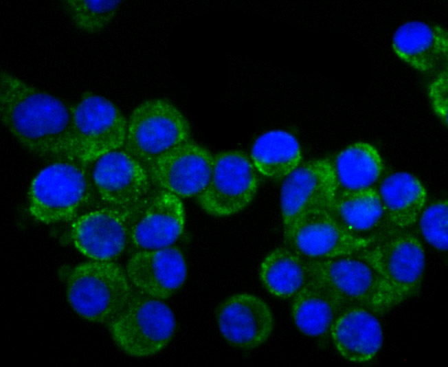
Fig2: ICC staining CRMP1 in N2A cells (green). The nuclear counter stain is DAPI (blue). Cells were fixed in paraformaldehyde, permeabilised with 0.25% Triton X100/PBS.Application
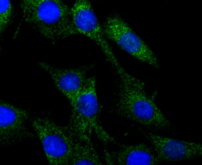
Fig3: ICC staining CRMP1 in SHG-44 cells (green). The nuclear counter stain is DAPI (blue). Cells were fixed in paraformaldehyde, permeabilised with 0.25% Triton X100/PBS.Application
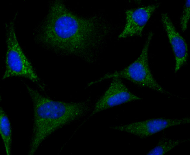
Fig4: ICC staining CRMP1 in SH-SY5Y cells (green). The nuclear counter stain is DAPI (blue). Cells were fixed in paraformaldehyde, permeabilised with 0.25% Triton X100/PBS.Application
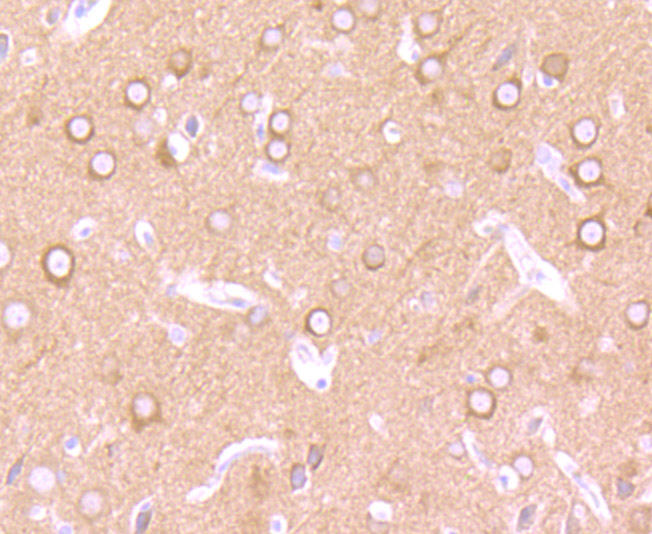
Fig5: Immunohistochemical analysis of paraffin-embedded rat brain tissue using anti-CRMP1 antibody. Counter stained with hematoxylin.Application
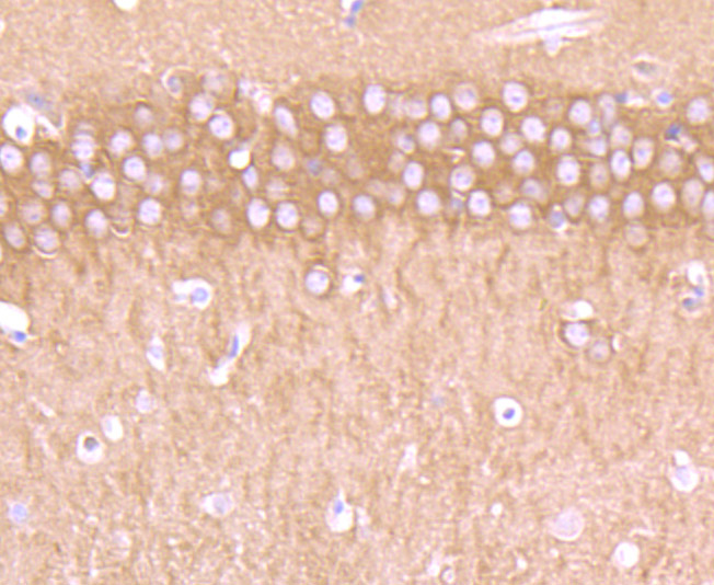
Fig6: Immunohistochemical analysis of paraffin-embedded mouse brain tissue using anti-CRMP1 antibody. Counter stained with hematoxylin.Application
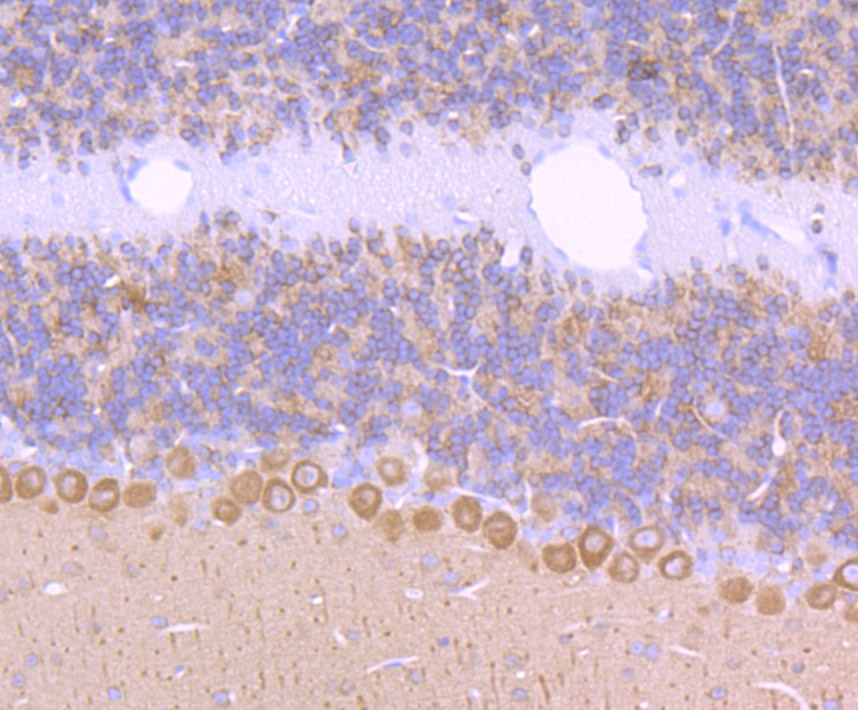
Fig7: Immunohistochemical analysis of paraffin-embedded mouse cerebellum tissue using anti-CRMP1 antibody. Counter stained with hematoxylin.Application
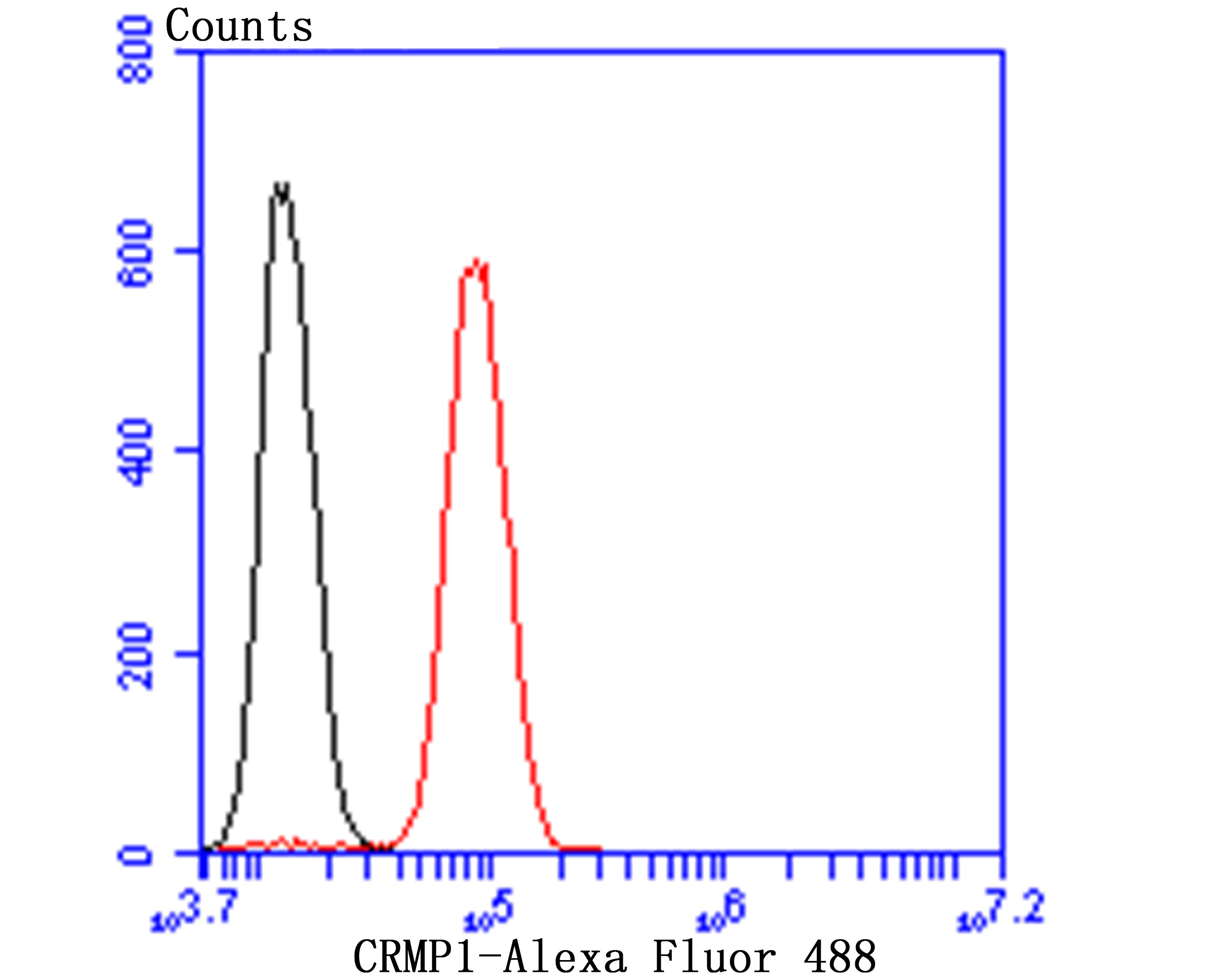
Fig8: Flow cytometric analysis of SH-SY-5Y cells with CRMP1 antibody at 1/50 dilution (red) compared with an unlabelled control (cells without incubation with primary antibody; black). Alexa Fluor 488-conjugated goat anti rabbit IgG was used as the secondary antibody.| Product Name | CRMP1 Recombinant Rabbit Monoclonal Antibody |
|---|---|
| Antibody Type | Primary Antibodies |
| Product description | Collapsin response mediator proteins (CRMPs), including CRMP-1 (DRP-1), CRMP-2 (DRP-2 or TOAD64), CRMP-3 (DRP-4), CRMP-4 (DRP-3) and CRMP-5 (DRP-5), mediate signal transduction after exposure of neural cells to the axon guidance molecule Semaphorin 3A (SEMA3A)/collapsin. CRMPs are present in the developing cerebral cortex and neocortical neurons and are responsive to SEMA3A. In the adult brain, the expression of CRMPs is dramatically downregulated. However, they remain expressed in structures that retain their capacity for differentiation and plasticity. CRMP-1 is also involved in cancer invasion and metastasis. |
| Immunogen | Synthetic peptide within Human CRMP1 aa 489-538 / 572. |
| Clonality | Monoclonal |
|---|---|
| Isotype | IgG |
| Host Species | Recombinant rabbit |
| Tested Applications | FCICC/IFIHCWB |
| WB:1:5000 ICC/IF:1:50-1:200 IHC:1:50-1:200 FC:1:50-1:100 |
|
| Species Reactivity | HumanMouseRat |
| Concentration | 1mg/ml |
| Purification | Protein A |
| Alternative Names | Collapsin response mediator protein 1 antibody CRMP 1 antibody CRMP-1 antibody Crmp1 antibody Dihydropyrimidinase like 1 antibody Dihydropyrimidinase related protein 1 antibody Dihydropyrimidinase-related protein 1 antibody DPYL1_HUMAN antibody DPYSL1 antibody DRP 1 antibody DRP-1 antibody DRP1 antibody ULIP-3 antibody Ulip3 antibody Unc-33-like phosphoprotein 3 antibody |
|---|---|
| Molecular Weight(MW) | 62kDa |
| Cellular Localization | Cytoplasm. Cytoskeleton. |
Application
Fig1: Western blot analysis of CRMP1 on different lysates with Rabbit anti-CRMP1 antibody at 1/5,000 dilution. Lane 1: Human brain tissue lysate, Lane 2: Mouse brain tissue lysate, Lane 3: Rat brain tissue lysate, Lysates/proteins at 20 µg/Lane. Exposure time: 24 seconds; 4-20% SDS-PAGE gel. Proteins were transferred to a PVDF membrane and blocked with 5% NFDM/TBST for 1 hour at room temperature. The primary antibody at 1/5,000 dilution was used in 5% NFDM/TBST at 4℃ overnight. Goat Anti-Rabbit IgG - HRP Secondary Antibody at 1:50,000 dilution was used for 1 hour at room temperature.
Application
Fig2: ICC staining CRMP1 in N2A cells (green). The nuclear counter stain is DAPI (blue). Cells were fixed in paraformaldehyde, permeabilised with 0.25% Triton X100/PBS.
Application
Fig3: ICC staining CRMP1 in SHG-44 cells (green). The nuclear counter stain is DAPI (blue). Cells were fixed in paraformaldehyde, permeabilised with 0.25% Triton X100/PBS.
Application
Fig4: ICC staining CRMP1 in SH-SY5Y cells (green). The nuclear counter stain is DAPI (blue). Cells were fixed in paraformaldehyde, permeabilised with 0.25% Triton X100/PBS.
Application
Fig5: Immunohistochemical analysis of paraffin-embedded rat brain tissue using anti-CRMP1 antibody. Counter stained with hematoxylin.
Application
Fig6: Immunohistochemical analysis of paraffin-embedded mouse brain tissue using anti-CRMP1 antibody. Counter stained with hematoxylin.
Application
Fig7: Immunohistochemical analysis of paraffin-embedded mouse cerebellum tissue using anti-CRMP1 antibody. Counter stained with hematoxylin.
Application
Fig8: Flow cytometric analysis of SH-SY-5Y cells with CRMP1 antibody at 1/50 dilution (red) compared with an unlabelled control (cells without incubation with primary antibody; black). Alexa Fluor 488-conjugated goat anti rabbit IgG was used as the secondary antibody.| Positive Control | Mouse brain tissue, N2A, SHG-44, SH-SY5Y, rat brain tissue, mouse cerebellum tissue. |
|---|---|
| Application Notes | WB:1:5000 ICC/IF:1:50-1:200 IHC:1:50-1:200 FC:1:50-1:100 |
| Form | Liquid |
|---|---|
| Storage Instructions | Store at +4℃ after thawing. Aliquot store at -20℃ or -80℃. Avoid repeated freeze / thaw cycles. |
| Storage Buffer | 1*TBS (pH7.4), 1%BSA, 40%Glycerol. Preservative: 0.05% Sodium Azide. |


