WB
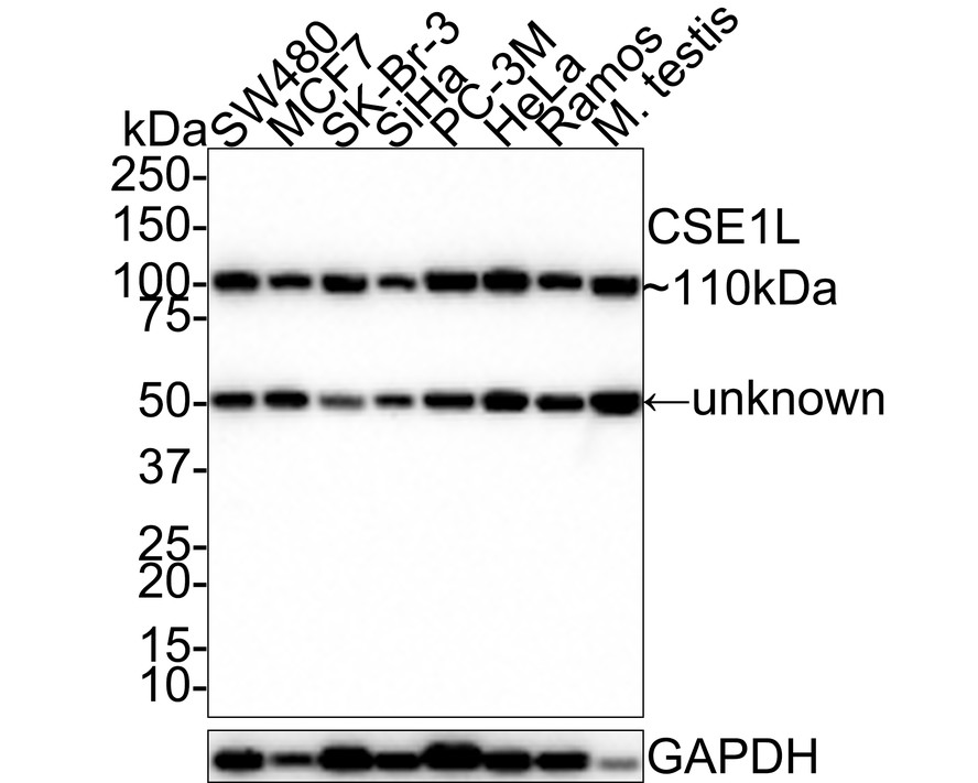
Western blot analysis of Cellular Apoptosis Susceptibility on different lysates with Rabbit anti-Cellular Apoptosis Susceptibility antibody at 1/1,000 dilution. Lane 1: SW480 cell lysate (20 µg/Lane), Lane 2: MCF7 cell lysate (20 µg/Lane), Lane 3: SK-Br-3 cell lysate (20 µg/Lane), Lane 4: SiHa cell lysate (20 µg/Lane), Lane 5: PC-3M cell lysate (20 µg/Lane), Lane 6: HeLa cell lysate (20 µg/Lane), Lane 7: Ramos cell lysate (20 µg/Lane), Lane 8: Mouse testis tissue lysate (40 µg/Lane), Exposure time: 30 seconds; 4-20% SDS-PAGE gel. Proteins were transferred to a PVDF membrane and blocked with 5% NFDM/TBST for 1 hour at room temperature. The primary antibody at 1/1,000 dilution was used in 5% NFDM/TBST at room temperature for 2 hours. Goat Anti-Rabbit IgG - HRP Secondary Antibody at 1:100,000 dilution was used for 1 hour at room temperature.IHC
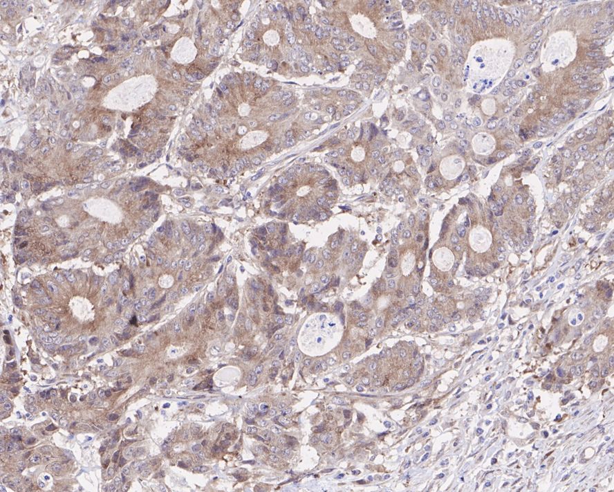
Immunohistochemical analysis of paraffin-embedded human colon carcinoma tissue with Rabbit anti-Cellular Apoptosis Susceptibility antibody at 1/200 dilution. The section was pre-treated using heat mediated antigen retrieval with sodium citrate buffer (pH 6.0) for 2 minutes. The tissues were blocked in 1% BSA for 20 minutes at room temperature, washed with ddH2O and PBS, and then probed with the primary antibody at 1/200 dilution for 1 hour at room temperature. The detection was performed using an HRP conjugated compact polymer system. DAB was used as the chromogen. Tissues were counterstained with hematoxylin and mounted with DPX.IHC
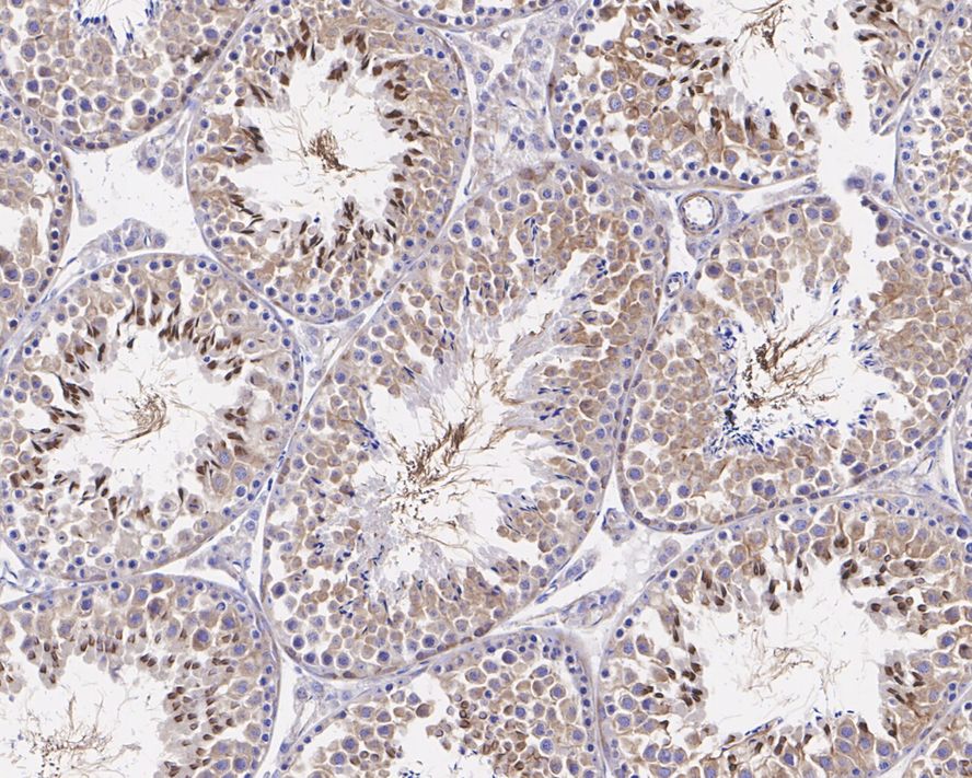
Immunohistochemical analysis of paraffin-embedded mouse testis tissue with Rabbit anti-Cellular Apoptosis Susceptibility antibody at 1/1,000 dilution. The section was pre-treated using heat mediated antigen retrieval with sodium citrate buffer (pH 6.0) for 2 minutes. The tissues were blocked in 1% BSA for 20 minutes at room temperature, washed with ddH2O and PBS, and then probed with the primary antibody at 1/1,000 dilution for 1 hour at room temperature. The detection was performed using an HRP conjugated compact polymer system. DAB was used as the chromogen. Tissues were counterstained with hematoxylin and mounted with DPX.ICC/IF
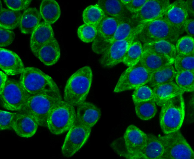
ICC staining Cellular Apoptosis Susceptibility in LOVO cells (green). The nuclear counter stain is DAPI (blue). Cells were fixed in paraformaldehyde, permeabilised with 0.25% Triton X100/PBS.FC
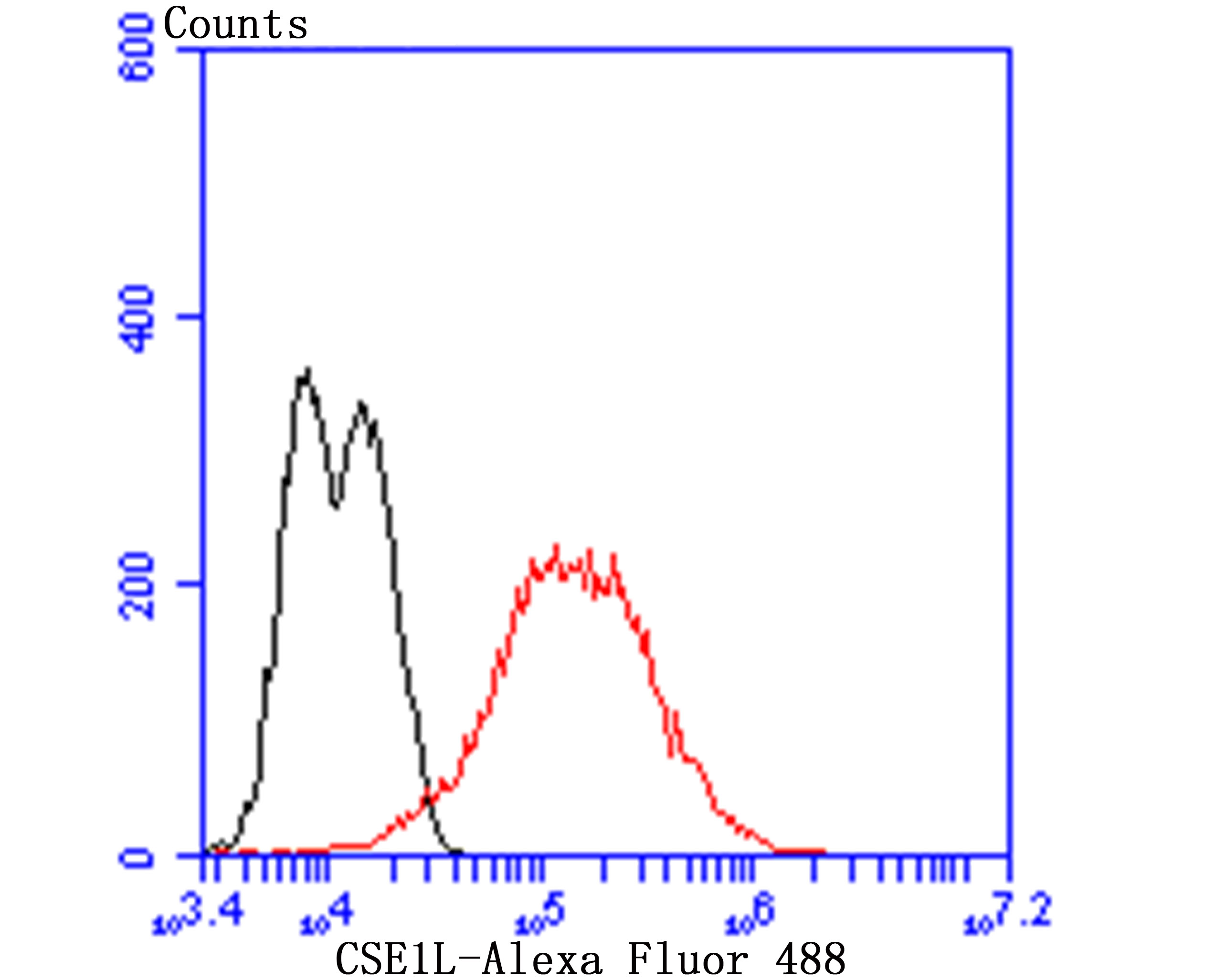
Flow cytometric analysis of LOVO cells with Cellular Apoptosis Susceptibility antibody at 1/100 dilution (red) compared with an unlabelled control (cells without incubation with primary antibody; black). Alexa Fluor 488-conjugated goat anti-rabbit IgG was used as the secondary antibody.| Product Name | Cellular Apoptosis Susceptibility Recombinant Rabbit Monoclonal Antibody |
|---|---|
| Antibody Type | Primary Antibodies |
| Product description | Cellular apoptosis susceptibility protein (CAS), also called Exportin 2, is a 971 amino acid member of the CSE1 family. CAS mediates Importin α re-export from the nucleus to the cytoplasm after import substrates have been released into the nucleoplasm. In the nucleus, CAS binds cooperatively to Importin α and to the GTPase Ran in its GTP-bound (active) form. This complex binds to nucleoporins as it docks to the nuclear pore complex. Once in the cytoplasm, the complex dissociates and Importin α is released and CAS returns to the nuclear compartment and the process begins anew. CAS can be detected highly in proliferating cells. Three isoforms of CAS have been named due to alternative splicing. Isoform 1 is the full length, 971 amino acid protein. Isoform 2 contains an alternative sequence for amino acids 190-195 and is missing amino acids 196-971. Isoform 3 contains an alternative sequence for amino acids 943-945 and is missing amino acids 946-971. |
| Immunogen | Recombinant protein within Human Cellular Apoptosis Susceptibility aa 860-971 / 971. |
| Clonality | monoclonal |
|---|---|
| Isotype | IgG |
| Host Species | Recombinant rabbit |
| Tested Applications | FCICC/IFIHCWB |
| WB:1:1000 IHC:1:200-1:1000 ICC/IF:1:50-1:200 FC:1:50-1:100 |
|
| Species Reactivity | HumanMouseRat |
| Concentration | 1mg/ml |
| Purification | Protein A |
| Alternative Names | CAS antibody Cellular apoptosis susceptibility protein antibody Chromosome segregation 1 (yeast homolog) like antibody Chromosome segregation 1 Like antibody Chromosome segregation 1 like protein antibody Chromosome segregation 1-like protein antibody Chromosome segregation gene CSE1 antibody CSE 1 antibody CSE 1 chromosome segregation 1 like antibody CSE 1 chromosome segregation 1 like protein antibody CSE 1L antibody CSE1 antibody CSE1 chromosome segregation 1 like (yeast) antibody CSE1 chromosome segregation 1 like antibody CSE1 chromosome segregation 1 like protein antibody CSE1L antibody Exp 2 antibody Exp2 antibody Exportin 2 antibody Exportin-2 antibody Exportin2 antibody Importin alpha re exporter antibody Importin-alpha re-exporter antibody MGC117283 antibody MGC130036 antibody MGC130037 antibody XPO 2 antibody XPO2 antibody XPO2_HUMAN antibody |
|---|---|
| Molecular Weight(MW) | 110 kDa |
| Cellular Localization | Cytoplasm. Nucleus. |
WB
Western blot analysis of Cellular Apoptosis Susceptibility on different lysates with Rabbit anti-Cellular Apoptosis Susceptibility antibody at 1/1,000 dilution. Lane 1: SW480 cell lysate (20 µg/Lane), Lane 2: MCF7 cell lysate (20 µg/Lane), Lane 3: SK-Br-3 cell lysate (20 µg/Lane), Lane 4: SiHa cell lysate (20 µg/Lane), Lane 5: PC-3M cell lysate (20 µg/Lane), Lane 6: HeLa cell lysate (20 µg/Lane), Lane 7: Ramos cell lysate (20 µg/Lane), Lane 8: Mouse testis tissue lysate (40 µg/Lane), Exposure time: 30 seconds; 4-20% SDS-PAGE gel. Proteins were transferred to a PVDF membrane and blocked with 5% NFDM/TBST for 1 hour at room temperature. The primary antibody at 1/1,000 dilution was used in 5% NFDM/TBST at room temperature for 2 hours. Goat Anti-Rabbit IgG - HRP Secondary Antibody at 1:100,000 dilution was used for 1 hour at room temperature.
IHC
Immunohistochemical analysis of paraffin-embedded human colon carcinoma tissue with Rabbit anti-Cellular Apoptosis Susceptibility antibody at 1/200 dilution. The section was pre-treated using heat mediated antigen retrieval with sodium citrate buffer (pH 6.0) for 2 minutes. The tissues were blocked in 1% BSA for 20 minutes at room temperature, washed with ddH2O and PBS, and then probed with the primary antibody at 1/200 dilution for 1 hour at room temperature. The detection was performed using an HRP conjugated compact polymer system. DAB was used as the chromogen. Tissues were counterstained with hematoxylin and mounted with DPX.
IHC
Immunohistochemical analysis of paraffin-embedded mouse testis tissue with Rabbit anti-Cellular Apoptosis Susceptibility antibody at 1/1,000 dilution. The section was pre-treated using heat mediated antigen retrieval with sodium citrate buffer (pH 6.0) for 2 minutes. The tissues were blocked in 1% BSA for 20 minutes at room temperature, washed with ddH2O and PBS, and then probed with the primary antibody at 1/1,000 dilution for 1 hour at room temperature. The detection was performed using an HRP conjugated compact polymer system. DAB was used as the chromogen. Tissues were counterstained with hematoxylin and mounted with DPX.
ICC/IF
ICC staining Cellular Apoptosis Susceptibility in LOVO cells (green). The nuclear counter stain is DAPI (blue). Cells were fixed in paraformaldehyde, permeabilised with 0.25% Triton X100/PBS.
FC
Flow cytometric analysis of LOVO cells with Cellular Apoptosis Susceptibility antibody at 1/100 dilution (red) compared with an unlabelled control (cells without incubation with primary antibody; black). Alexa Fluor 488-conjugated goat anti-rabbit IgG was used as the secondary antibody.| Positive Control | Mouse testis tissue, SiHa, SK-BR-3, PC-3M, LOVO, SH-SY5Y, human colon cancer tissue, LOVO. |
|---|---|
| Application Notes | WB:1:1000 IHC:1:200-1:1000 ICC/IF:1:50-1:200 FC:1:50-1:100 |
| Form | Liquid |
|---|---|
| Storage Instructions | Store at +4℃ after thawing. Aliquot store at -20℃ or -80℃. Avoid repeated freeze / thaw cycles. |
| Storage Buffer | 1*TBS (pH7.4), 1%BSA, 40%Glycerol. Preservative: 0.05% Sodium Azide. |


