Application
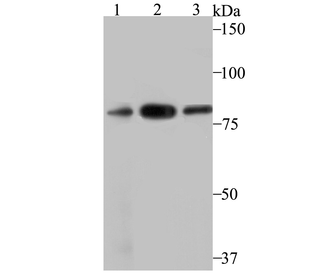
Fig1: Western blot analysis of VPS35 on different lysates using anti-VPS35 antibody at 1/500 dilution. Positive control: Lane 1: PC-3M Lane 2: U937 Lane 3: SH-SY-5YApplication
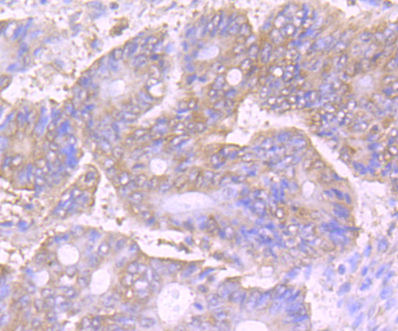
Fig2: Immunohistochemical analysis of paraffin-embedded human colon cancer tissue using anti-VPS35 antibody. Counter stained with hematoxylin.Application
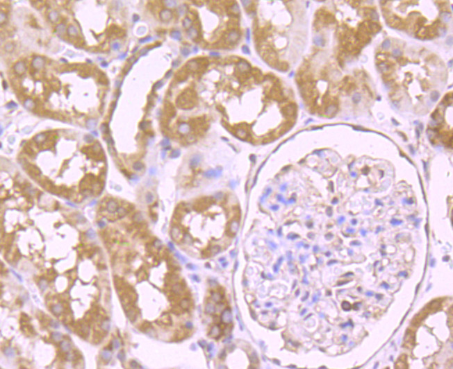
Fig3: Immunohistochemical analysis of paraffin-embedded human kidney tissue using anti-VPS35 antibody. Counter stained with hematoxylin.Application
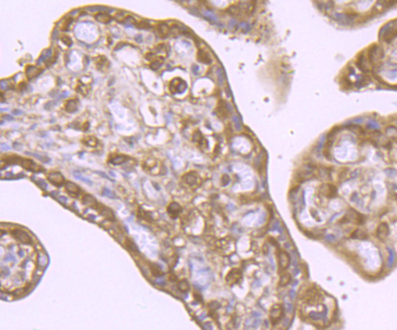
Fig4: Immunohistochemical analysis of paraffin-embedded human placenta tissue using anti-VPS35 antibody. Counter stained with hematoxylin.Application
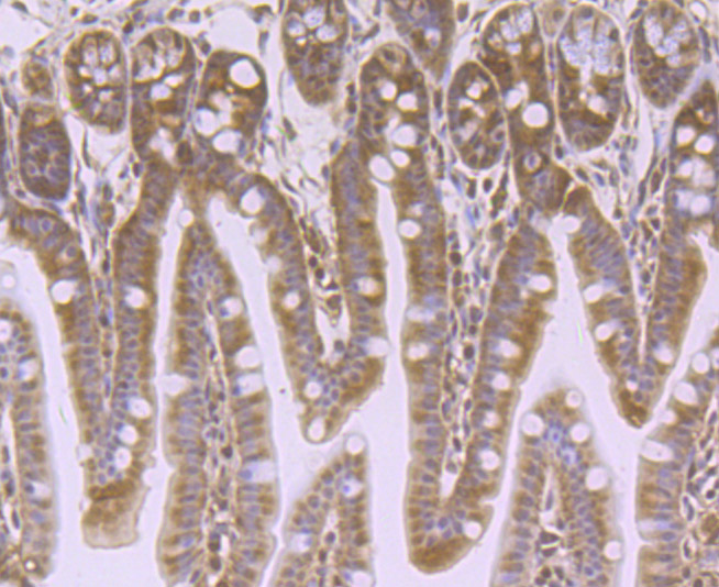
Fig5: Immunohistochemical analysis of paraffin-embedded mouse colon tissue using anti-VPS35 antibody. Counter stained with hematoxylin.Application
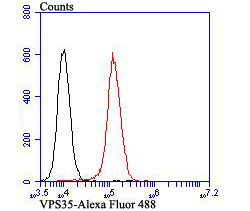
Fig6: Flow cytometric analysis of 293T cells with VPS35 antibody at 1/100 dilution (red) compared with an unlabelled control (cells without incubation with primary antibody; black). Alexa Fluor 488-conjugated goat anti-rabbit IgG was used as the secondary antibody.| Product Name | VPS35 [JB33-82] |
|---|---|
| Antibody Type | Primary Antibodies |
| Antigen Alias | DKFZp434E1211 antibody DKFZp434P1672 antibody FLJ10752 antibody FLJ13588 antibody FLJ20388 antibody hVPS35 antibody Maternal embryonic 3 antibody Maternal-embryonic 3 antibody MEM 3 antibody MEM3 antibody PARK17 antibody TCCCTA00141 antibody Vacuolar protein sorting 35 (yeast) antibody Vacuolar protein sorting 35 antibody Vacuolar protein sorting 35 homolog antibody Vacuolar protein sorting associated protein 35 antibody Vacuolar protein sorting-associated protein 35 antibody Vesicle protein sorting 35 antibody VPS 35 antibody VPS35 antibody VPS35_HUMAN antibody |
| Product description | Vacuolar sorting proteins (VPSs) are required for proper trafficking of endocytic and biosynthetic proteins to the vacuole and play an important role in the budding process of cells. VPS35 (vacuolar protein sorting 35), also known as MEM3, is the 796 amino acid human homolog of the S. cerevisiae Vps35 protein. Localized to the cytoplasm and to the peripheral membrane, VPS35 is an essential component of the retromer complex which is involved in retrieval of lysosomal enzyme receptors from endosomes to the trans-Golgi network. VPS35 is expressed ubiquitously with highest expression in heart, placenta, brain, testis, kidney, colon, ovary and spleen. In addition to its crucial role in the retromer complex, VPS35 is part of a subcomplex that is required to regulate transcytosis of the polymeric immunoglobulin receptor from the basolateral to the apical surface of epithelial cells and hepatocytes. |
| Immunogen | Recombinant protein |
| Clonality | Monoclonal |
|---|---|
| Isotype | IgG |
| Host Species | Recombinant rabbit |
| Tested Applications | WBIHCFC |
| WB:1:500-1:1,000 IHC:1:100-1:300 FC:1:50-1:100 |
|
| Species Reactivity | HumanMouse |
| Concentration | 1mg/ml |
| Alternative Names | DKFZp434E1211 antibody DKFZp434P1672 antibody FLJ10752 antibody FLJ13588 antibody FLJ20388 antibody hVPS35 antibody Maternal embryonic 3 antibody Maternal-embryonic 3 antibody MEM 3 antibody MEM3 antibody PARK17 antibody TCCCTA00141 antibody Vacuolar protein sorting 35 (yeast) antibody Vacuolar protein sorting 35 antibody Vacuolar protein sorting 35 homolog antibody Vacuolar protein sorting associated protein 35 antibody Vacuolar protein sorting-associated protein 35 antibody Vesicle protein sorting 35 antibody VPS 35 antibody VPS35 antibody VPS35_HUMAN antibody |
|---|---|
| Molecular Weight(MW) | 91 kDa |
| Cellular Localization | Cytoplasm. Membrane. Endosome. |
Application
Fig1: Western blot analysis of VPS35 on different lysates using anti-VPS35 antibody at 1/500 dilution. Positive control: Lane 1: PC-3M Lane 2: U937 Lane 3: SH-SY-5Y
Application
Fig2: Immunohistochemical analysis of paraffin-embedded human colon cancer tissue using anti-VPS35 antibody. Counter stained with hematoxylin.
Application
Fig3: Immunohistochemical analysis of paraffin-embedded human kidney tissue using anti-VPS35 antibody. Counter stained with hematoxylin.
Application
Fig4: Immunohistochemical analysis of paraffin-embedded human placenta tissue using anti-VPS35 antibody. Counter stained with hematoxylin.
Application
Fig5: Immunohistochemical analysis of paraffin-embedded mouse colon tissue using anti-VPS35 antibody. Counter stained with hematoxylin.
Application
Fig6: Flow cytometric analysis of 293T cells with VPS35 antibody at 1/100 dilution (red) compared with an unlabelled control (cells without incubation with primary antibody; black). Alexa Fluor 488-conjugated goat anti-rabbit IgG was used as the secondary antibody.| Positive Control | PC-3M, U937, SH-SY-5Y, human colon cancer tissue, human kidney tissue, human placenta tissue, mouse colon tissue, 293T. |
|---|---|
| Application Notes | WB:1:500-1:1,000 IHC:1:100-1:300 FC:1:50-1:100 |
| Form | Liquid |
|---|---|
| Storage Instructions | Store at +4℃ after thawing. Aliquot store at -20℃ or -80℃. Avoid repeated freeze / thaw cycles. |
| Storage Buffer | 1*TBS (pH7.4), 1%BSA, 40%Glycerol. Preservative: 0.05% Sodium Azide. |


