Application
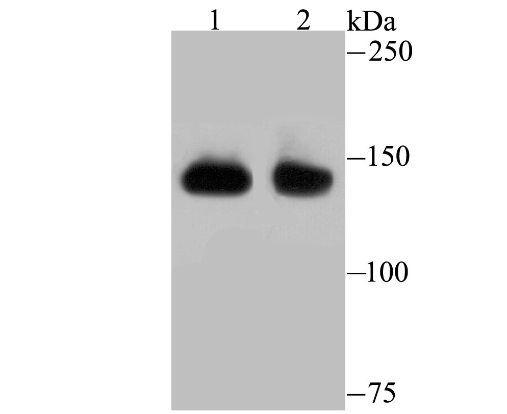
Fig1: Western blot analysis of SF3B1 on different lysates using anti-SF3B1 antibody at 1/500 dilution. Positive control: Lane 1: Mouse thymus Lane 2: HelaApplication
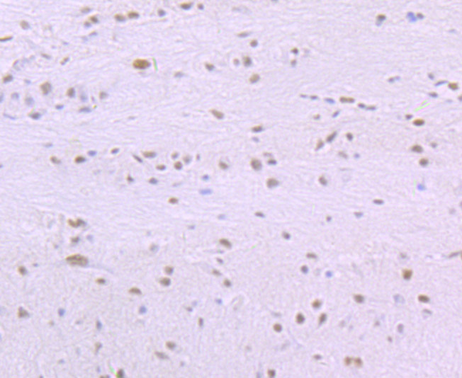
Fig2: Immunohistochemical analysis of paraffin-embedded rat brain tissue using anti-SF3B1 antibody. Counter stained with hematoxylin.Application
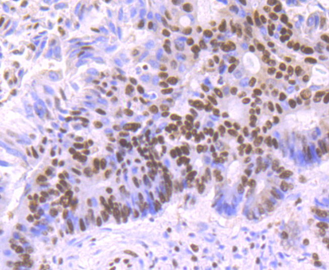
Fig3: Immunohistochemical analysis of paraffin-embedded human colon cancer tissue using anti-SF3B1 antibody. Counter stained with hematoxylin.Application
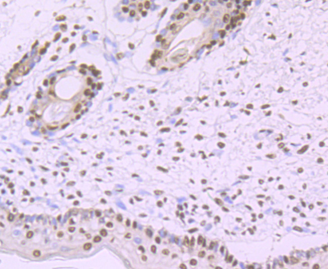
Fig4: Immunohistochemical analysis of paraffin-embedded human skin tissue using anti-SF3B1 antibody. Counter stained with hematoxylin.Application
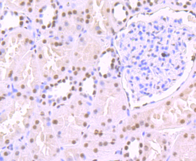
Fig5: Immunohistochemical analysis of paraffin-embedded human kidney tissue using anti-SF3B1 antibody. Counter stained with hematoxylin.ICC/IF
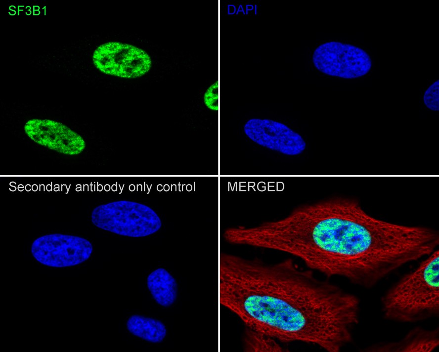
Fig6: Immunocytochemistry analysis of HeLa cells labeling SF3B1 with Rabbit anti-SF3B1 antibody at 1/100 dilution. Cells were fixed in 4% paraformaldehyde for 15 minutes at room temperature, permeabilized with 0.1% Triton X-100 in PBS for 15 minutes at room temperature, then blocked with 1% BSA in 10% negative goat serum for 1 hour at room temperature. Cells were then incubated with Rabbit anti-SF3B1 antibody at 1/100 dilution in 1% BSA in PBST overnight at 4 ℃. Goat Anti-Rabbit IgG H&L (488) was used as the secondary antibody at 1/1,000 dilution. PBS instead of the primary antibody was used as the secondary antibody only control. Nuclear DNA was labelled in blue with DAPI. Beta tubulin (red) was stained at 1/100 dilution overnight at +4℃. Goat Anti-Mouse IgG H&L (594) was used as the secondary antibody at 1/1,000 dilution| Product Name | SF3B1 Recombinant Rabbit Monoclonal Antibody |
|---|---|
| Antibody Type | Primary Antibodies |
| Product description | Involved in pre-mRNA splicing as a component of the splicing factor SF3B complex. SF3B complex is required for 'A' complex assembly formed by the stable binding of U2 snRNP to the branchpoint sequence (BPS) in pre-mRNA. Sequence independent binding of SF3A/SF3B complex upstream of the branch site is essential, it may anchor U2 snRNP to the pre-mRNA. May also be involved in the assembly of the 'E' complex. Belongs also to the minor U12-dependent spliceosome, which is involved in the splicing of rare class of nuclear pre-mRNA intron. |
| Immunogen | Recombinant protein within Human SF3B1 aa 1-105 / 1,304. |
| Clonality | monoclonal |
|---|---|
| Isotype | IgG |
| Host Species | Recombinant rabbit |
| Tested Applications | ICC/IFIHCWB |
| WB:1:500-1:2,000 IHC:1:50-1:200 ICC/IF:1:100 |
|
| Species Reactivity | HumanMouseRat |
| Concentration | 1mg/ml |
| Purification | Protein A |
| Alternative Names | Hsh 155 antibody MDS antibody Pre mRNA processing 10 antibody Pre mRNA splicing factor SF3b 155 kDa subunit antibody PRP 10 antibody PRP10 antibody PRPF 10 antibody PRPF10 antibody SAP 155 antibody SAP155 antibody SF3B 1 antibody SF3b155 antibody Spliceosome associated protein 155 antibody Splicing factor 3b subunit 1 155kDa antibody Splicing factor 3B subunit 1 antibody |
|---|---|
| Molecular Weight(MW) | 146kDa |
| Cellular Localization | Nucleus. |
Application
Fig1: Western blot analysis of SF3B1 on different lysates using anti-SF3B1 antibody at 1/500 dilution. Positive control: Lane 1: Mouse thymus Lane 2: Hela
Application
Fig2: Immunohistochemical analysis of paraffin-embedded rat brain tissue using anti-SF3B1 antibody. Counter stained with hematoxylin.
Application
Fig3: Immunohistochemical analysis of paraffin-embedded human colon cancer tissue using anti-SF3B1 antibody. Counter stained with hematoxylin.
Application
Fig4: Immunohistochemical analysis of paraffin-embedded human skin tissue using anti-SF3B1 antibody. Counter stained with hematoxylin.
Application
Fig5: Immunohistochemical analysis of paraffin-embedded human kidney tissue using anti-SF3B1 antibody. Counter stained with hematoxylin.
ICC/IF
Fig6: Immunocytochemistry analysis of HeLa cells labeling SF3B1 with Rabbit anti-SF3B1 antibody at 1/100 dilution. Cells were fixed in 4% paraformaldehyde for 15 minutes at room temperature, permeabilized with 0.1% Triton X-100 in PBS for 15 minutes at room temperature, then blocked with 1% BSA in 10% negative goat serum for 1 hour at room temperature. Cells were then incubated with Rabbit anti-SF3B1 antibody at 1/100 dilution in 1% BSA in PBST overnight at 4 ℃. Goat Anti-Rabbit IgG H&L (488) was used as the secondary antibody at 1/1,000 dilution. PBS instead of the primary antibody was used as the secondary antibody only control. Nuclear DNA was labelled in blue with DAPI. Beta tubulin (red) was stained at 1/100 dilution overnight at +4℃. Goat Anti-Mouse IgG H&L (594) was used as the secondary antibody at 1/1,000 dilution| Application Notes | WB:1:500-1:2,000 IHC:1:50-1:200 ICC/IF:1:100 |
|---|
| Form | Liquid |
|---|---|
| Storage Instructions | Store at +4℃ after thawing. Aliquot store at -20℃ or -80℃. Avoid repeated freeze / thaw cycles. |
| Storage Buffer | 1*TBS (pH7.4), 1%BSA, 40%Glycerol. Preservative: 0.05% Sodium Azide. |


