Application
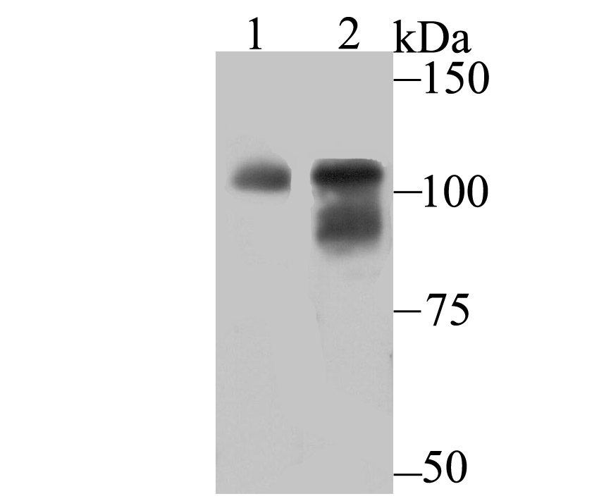
Fig1: Western blot analysis of OGT on Hela (1) and SiHa (2) cell lysate using anti-OGT antibody at 1/500 dilution.Application
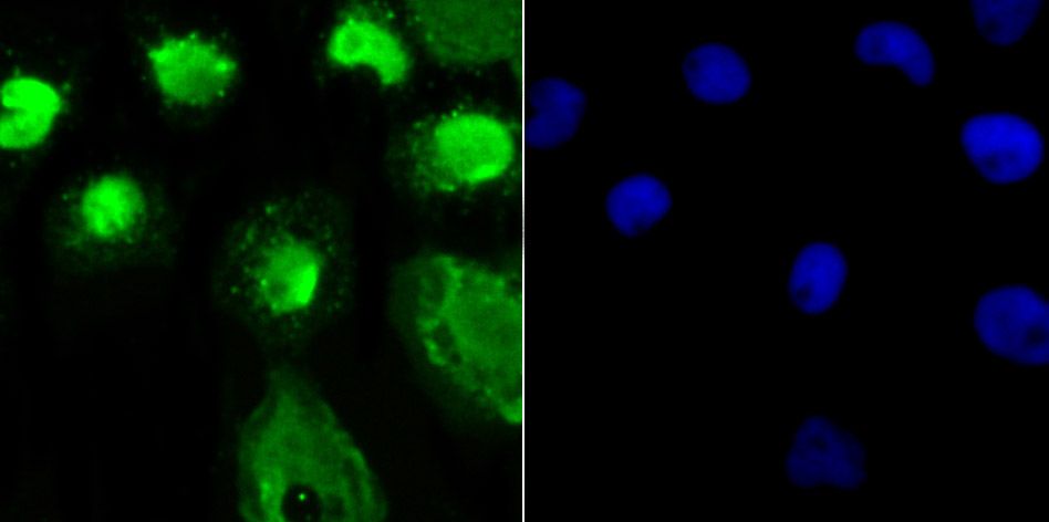
Fig2: ICC staining OGT in A431 cells (green). The nuclear counter stain is DAPI (blue). Cells were fixed in paraformaldehyde, permeabilised with 0.25% Triton X100/PBS.Application
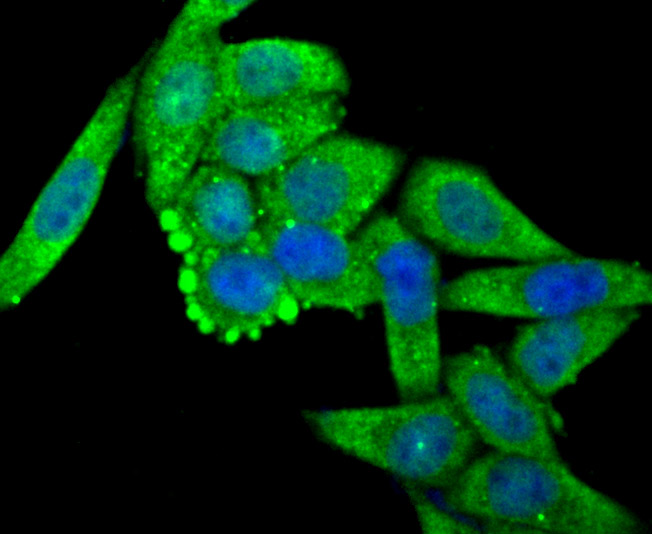
Fig3: ICC staining OGT in Hela cells (green). The nuclear counter stain is DAPI (blue). Cells were fixed in paraformaldehyde, permeabilised with 0.25% Triton X100/PBS.Application
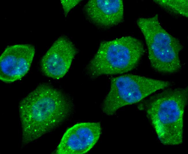
Fig4: ICC staining OGT in PC-3M cells (green). The nuclear counter stain is DAPI (blue). Cells were fixed in paraformaldehyde, permeabilised with 0.25% Triton X100/PBS.Application
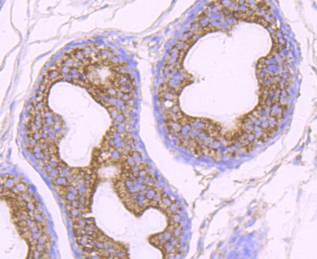
Fig5: Immunohistochemical analysis of paraffin-embedded rat epididymis tissue using anti-OGT antibody. Counter stained with hematoxylin.Application
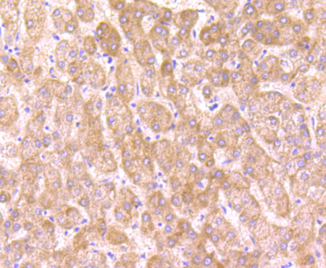
Fig6: Immunohistochemical analysis of paraffin-embedded human liver tissue using anti-OGT antibody. Counter stained with hematoxylin.Application
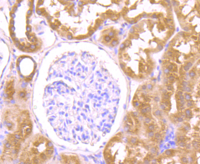
Fig7: Immunohistochemical analysis of paraffin-embedded human kidney tissue using anti-OGT antibody. Counter stained with hematoxylin.Application
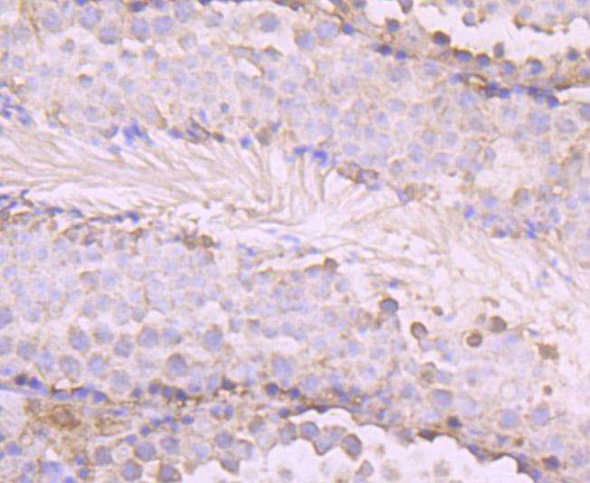
Fig8: Immunohistochemical analysis of paraffin-embedded mouse testis tissue using anti-OGT antibody. Counter stained with hematoxylin.Application
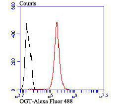
Fig9: Flow cytometric analysis of Hela cells with OGT antibody at 1/100 dilution (red) compared with an unlabelled control (cells without incubation with primary antibody; black). Alexa Fluor 488-conjugated goat anti rabbit IgG was used as the secondary antibody| Product Name | OGT [JB44-39] |
|---|---|
| Antibody Type | Primary Antibodies |
| Antigen Alias | FLJ23071 antibody GlcNAc transferase antibody HRNT1 antibody MGC22921 antibody O GlcNAc antibody O GlcNAc transferase p110 subunit antibody O GlcNAc transferase subunit p110 antibody O linked N acetylglucosamine (GlcNAc) transferase (UDP N acetylglucosamine:polypeptide N acetylglucosaminyl transferase) antibody O linked N acetylglucosamine (GlcNAc) transferase antibody O linked N acetylglucosamine transferase 110 kDa subunit antibody O-GlcNAc transferase subunit p110 antibody O-linked N-acetylglucosamine transferase 110 kDa subunit antibody ogt antibody OGT1_HUMAN antibody UDP N acetylglucosamine peptide N acetylglucosaminyltransferase 110 kDa subunit antibody UDP N acetylglucosamine peptide N acetylglucosaminyltransferase GlcNAc transferase antibody UDP-N-acetylglucosaminepeptide N-acetylglucosaminyltransferase 110 kDa subunit antibody UDP-N-acetylglucosamine:polypeptide-N-acetylglucosaminyl transferase antibody Uridinediphospho N acetylglucosamine:polypeptide beta N acetylglucosaminyl transferase antibody |
| Product description | O-linked N-acetylglucosamine (O-GlcNAc) transferase (also designated OGT) catalyzes the addition of a single N-acetylglucosamine in O-glycosidic linkage to serine or threonine residues. Since both phosphorylation and glycosylation compete for similar serine or threonine residues, the two processes may compete for sites, or they may alter the substrate specificity of nearby sites by steric or electrostatic effects. O-GlcNAc transferase has been purified from rat liver. It exists as a heterotrimeric complex with two subunits of the same molecular mass and one shorter subunit. Both polypeptides are related; the short subunit band is either a proteolytic product of the polypeptide or the product of an alternative translation start site. O-GlcNAc transferase is expressed as multiple transcripts that are present in different amounts in various human tissues, with the highest levels of expression in pancreas. Immunofluorescence of human cells expressing rat O-GlcNAc transferase indicated that it is present in both the nucleus and cytosol. HeLa cells expressing O-GlcNAc transferase do not survive well during prolonged incubations, suggesting that this protein may be toxic to the cells. |
| Immunogen | Recombinant protein |
| Clonality | Monoclonal |
|---|---|
| Isotype | IgG |
| Host Species | Recombinant rabbit |
| Tested Applications | WBICCIFIHCFC |
| WB:1:500 ICC:1:500-1:1,000 IHC:1:50-1:200 FC:1:50-1:100 |
|
| Species Reactivity | HumanMouseRat |
| Concentration | 1mg/ml |
| Alternative Names | FLJ23071 antibody GlcNAc transferase antibody HRNT1 antibody MGC22921 antibody O GlcNAc antibody O GlcNAc transferase p110 subunit antibody O GlcNAc transferase subunit p110 antibody O linked N acetylglucosamine (GlcNAc) transferase (UDP N acetylglucosamine:polypeptide N acetylglucosaminyl transferase) antibody O linked N acetylglucosamine (GlcNAc) transferase antibody O linked N acetylglucosamine transferase 110 kDa subunit antibody O-GlcNAc transferase subunit p110 antibody O-linked N-acetylglucosamine transferase 110 kDa subunit antibody ogt antibody OGT1_HUMAN antibody UDP N acetylglucosamine peptide N acetylglucosaminyltransferase 110 kDa subunit antibody UDP N acetylglucosamine peptide N acetylglucosaminyltransferase GlcNAc transferase antibody UDP-N-acetylglucosaminepeptide N-acetylglucosaminyltransferase 110 kDa subunit antibody UDP-N-acetylglucosamine:polypeptide-N-acetylglucosaminyl transferase antibody Uridinediphospho N acetylglucosamine:polypeptide beta N acetylglucosaminyl transferase antibody |
|---|---|
| Molecular Weight(MW) | 117 kDa |
| Cellular Localization | Nucleus. Cytoplasm. Membrane. |
Application
Fig1: Western blot analysis of OGT on Hela (1) and SiHa (2) cell lysate using anti-OGT antibody at 1/500 dilution.
Application
Fig2: ICC staining OGT in A431 cells (green). The nuclear counter stain is DAPI (blue). Cells were fixed in paraformaldehyde, permeabilised with 0.25% Triton X100/PBS.
Application
Fig3: ICC staining OGT in Hela cells (green). The nuclear counter stain is DAPI (blue). Cells were fixed in paraformaldehyde, permeabilised with 0.25% Triton X100/PBS.
Application
Fig4: ICC staining OGT in PC-3M cells (green). The nuclear counter stain is DAPI (blue). Cells were fixed in paraformaldehyde, permeabilised with 0.25% Triton X100/PBS.
Application
Fig5: Immunohistochemical analysis of paraffin-embedded rat epididymis tissue using anti-OGT antibody. Counter stained with hematoxylin.
Application
Fig6: Immunohistochemical analysis of paraffin-embedded human liver tissue using anti-OGT antibody. Counter stained with hematoxylin.
Application
Fig7: Immunohistochemical analysis of paraffin-embedded human kidney tissue using anti-OGT antibody. Counter stained with hematoxylin.
Application
Fig8: Immunohistochemical analysis of paraffin-embedded mouse testis tissue using anti-OGT antibody. Counter stained with hematoxylin.
Application
Fig9: Flow cytometric analysis of Hela cells with OGT antibody at 1/100 dilution (red) compared with an unlabelled control (cells without incubation with primary antibody; black). Alexa Fluor 488-conjugated goat anti rabbit IgG was used as the secondary antibody| Positive Control | Hela, SiHa, A431, PC-3M, rat epididymis tissue, human liver tissue, human kidney tissue, mouse testis tissue. |
|---|---|
| Application Notes | WB:1:500 ICC:1:500-1:1,000 IHC:1:50-1:200 FC:1:50-1:100 |
| Form | Liquid |
|---|---|
| Storage Instructions | Store at +4℃ after thawing. Aliquot store at -20℃ or -80℃. Avoid repeated freeze / thaw cycles. |
| Storage Buffer | 1*TBS (pH7.4), 1%BSA, 40%Glycerol. Preservative: 0.05% Sodium Azide. |


