Application
Fig1: Western blot analysis of SET on K562 cell lysates using anti-SET at 1/500 dilution.Application
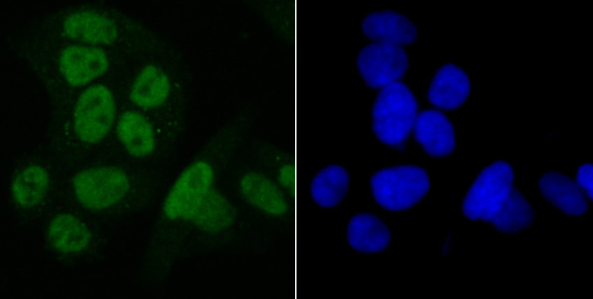
Fig2: ICC staining SET in Hela cells (green). The nuclear counter stain is DAPI (blue). Cells were fixed in paraformaldehyde, permeabilised with 0.25% Triton X100/PBS.Application
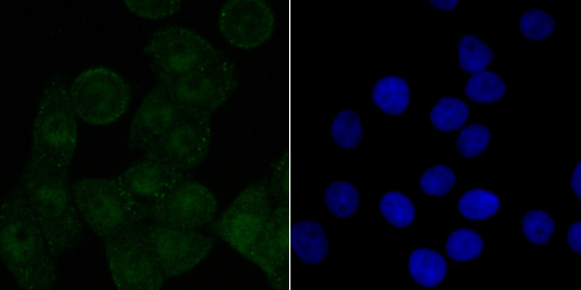
Fig3: ICC staining SET in LOVO cells (green). The nuclear counter stain is DAPI (blue). Cells were fixed in paraformaldehyde, permeabilised with 0.25% Triton X100/PBS.Application
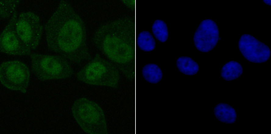
Fig4: ICC staining SET in MCF-7 cells (green). The nuclear counter stain is DAPI (blue). Cells were fixed in paraformaldehyde, permeabilised with 0.25% Triton X100/PBS.Application
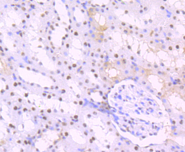
Fig5: Immunohistochemical analysis of paraffin-embedded rat kidney tissue using anti-SET antibody. Counter stained with hematoxylin.Application
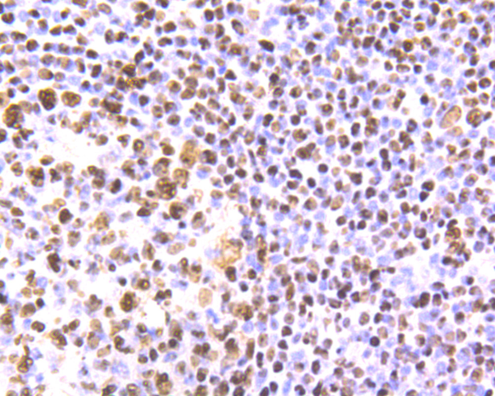
Fig6: Immunohistochemical analysis of paraffin-embedded human tonsil tissue using anti-SET antibody. Counter stained with hematoxylin.Application
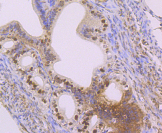
Fig7: Immunohistochemical analysis of paraffin-embedded mouse fallopian tube tissue using anti-SET antibody. Counter stained with hematoxylin.Application
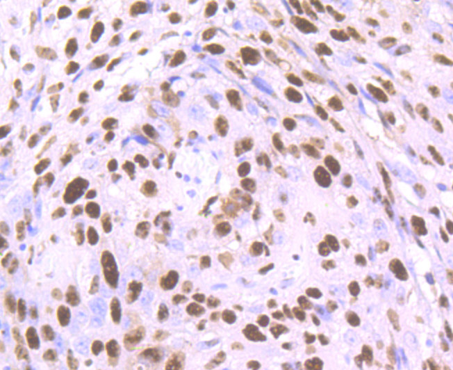
Fig8: Immunohistochemical analysis of paraffin-embedded human lung cancer tissue using anti-SET antibody. Counter stained with hematoxylin.Application
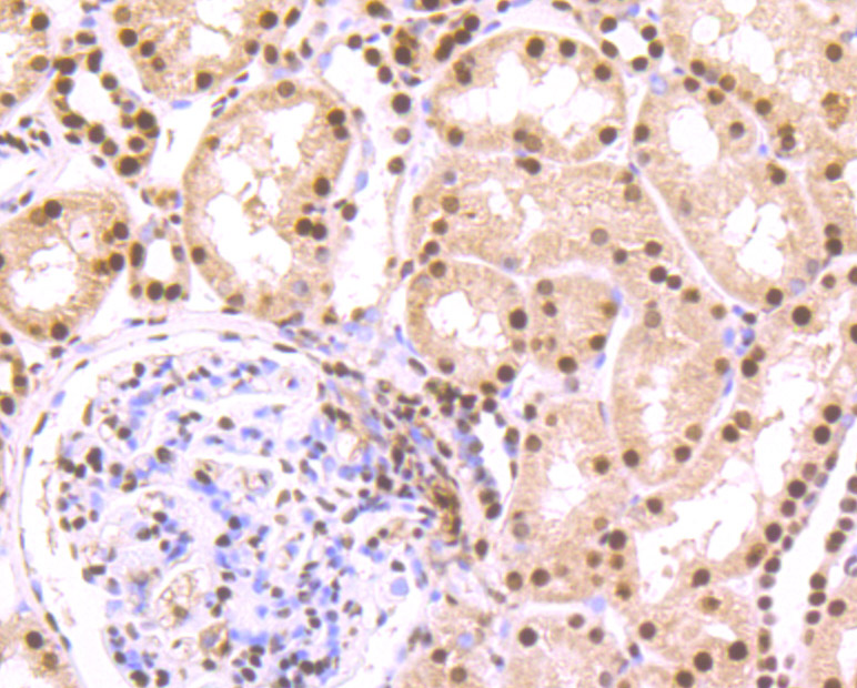
Fig9: Immunohistochemical analysis of paraffin-embedded human kidney tissue using anti-SET antibody. Counter stained with hematoxylin.| Product Name | SET [JB60-34] |
|---|---|
| Antibody Type | Primary Antibodies |
| Antigen Alias | 2PP2A antibody HLA DR associated protein II antibody HLA-DR-associated protein II antibody I 2PP2A antibody I-2PP2A antibody I2PP2A antibody IGAAD antibody Inhibitor of granzyme A activated DNase antibody Inhibitor of granzyme A-activated DNase antibody Inhibitor of GzmA-activated DNase antibody inhibitor-2 of protein phosphatase-2A antibody IPP2A2 antibody PHAPII antibody Phosphatase 2A inhibitor I2PP2A antibody protein phosphatase type 2A inhibitor antibody Protein SET antibody Set antibody SET nuclear oncogene antibody SET translocation antibody SET translocation (myeloid leukemia-associated) antibody SET_HUMAN antibody TAF I antibody TAF IBETA antibody TAF-I antibody TAFI antibody Template activating factor I antibody Template-activating factor I antibody Template-Activating Factor-I, chromatin remodelling factor antibody |
| Product description | Multitasking protein, involved in apoptosis, transcription, nucleosome assembly and histone chaperoning. Isoform 2 anti-apoptotic activity is mediated by inhibition of the GZMA-activated DNase, NME1. In the course of cytotoxic T-lymphocyte (CTL)-induced apoptosis, GZMA cleaves SET, disrupting its binding to NME1 and releasing NME1 inhibition. Isoform 1 and isoform 2 are potent inhibitors of protein phosphatase 2A. Isoform 1 and isoform 2 inhibit EP300/CREBBP and PCAF-mediated acetylation of histones (HAT) and nucleosomes, most probably by masking the accessibility of lysines of histones to the acetylases. The predominant target for inhibition is histone H4. HAT inhibition leads to silencing of HAT-dependent transcription and prevents active demethylation of DNA. Both isoforms stimulate DNA replication of the adenovirus genome complexed with viral core proteins; however, isoform 2 specific activity is higher. |
| Immunogen | Recombinant protein |
| Clonality | Monoclonal |
|---|---|
| Isotype | IgG |
| Host Species | Recombinant rabbit |
| Tested Applications | WBICCIFIHCIPFC |
| WB:1:500-1:2,000 ICC:1:50-1:200 IHC:1:50-1:200 IP:1:10-1:50 FC:1:50-1:100 |
|
| Species Reactivity | HumanMouseRat |
| Concentration | 1mg/ml |
| Alternative Names | 2PP2A antibody
HLA DR associated protein II antibody
HLA-DR-associated protein II antibody
I 2PP2A antibody
I-2PP2A antibody
I2PP2A antibody
IGAAD antibody
Inhibitor of granzyme A activated DNase antibody
Inhibitor of granzyme A-activated DNase antibody
Inhibitor of GzmA-activated DNase antibody
inhibitor-2 of protein phosphatase-2A antibody
IPP2A2 antibody
PHAPII antibody
Phosphatase 2A inhibitor I2PP2A antibody
protein phosphatase type 2A inhibitor antibody
Protein SET antibody
Set antibody
SET nuclear oncogene antibody
SET translocation antibody
SET translocation (myeloid leukemia-associated) antibody
SET_HUMAN antibody
TAF I antibody
TAF IBETA antibody
TAF-I antibody
TAFI antibody
Template activating factor I antibody
Template-activating factor I antibody
Template-Activating Factor-I chromatin remodelling factor antibody |
|---|---|
| Molecular Weight(MW) | 37 kDa |
| Cellular Localization | Nucleus. Cytoplasm. Endoplasmic reticulum. |
Application
Fig1: Western blot analysis of SET on K562 cell lysates using anti-SET at 1/500 dilution.
Application
Fig2: ICC staining SET in Hela cells (green). The nuclear counter stain is DAPI (blue). Cells were fixed in paraformaldehyde, permeabilised with 0.25% Triton X100/PBS.
Application
Fig3: ICC staining SET in LOVO cells (green). The nuclear counter stain is DAPI (blue). Cells were fixed in paraformaldehyde, permeabilised with 0.25% Triton X100/PBS.
Application
Fig4: ICC staining SET in MCF-7 cells (green). The nuclear counter stain is DAPI (blue). Cells were fixed in paraformaldehyde, permeabilised with 0.25% Triton X100/PBS.
Application
Fig5: Immunohistochemical analysis of paraffin-embedded rat kidney tissue using anti-SET antibody. Counter stained with hematoxylin.
Application
Fig6: Immunohistochemical analysis of paraffin-embedded human tonsil tissue using anti-SET antibody. Counter stained with hematoxylin.
Application
Fig7: Immunohistochemical analysis of paraffin-embedded mouse fallopian tube tissue using anti-SET antibody. Counter stained with hematoxylin.
Application
Fig8: Immunohistochemical analysis of paraffin-embedded human lung cancer tissue using anti-SET antibody. Counter stained with hematoxylin.
Application
Fig9: Immunohistochemical analysis of paraffin-embedded human kidney tissue using anti-SET antibody. Counter stained with hematoxylin.| Positive Control | K562, Hela, LOVO, MCF-7, human tonsil tissue, human kidney tissue, human lung cancer tissue, mouse fallopian tube tissue, rat kidney tissue. |
|---|---|
| Application Notes | WB:1:500-1:2,000 ICC:1:50-1:200 IHC:1:50-1:200 IP:1:10-1:50 FC:1:50-1:100 |
| Form | Liquid |
|---|---|
| Storage Instructions | Store at +4℃ after thawing. Aliquot store at -20℃ or -80℃. Avoid repeated freeze / thaw cycles. |
| Storage Buffer | 1*TBS (pH7.4), 1%BSA, 40%Glycerol. Preservative: 0.05% Sodium Azide. |


