WB
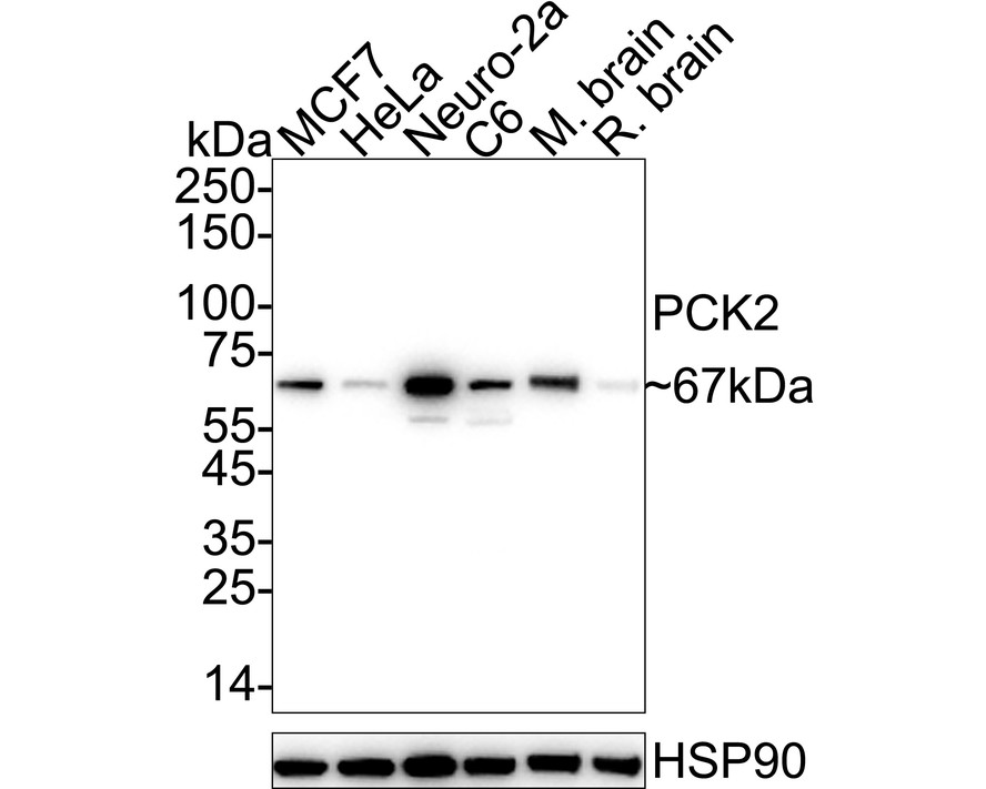
Western blot analysis of PCK2 on different lysates with Rabbit anti-PCK2 antibody at 1/2,000 dilution. Lane 1: MCF7 cell lysate (20 µg/Lane), Lane 2: HeLa cell lysate (20 µg/Lane), Lane 3: Neuro-2a cell lysate (20 µg/Lane), Lane 4: C6 cell lysate (20 µg/Lane), Lane 5: Mouse brain tissue lysate (40 µg/Lane), Lane 6: Rat brain tissue lysate (40 µg/Lane), Exposure time: 20 seconds; 4-20% SDS-PAGE gel. Proteins were transferred to a PVDF membrane and blocked with 5% NFDM/TBST for 1 hour at room temperature. The primary antibody at 1/2,000 dilution was used in 5% NFDM/TBST at 4℃ overnight. Goat Anti-Rabbit IgG - HRP Secondary Antibody at 1/50,000 dilution was used for 1 hour at room temperature.IHC
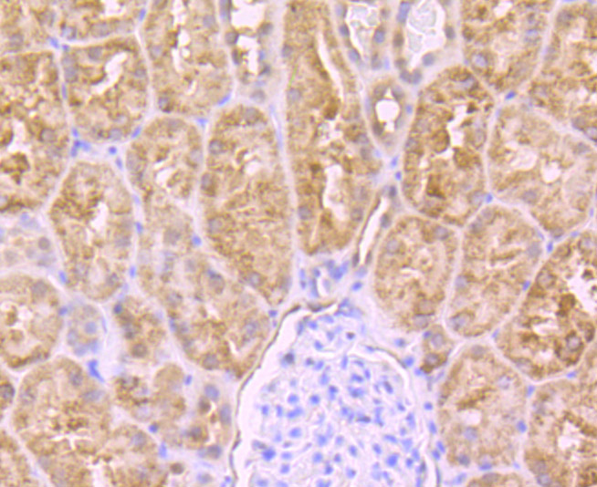
Immunohistochemical analysis of paraffin-embedded human kidney tissue using anti-PCK2 antibody. Counter stained with hematoxylin.IHC
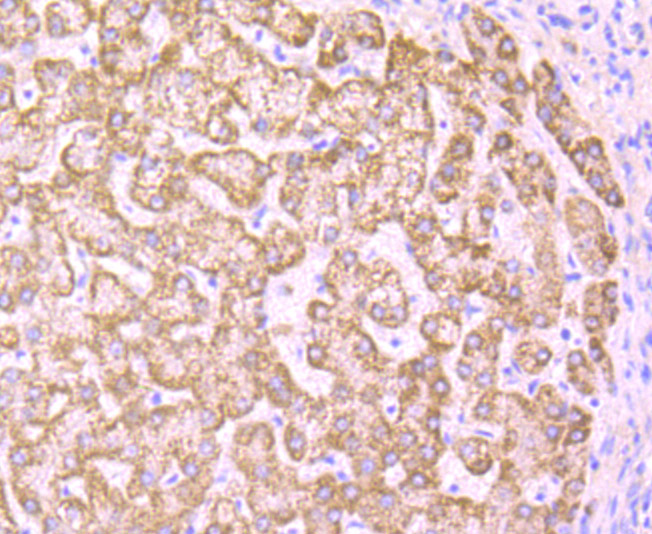
Immunohistochemical analysis of paraffin-embedded human liver tissue using anti-PCK2 antibody. Counter stained with hematoxylin.IHC
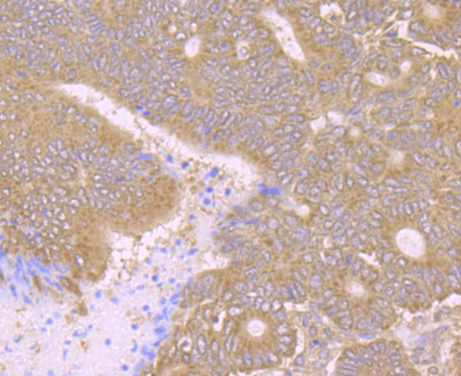
Immunohistochemical analysis of paraffin-embedded human colon cancer tissue using anti-PCK2 antibody. Counter stained with hematoxylin.ICC/IF
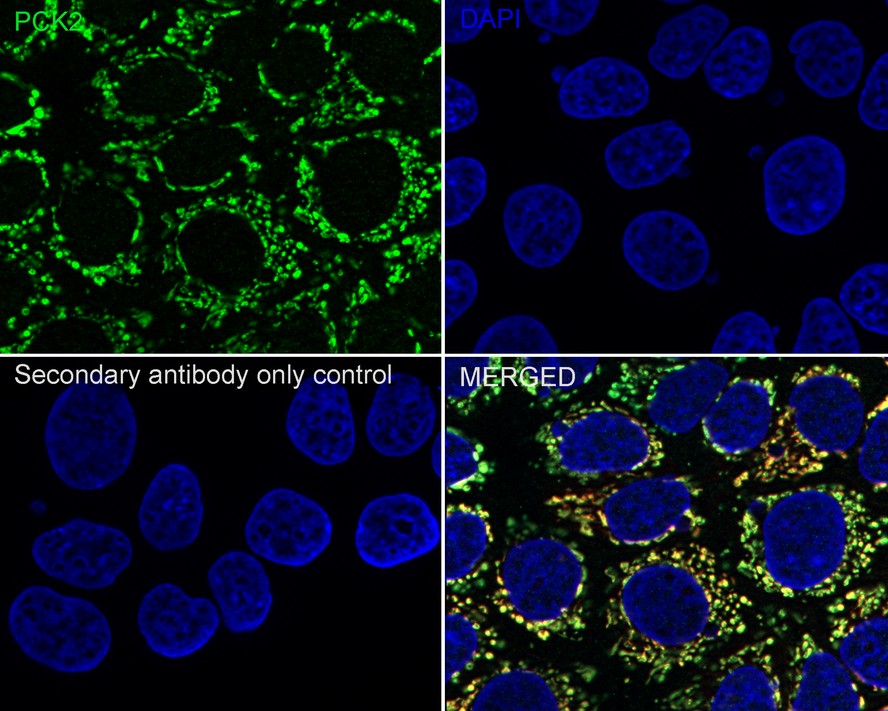
Immunocytochemistry analysis of MCF7 cells labeling PCK2 with Rabbit anti-PCK2 antibody at 1/100 dilution. Cells were fixed in 4% paraformaldehyde for 15 minutes at room temperature, permeabilized with 0.1% Triton X-100 in PBS for 15 minutes at room temperature, then blocked with 1% BSA in 10% negative goat serum for 1 hour at room temperature. Cells were then incubated with Rabbit anti-PCK2 antibody at 1/100 dilution in 1% BSA in PBST overnight at 4 ℃. Goat Anti-Rabbit IgG H&L (488) was used as the secondary antibody at 1/1,000 dilution. PBS instead of the primary antibody was used as the secondary antibody only control. Counterstained with Mitotracker. Nuclear DNA was labelled in blue with DAPI.FC
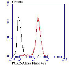
Flow cytometric analysis of MCF-7 cells with PCK2 antibody at 1/100 dilution (red) compared with an unlabelled control (cells without incubation with primary antibody; black). Alexa Fluor 488-conjugated goat anti rabbit IgG was used as the secondary antibody.| Product Name | PCK2 Recombinant Rabbit Monoclonal Antibody |
|---|---|
| Antibody Type | Primary Antibodies |
| Product description | Normal adjustment to changes in blood glucose levels depends on Insulin signaling as well as enzymes involved in the regulation of gluconeogenesis. Pathological changes to this process are central to the type 2 diabetes phenotype. Phosphoenolpyruvate carboxykinase (PEPCK) plays an important role in this process by stimulating hepatic glucose production. PEPCK expression increases in response to glucagon and glucocorticoids, while Insulin suppresses expression. Modulation of the signals governing PEPCK levels present a potential therapeutic approach to the treatment of Insulin resistance and consequently obesity. The cytosolic form of PEPCK, known as PEPCK-C, and the mitochondrial form, known as PEPCK-M, are encoded by two different nuclear genes in mouse, human and chicken. |
| Immunogen | Recombinant protein within C-terminal Human PCK2 . |
| Clonality | Monoclonal |
|---|---|
| Isotype | IgG |
| Host Species | Recombinant rabbit |
| Tested Applications | FCICC/IFIHCWB |
| WB:1:1000-1:2000 IHC:1:50-1:200 ICC/IF:1:100 FC:1:50-1:100 |
|
| Species Reactivity | HumanMouseRat |
| Concentration | 1mg/ml |
| Purification | Protein A |
| Alternative Names | EC 4.1.1.32 antibody GTP mitochondrial precursor antibody HGNC:8725 antibody mitochondrial antibody Mitochondrial phosphoenolpyruvate carboxykinase 2 antibody OTTHUMP00000164700 antibody PCK2 antibody PCKGM_HUMAN antibody PE antibody PEP carboxykinase antibody PEPCK antibody PEPCK deficiency mitochondrial antibody PEPCK M antibody PEPCK-M antibody PEPCK2 antibody Phosphoenolpyruvate carboxykinase [GTP] antibody Phosphoenolpyruvate carboxykinase 2 (mitochondrial) antibody Phosphoenolpyruvate carboxykinase 2 mitochondrial antibody Phosphoenolpyruvate carboxylase antibody Phosphopyruvate carboxylase antibody |
|---|---|
| Molecular Weight(MW) | 71 kDa(Observed band size: 67 kDa) |
| Cellular Localization | Mitochondrion. |
WB
Western blot analysis of PCK2 on different lysates with Rabbit anti-PCK2 antibody at 1/2,000 dilution. Lane 1: MCF7 cell lysate (20 µg/Lane), Lane 2: HeLa cell lysate (20 µg/Lane), Lane 3: Neuro-2a cell lysate (20 µg/Lane), Lane 4: C6 cell lysate (20 µg/Lane), Lane 5: Mouse brain tissue lysate (40 µg/Lane), Lane 6: Rat brain tissue lysate (40 µg/Lane), Exposure time: 20 seconds; 4-20% SDS-PAGE gel. Proteins were transferred to a PVDF membrane and blocked with 5% NFDM/TBST for 1 hour at room temperature. The primary antibody at 1/2,000 dilution was used in 5% NFDM/TBST at 4℃ overnight. Goat Anti-Rabbit IgG - HRP Secondary Antibody at 1/50,000 dilution was used for 1 hour at room temperature.
IHC
Immunohistochemical analysis of paraffin-embedded human kidney tissue using anti-PCK2 antibody. Counter stained with hematoxylin.
IHC
Immunohistochemical analysis of paraffin-embedded human liver tissue using anti-PCK2 antibody. Counter stained with hematoxylin.
IHC
Immunohistochemical analysis of paraffin-embedded human colon cancer tissue using anti-PCK2 antibody. Counter stained with hematoxylin.
ICC/IF
Immunocytochemistry analysis of MCF7 cells labeling PCK2 with Rabbit anti-PCK2 antibody at 1/100 dilution. Cells were fixed in 4% paraformaldehyde for 15 minutes at room temperature, permeabilized with 0.1% Triton X-100 in PBS for 15 minutes at room temperature, then blocked with 1% BSA in 10% negative goat serum for 1 hour at room temperature. Cells were then incubated with Rabbit anti-PCK2 antibody at 1/100 dilution in 1% BSA in PBST overnight at 4 ℃. Goat Anti-Rabbit IgG H&L (488) was used as the secondary antibody at 1/1,000 dilution. PBS instead of the primary antibody was used as the secondary antibody only control. Counterstained with Mitotracker. Nuclear DNA was labelled in blue with DAPI.
FC
Flow cytometric analysis of MCF-7 cells with PCK2 antibody at 1/100 dilution (red) compared with an unlabelled control (cells without incubation with primary antibody; black). Alexa Fluor 488-conjugated goat anti rabbit IgG was used as the secondary antibody.| Application Notes | WB:1:1000-1:2000 IHC:1:50-1:200 ICC/IF:1:100 FC:1:50-1:100 |
|---|
| Form | Liquid |
|---|---|
| Storage Instructions | Store at +4℃ after thawing. Aliquot store at -20℃ or -80℃. Avoid repeated freeze / thaw cycles. |
| Storage Buffer | 1*TBS (pH7.4), 1%BSA, 40%Glycerol. Preservative: 0.05% Sodium Azide. |


