Application
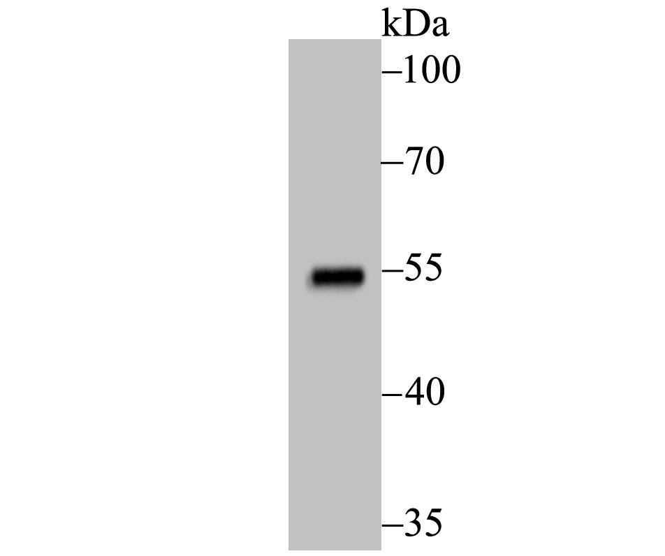
Fig1: Western blot analysis of Mutant p53 on 293 cell using anti-Mutant p53 antibody at 1/500 dilution.Application
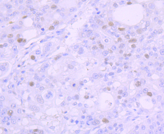
Fig2: Immunohistochemical analysis of paraffin-embedded human stomach cancer tissue using anti-Mutant p53 antibody. Counter stained with hematoxylin.Application
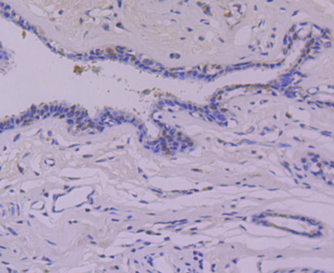
Fig3: Immunohistochemical analysis of paraffin-embedded human breast cancer tissue using anti-Mutant p53 antibody. Counter stained with hematoxylin.Application
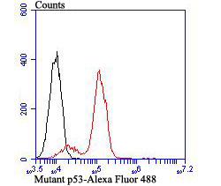
Fig4: Flow cytometric analysis of Daudi cells with Mutant p53 antibody at 1/100 dilution (red) compared with an unlabelled control (cells without incubation with primary antibody; black). Alexa Fluor 488-conjugated goat anti-rabbit IgG was used as the secondary antibody.| Product Name | Mutant p53 [JB42-26] |
|---|---|
| Antibody Type | Primary Antibodies |
| Antigen Alias | Antigen NY-CO-13 antibody BCC7 antibody Cellular tumor antigen p53 antibody LFS1 antibody Mutant tumor protein 53 antibody p53 tumor suppressor antibody P53_HUMAN antibody Phosphoprotein p53 antibody Tp53 antibody Transformation related protein 53 antibody TRP53 antibody Tumor protein p53 antibody Tumor suppressor p53 antibody |
| Product description | Acts as a tumor suppressor in many tumor types; induces growth arrest or apoptosis depending on the physiological circumstances and cell type. Involved in cell cycle regulation as a trans-activator that acts to negatively regulate cell division by controlling a set of genes required for this process. One of the activated genes is an inhibitor of cyclin-dependent kinases. Apoptosis induction seems to be mediated either by stimulation of BAX and FAS antigen expression, or by repression of Bcl-2 expression. In cooperation with mitochondrial PPIF is involved in activating oxidative stress-induced necrosis; the function is largely independent of transcription. Induces the transcription of long intergenic non-coding RNA p21 (lincRNA-p21) and lincRNA-Mkln1. LincRNA-p21 participates in TP53-dependent transcriptional repression leading to apoptosis and seem to have to effect on cell-cycle regulation. Implicated in Notch signaling cross-over. Prevents CDK7 kinase activity when associated to CAK complex in response to DNA damage, thus stopping cell cycle progression. |
| Immunogen | Recombinant protein |
| Clonality | Monoclonal |
|---|---|
| Isotype | IgG |
| Host Species | Recombinant rabbit |
| Tested Applications | WBIHCFCIPICCIF |
| WB:1:500 IHC:1:100 FC:1:50-1:100 ICC:1:100-1:500 |
|
| Species Reactivity | Human |
| Concentration | 1mg/ml |
| Alternative Names | Antigen NY-CO-13 antibody BCC7 antibody Cellular tumor antigen p53 antibody LFS1 antibody Mutant tumor protein 53 antibody p53 tumor suppressor antibody P53_HUMAN antibody Phosphoprotein p53 antibody Tp53 antibody Transformation related protein 53 antibody TRP53 antibody Tumor protein p53 antibody Tumor suppressor p53 antibody |
|---|---|
| Molecular Weight(MW) | 53 kDa |
| Cellular Localization | Cytoplasm. Nucles. |
| SwissProt ID | P04637 |
|---|
Application
Fig1: Western blot analysis of Mutant p53 on 293 cell using anti-Mutant p53 antibody at 1/500 dilution.
Application
Fig2: Immunohistochemical analysis of paraffin-embedded human stomach cancer tissue using anti-Mutant p53 antibody. Counter stained with hematoxylin.
Application
Fig3: Immunohistochemical analysis of paraffin-embedded human breast cancer tissue using anti-Mutant p53 antibody. Counter stained with hematoxylin.
Application
Fig4: Flow cytometric analysis of Daudi cells with Mutant p53 antibody at 1/100 dilution (red) compared with an unlabelled control (cells without incubation with primary antibody; black). Alexa Fluor 488-conjugated goat anti-rabbit IgG was used as the secondary antibody.| Positive Control | 293, Daudi, human breast cancer tissue, human stomach cancer tissue. |
|---|---|
| Application Notes | WB:1:500 IHC:1:100 FC:1:50-1:100 ICC:1:100-1:500 |
| Form | Liquid |
|---|---|
| Storage Instructions | Store at +4℃ after thawing. Aliquot store at -20℃ or -80℃. Avoid repeated freeze / thaw cycles. |
| Storage Buffer | 1*TBS (pH7.4), 1%BSA, 40%Glycerol. Preservative: 0.05% Sodium Azide. |


