Application
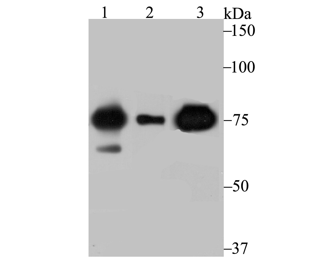
Fig1: Western blot analysis of Cytochrome P450 Reductase on SK-Br-3 (1) , A549 (2) and mouse lung (3) lysate using anti-Cytochrome P450 Reductase antibody at 1/500 dilution.Application
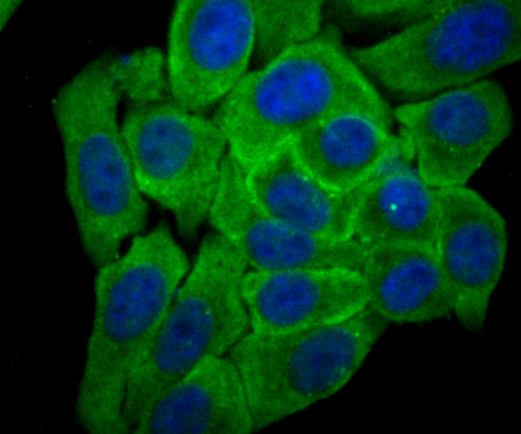
Fig2: ICC staining Cytochrome P450 Reductase in HepG2 cells (green). The nuclear counter stain is DAPI (blue). Cells were fixed in paraformaldehyde, permeabilised with 0.25% Triton X100/PBS.Application
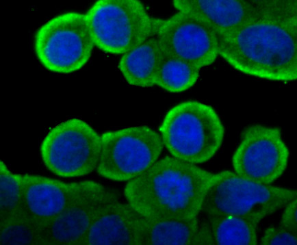
Fig3: ICC staining Cytochrome P450 Reductase in LOVO cells (green). The nuclear counter stain is DAPI (blue). Cells were fixed in paraformaldehyde, permeabilised with 0.25% Triton X100/PBS.Application
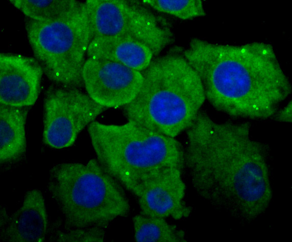
Fig4: ICC staining Cytochrome P450 Reductase in A549 cells (green). The nuclear counter stain is DAPI (blue). Cells were fixed in paraformaldehyde, permeabilised with 0.25% Triton X100/PBS.Application
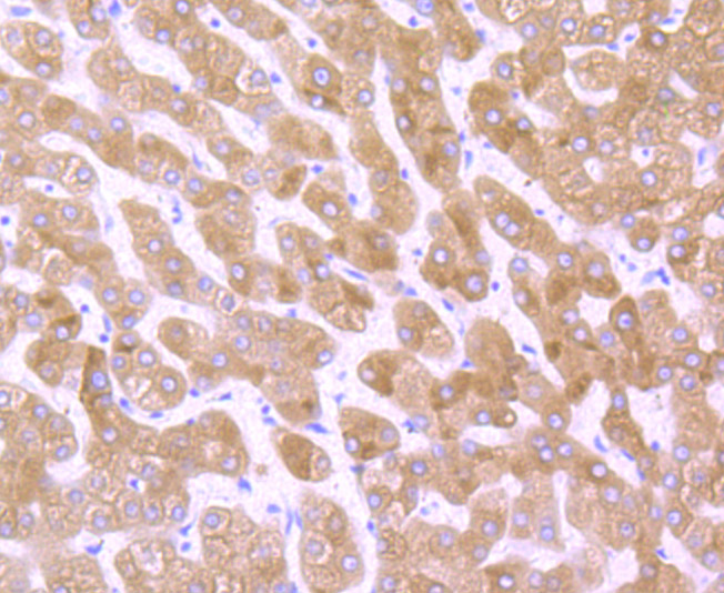
Fig5: Immunohistochemical analysis of paraffin-embedded human liver tissue using anti-Cytochrome P450 Reductase antibody. Counter stained with hematoxylin.Application
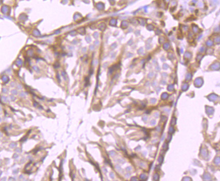
Fig6: Immunohistochemical analysis of paraffin-embedded mouse testis tissue using anti-Cytochrome P450 Reductase antibody. Counter stained with hematoxylin.Application
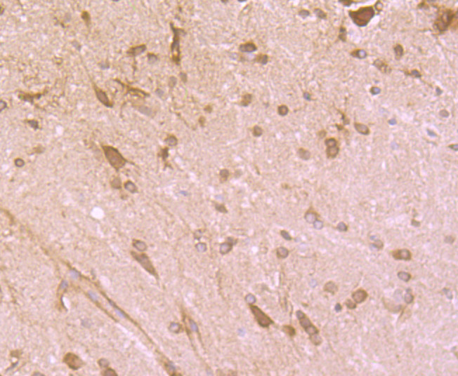
Fig7: Immunohistochemical analysis of paraffin-embedded rat brain tissue using anti-Cytochrome P450 Reductase antibody. Counter stained with hematoxylin.Application
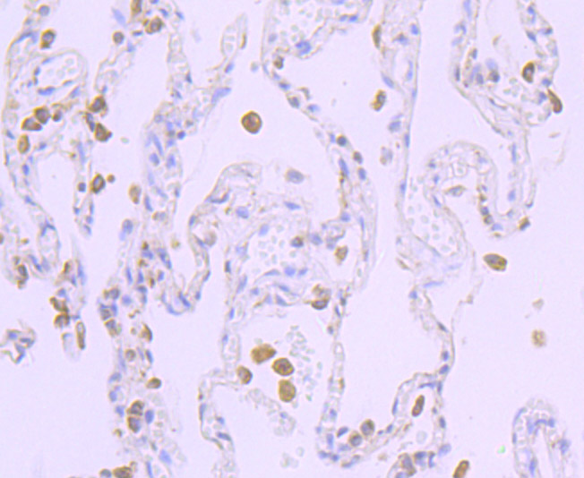
Fig8: Immunohistochemical analysis of paraffin-embedded human lung cancer tissue using anti-Cytochrome P450 Reductase antibody. Counter stained with hematoxylin.Application
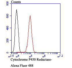
Fig9: Flow cytometric analysis of HepG2 cells with Cytochrome P450 Reductase antibody at 1/100 dilution (red) compared with an unlabelled control (cells without incubation with primary antibody; black). Alexa Fluor 488-conjugated goat anti-rabbit IgG was used as the secondary antibody.| Product Name | Cytochrome P450 Reductase [JB54-31] |
|---|---|
| Antibody Type | Primary Antibodies |
| Antigen Alias | CPR antibody CYPOR antibody Cytochrome p450 oxidoreductase antibody DKFZp686G04235 antibody FLJ26468 antibody NADPH Cytochrome P450 Reductase antibody NADPH dependent cytochrome P450 reductase antibody NADPHcytochrome P450 reductase antibody NCPR_HUMAN antibody P450 (cytochrome) oxidoreductase antibody P450 Cytochrome Oxidoreductase antibody P450R antibody por antibody |
| Product description | P450 enzymes constitute a family of monooxygenase enzymes that are involved in the metabolism of a wide array of endogenous and xenobiotic compounds. Several P450 enzymes have been classified by sequence similarities as members of the CYP1A and CYP2A subfamilies. CYPOR, also known as cytochrome P450 reductase and NADPH cytochrome P450 reductase, is a microsomal enzyme responsible for the transfer of electrons from NADPH to cytochrome P450 enzymes during the P450 catalytic cycle. CYPOR is localized to the endoplasmic reticulum, where it is also able to transfer electrons to heme oxygenase and cytochrome b5. CYPOR is structurally related to two separate flavoprotein families, ferredoxin nucleotide reductase (FNR) and flavodoxin. Electron transfer of CYPOR requires the binding of two flavin cofactors, FAD and FMN, to the FNR and flavodoxin domains, respectively. |
| Immunogen | Recombinant protein |
| Clonality | Monoclonal |
|---|---|
| Isotype | IgG |
| Host Species | Recombinant rabbit |
| Tested Applications | WBICCIFIHCFC |
| WB:1:500-1:1,000 ICC:1:50-1:200 IHC:1:50-1:200 FC:1:50-1:100 |
|
| Species Reactivity | HumanMouseRat |
| Concentration | 1mg/ml |
| Alternative Names | CPR antibody CYPOR antibody Cytochrome p450 oxidoreductase antibody DKFZp686G04235 antibody FLJ26468 antibody NADPH Cytochrome P450 Reductase antibody NADPH dependent cytochrome P450 reductase antibody NADPHcytochrome P450 reductase antibody NCPR_HUMAN antibody P450 (cytochrome) oxidoreductase antibody P450 Cytochrome Oxidoreductase antibody P450R antibody por antibody |
|---|---|
| Molecular Weight(MW) | 77 kDa |
| Cellular Localization | Endoplasmic reticulum. |
Application
Fig1: Western blot analysis of Cytochrome P450 Reductase on SK-Br-3 (1) , A549 (2) and mouse lung (3) lysate using anti-Cytochrome P450 Reductase antibody at 1/500 dilution.
Application
Fig2: ICC staining Cytochrome P450 Reductase in HepG2 cells (green). The nuclear counter stain is DAPI (blue). Cells were fixed in paraformaldehyde, permeabilised with 0.25% Triton X100/PBS.
Application
Fig3: ICC staining Cytochrome P450 Reductase in LOVO cells (green). The nuclear counter stain is DAPI (blue). Cells were fixed in paraformaldehyde, permeabilised with 0.25% Triton X100/PBS.
Application
Fig4: ICC staining Cytochrome P450 Reductase in A549 cells (green). The nuclear counter stain is DAPI (blue). Cells were fixed in paraformaldehyde, permeabilised with 0.25% Triton X100/PBS.
Application
Fig5: Immunohistochemical analysis of paraffin-embedded human liver tissue using anti-Cytochrome P450 Reductase antibody. Counter stained with hematoxylin.
Application
Fig6: Immunohistochemical analysis of paraffin-embedded mouse testis tissue using anti-Cytochrome P450 Reductase antibody. Counter stained with hematoxylin.
Application
Fig7: Immunohistochemical analysis of paraffin-embedded rat brain tissue using anti-Cytochrome P450 Reductase antibody. Counter stained with hematoxylin.
Application
Fig8: Immunohistochemical analysis of paraffin-embedded human lung cancer tissue using anti-Cytochrome P450 Reductase antibody. Counter stained with hematoxylin.
Application
Fig9: Flow cytometric analysis of HepG2 cells with Cytochrome P450 Reductase antibody at 1/100 dilution (red) compared with an unlabelled control (cells without incubation with primary antibody; black). Alexa Fluor 488-conjugated goat anti-rabbit IgG was used as the secondary antibody.| Positive Control | SK-Br-3, A549, mouse lung tissue lysate, rat brain tissue, human lung cancer tissue, human liver tissue, mouse testis tissue, HepG2. |
|---|---|
| Application Notes | WB:1:500-1:1,000 ICC:1:50-1:200 IHC:1:50-1:200 FC:1:50-1:100 |
| Form | Liquid |
|---|---|
| Storage Instructions | Store at +4℃ after thawing. Aliquot store at -20℃ or -80℃. Avoid repeated freeze / thaw cycles. |
| Storage Buffer | 1*TBS (pH7.4), 1%BSA, 40%Glycerol. Preservative: 0.05% Sodium Azide. |


