WB
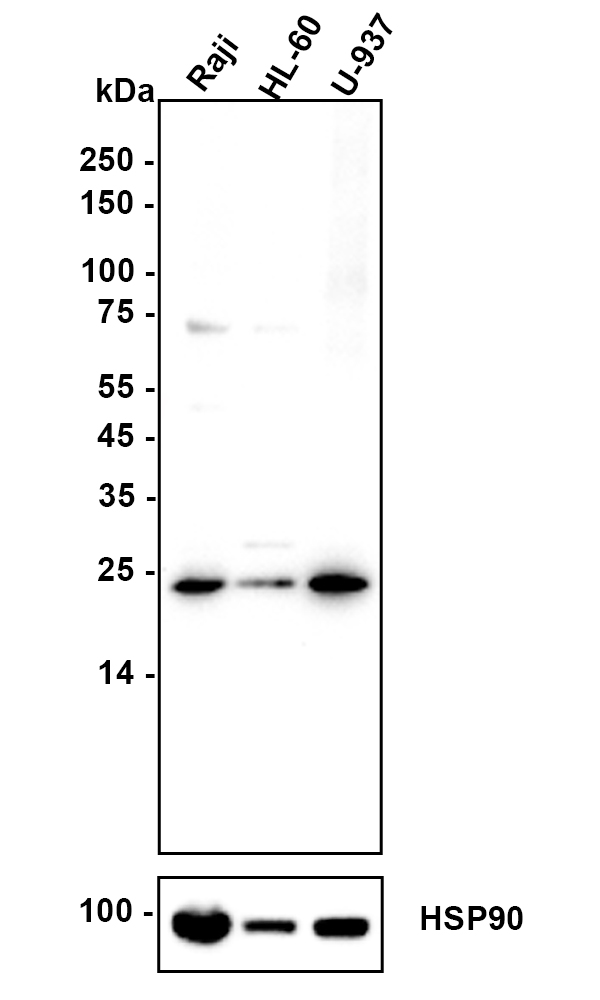
Western blot analysis of Proteasome 20S LMP7 on different lysates with Rabbit anti-Proteasome 20S LMP7 antibody at 1/50,000 dilution. Lane 1: Raji cell lysate, Lane 2: HL-60 cell lysate, Lane 3: U-937 cell lysate, Lysates/proteins at 20 µg/Lane. Exposure time: 3 minute; 4-20% SDS-PAGE gel. Proteins were transferred to a PVDF membrane and blocked with 5% NFDM/TBST for 1 hour at room temperature. The primary antibody at 1/50,000 dilution was used in 5% NFDM/TBST at 4℃ overnight. Goat Anti-Rabbit IgG - HRP Secondary Antibody at 1:50,000 dilution was used for 1 hour at room temperature.IHC
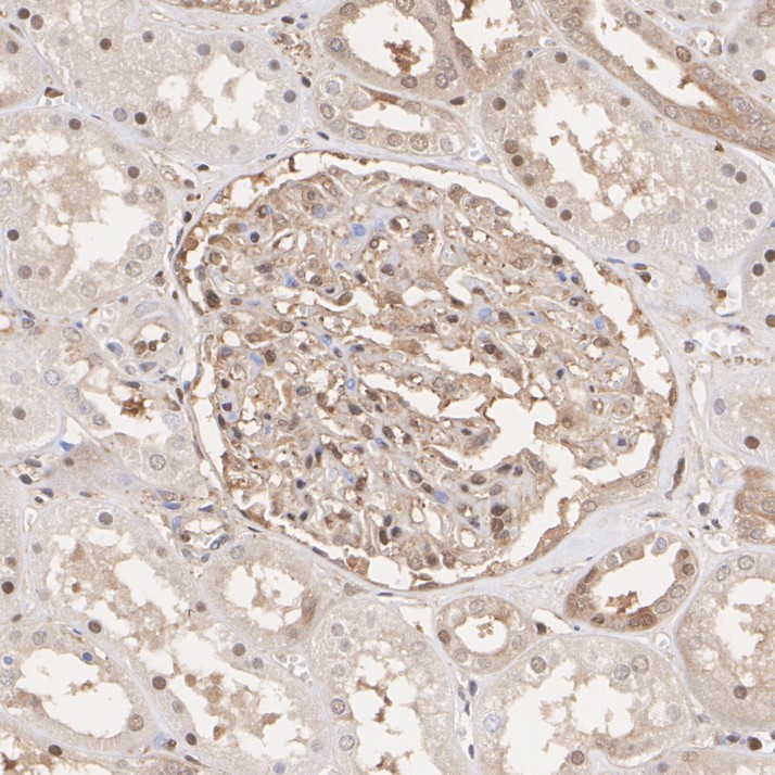
Immunohistochemical analysis of paraffin-embedded human kidney tissue with Rabbit anti-Proteasome 20S LMP7 antibody at 1/1,000 dilution. The section was pre-treated using heat mediated antigen retrieval with sodium citrate buffer (pH 6.0) (high pressure) for 2 minutes. The tissues were blocked in 1% BSA for 20 minutes at room temperature, washed with ddH2O and PBS, and then probed with the primary antibody at 1/1,000 dilution for 1 hour at room temperature. The detection was performed using an HRP conjugated compact polymer system. DAB was used as the chromogen. Tissues were counterstained with hematoxylin and mounted with DPX.IHC
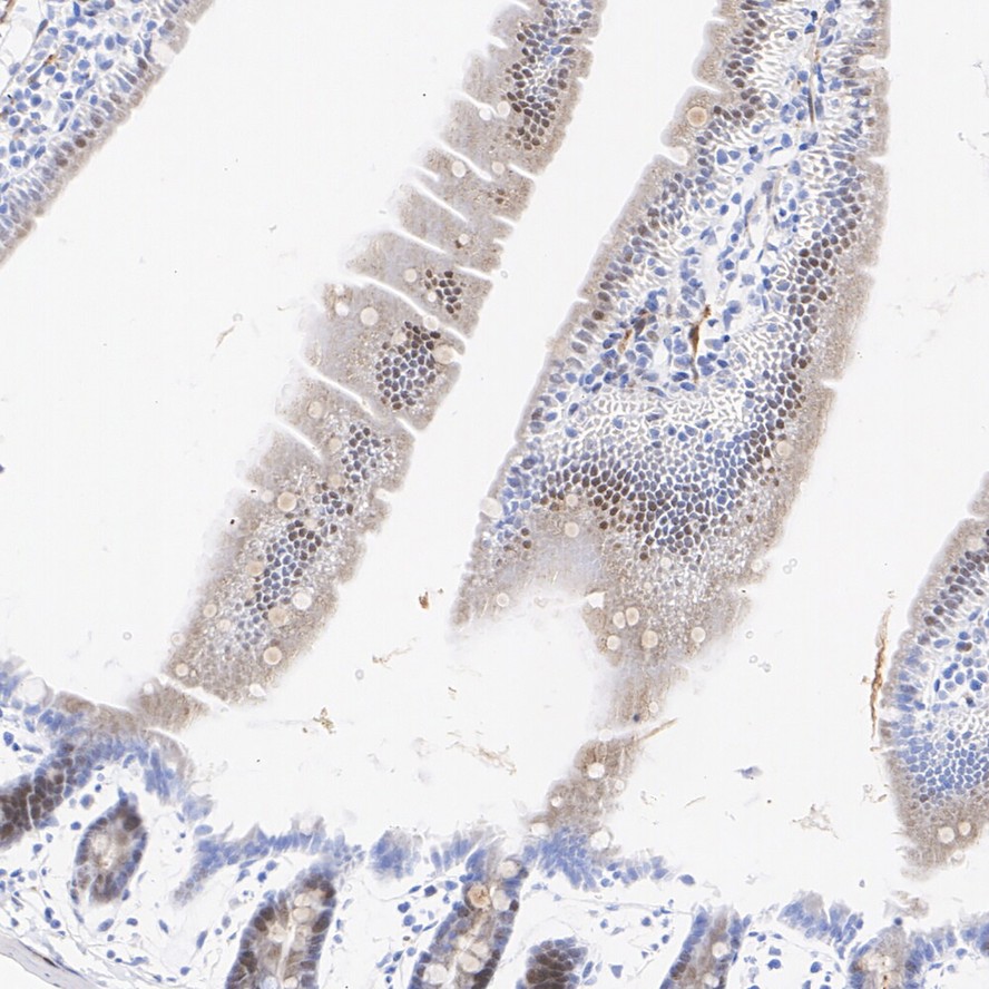
Immunohistochemical analysis of paraffin-embedded mouse small intestine tissue with Rabbit anti-Proteasome 20S LMP7 antibody at 1/1,000 dilution. The section was pre-treated using heat mediated antigen retrieval with sodium citrate buffer (pH 6.0) (high pressure) for 2 minutes. The tissues were blocked in 1% BSA for 20 minutes at room temperature, washed with ddH2O and PBS, and then probed with the primary antibody at 1/1,000 dilution for 1 hour at room temperature. The detection was performed using an HRP conjugated compact polymer system. DAB was used as the chromogen. Tissues were counterstained with hematoxylin and mounted with DPX.IHC
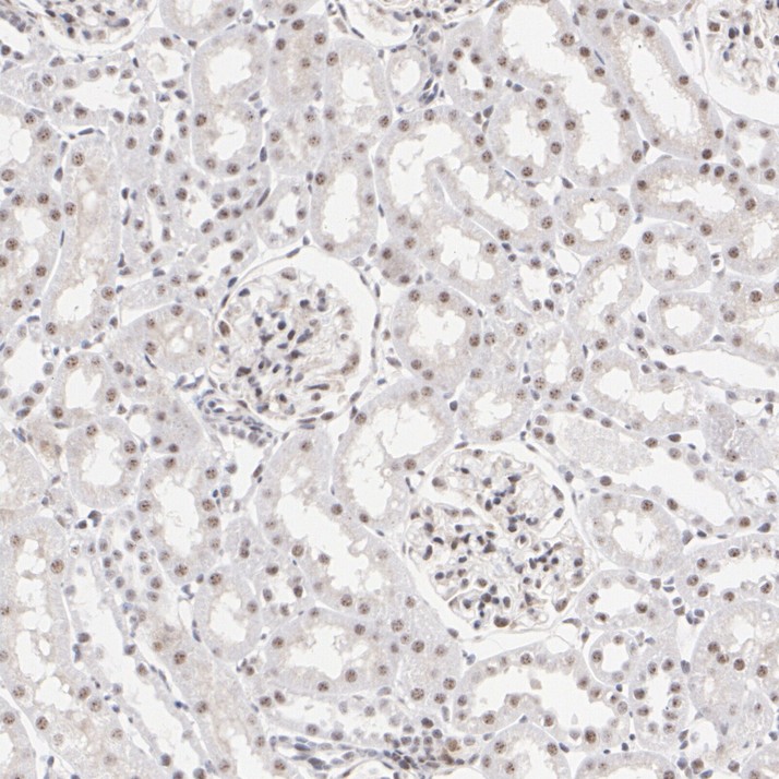
Immunohistochemical analysis of paraffin-embedded rat kidney tissue with Rabbit anti-Proteasome 20S LMP7 antibody at 1/200 dilution. The section was pre-treated using heat mediated antigen retrieval with sodium citrate buffer (pH 6.0) (high pressure) for 2 minutes. The tissues were blocked in 1% BSA for 20 minutes at room temperature, washed with ddH2O and PBS, and then probed with the primary antibody at 1/200 dilution for 1 hour at room temperature. The detection was performed using an HRP conjugated compact polymer system. DAB was used as the chromogen. Tissues were counterstained with hematoxylin and mounted with DPX.ICC/IF
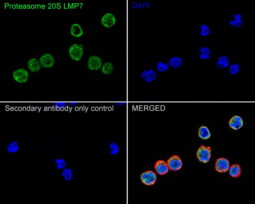
Immunocytochemistry analysis of U-937 cells labeling Proteasome 20S LMP7 with Rabbit anti-Proteasome 20S LMP7 antibody at 1/100 dilution. Cells were fixed in 4% paraformaldehyde for 15 minutes at room temperature, permeabilized with 0.1% Triton X-100 in PBS for 15 minutes at room temperature, then blocked with 1% BSA in 10% negative goat serum for 1 hour at room temperature. Cells were then incubated with Rabbit anti-Proteasome 20S LMP7 antibody at 1/100 dilution in 1% BSA in PBST overnight at 4 ℃. Goat Anti-Rabbit IgG H&L (488) was used as the secondary antibody at 1/1,000 dilution. PBS instead of the primary antibody was used as the secondary antibody only control. Nuclear DNA was labelled in blue with DAPI. Beta tubulin (red) was stained at 1/100 dilution overnight at +4℃. Goat Anti-Mouse IgG H&L (594) was used as the secondary antibody at 1/1,000 dilution.FC
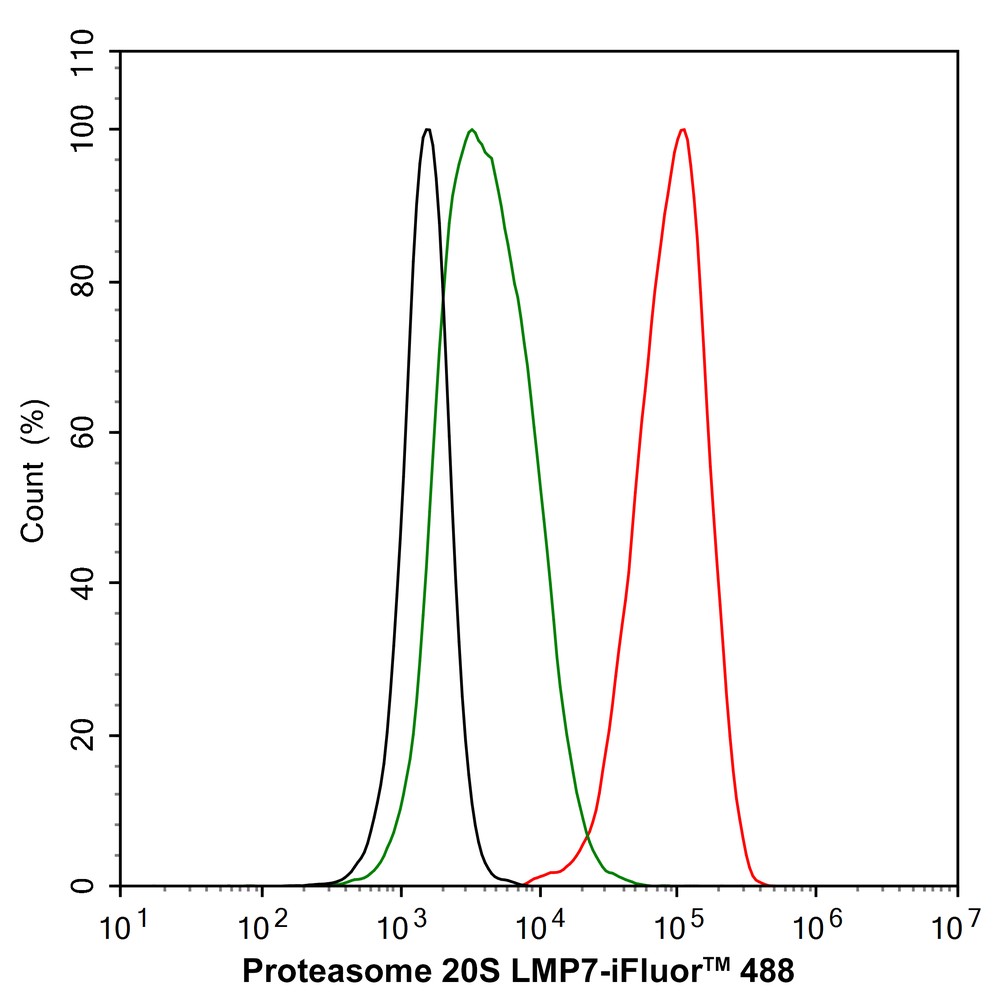
Flow cytometric analysis of U-937 cells labeling Proteasome 20S LMP7. Cells were fixed and permeabilized. Then stained with the primary antibody (1μg/mL) (red) compared with Rabbit IgG Isotype Control (green). After incubation of the primary antibody at +4℃ for an hour, the cells were stained with a iFluor™ 488 conjugate-Goat anti-Rabbit IgG Secondary antibody at 1/1,000 dilution for 30 minutes at +4℃. Unlabelled sample was used as a control (cells without incubation with primary antibody; black).| Product Name | Proteasome 20S LMP7 Recombinant Rabbit Monoclonal Antibody |
|---|---|
| Antibody Type | Primary Antibodies |
| Product description | The eukaryotic multi-catalytic proteinase complex, otherwise known as the proteasome, is present in both the nucleus and cytoplasm of cells and contains at least 15 nonidentical subunits, which form a highly ordered ring-shaped structure. The proteasome is involved in an ATP/Ubiquitin-dependent proteolytic pathway and expresses at least five distinct proteolytic activities, including the cleavage of peptides after branched-chain amino acids or bulky hydrophobic amino acids. Two components of the proteasome are the low molecular mass proteins LMP2 and LMP7, which are thought to connect the proteasome to the MHC class-I antigen-processing pathway. Upon stimulation with IFN-γ, LMP2 and LMP7 displace housekeeping subunits in the proteasome and activate cytotoxic T cells (CTLs). LMP2 and LMP7 are produced as precursor proteins, which are processed to subunits that have the ability to complex with the proteasome. LMP2 is expressed as two alternatively spliced forms, LMP2.l and LMP2.s, in lymphoblastoid cell lines and in fibroblasts after IFN-γ stimulation. LMP7 is also expressed as two forms, LMP7A and LMP7B, also designated LMP7-E1 and E2, in several tissues. |
| Immunogen | Recombinant protein within Human Proteasome 20S LMP7 aa 160-276 / 276. |
| Clonality | Monoclonal |
|---|---|
| Isotype | IgG |
| Host Species | Recombinant rabbit |
| Tested Applications | FCICC/IFIHCWB |
| WB:1:500-1:1000 IHC:1:200-1:1000 ICC/IF:1:100 FC:1:1000 |
|
| Species Reactivity | HumanMouseRat |
| Concentration | 1mg/ml |
| Purification | Protein A |
| Alternative Names | ALDD antibody
D6S216 antibody
D6S216E antibody
Large multifunctional peptidase 7 antibody
Large multifunctional protease 7 antibody
LMP 7 antibody
LMP7 antibody
Low molecular mass protein 7 antibody
Low molecular weight protein 7 antibody
Macropain subunit C13 antibody
MGC1491 antibody
Multicatalytic endopeptidase complex subunit C13 antibody
NKJO antibody
OTTHUMP00000062981 antibody
Protease component C13 antibody
Proteasome (prosome macropain) subunit beta type 8 antibody
Proteasome (prosome macropain) subunit beta type 8 (large multifunctional peptidase 7) antibody Proteasome beta 8 subunit antibody Proteasome catalytic subunit 3i antibody Proteasome component C13 antibody Proteasome related gene 7 antibody Proteasome subunit beta 5i antibody Proteasome subunit beta 8 antibody Proteasome subunit beta type 8 antibody Proteasome subunit beta type antibody Proteasome subunit beta type-8 antibody Proteasome subunit beta-5i antibody Proteasome subunit Y2 antibody PSB8_HUMAN antibody PSMB 8 antibody PSMB5i antibody PSMB8 antibody Really interesting new gene 10 protein antibody RING 10 antibody RING10 antibody Y2 antibody |
|---|---|
| Molecular Weight(MW) | 23 kDa |
| Cellular Localization | Nucleus. Cytoplasm. |
WB
Western blot analysis of Proteasome 20S LMP7 on different lysates with Rabbit anti-Proteasome 20S LMP7 antibody at 1/50,000 dilution. Lane 1: Raji cell lysate, Lane 2: HL-60 cell lysate, Lane 3: U-937 cell lysate, Lysates/proteins at 20 µg/Lane. Exposure time: 3 minute; 4-20% SDS-PAGE gel. Proteins were transferred to a PVDF membrane and blocked with 5% NFDM/TBST for 1 hour at room temperature. The primary antibody at 1/50,000 dilution was used in 5% NFDM/TBST at 4℃ overnight. Goat Anti-Rabbit IgG - HRP Secondary Antibody at 1:50,000 dilution was used for 1 hour at room temperature.
IHC
Immunohistochemical analysis of paraffin-embedded human kidney tissue with Rabbit anti-Proteasome 20S LMP7 antibody at 1/1,000 dilution. The section was pre-treated using heat mediated antigen retrieval with sodium citrate buffer (pH 6.0) (high pressure) for 2 minutes. The tissues were blocked in 1% BSA for 20 minutes at room temperature, washed with ddH2O and PBS, and then probed with the primary antibody at 1/1,000 dilution for 1 hour at room temperature. The detection was performed using an HRP conjugated compact polymer system. DAB was used as the chromogen. Tissues were counterstained with hematoxylin and mounted with DPX.
IHC
Immunohistochemical analysis of paraffin-embedded mouse small intestine tissue with Rabbit anti-Proteasome 20S LMP7 antibody at 1/1,000 dilution. The section was pre-treated using heat mediated antigen retrieval with sodium citrate buffer (pH 6.0) (high pressure) for 2 minutes. The tissues were blocked in 1% BSA for 20 minutes at room temperature, washed with ddH2O and PBS, and then probed with the primary antibody at 1/1,000 dilution for 1 hour at room temperature. The detection was performed using an HRP conjugated compact polymer system. DAB was used as the chromogen. Tissues were counterstained with hematoxylin and mounted with DPX.
IHC
Immunohistochemical analysis of paraffin-embedded rat kidney tissue with Rabbit anti-Proteasome 20S LMP7 antibody at 1/200 dilution. The section was pre-treated using heat mediated antigen retrieval with sodium citrate buffer (pH 6.0) (high pressure) for 2 minutes. The tissues were blocked in 1% BSA for 20 minutes at room temperature, washed with ddH2O and PBS, and then probed with the primary antibody at 1/200 dilution for 1 hour at room temperature. The detection was performed using an HRP conjugated compact polymer system. DAB was used as the chromogen. Tissues were counterstained with hematoxylin and mounted with DPX.
ICC/IF
Immunocytochemistry analysis of U-937 cells labeling Proteasome 20S LMP7 with Rabbit anti-Proteasome 20S LMP7 antibody at 1/100 dilution. Cells were fixed in 4% paraformaldehyde for 15 minutes at room temperature, permeabilized with 0.1% Triton X-100 in PBS for 15 minutes at room temperature, then blocked with 1% BSA in 10% negative goat serum for 1 hour at room temperature. Cells were then incubated with Rabbit anti-Proteasome 20S LMP7 antibody at 1/100 dilution in 1% BSA in PBST overnight at 4 ℃. Goat Anti-Rabbit IgG H&L (488) was used as the secondary antibody at 1/1,000 dilution. PBS instead of the primary antibody was used as the secondary antibody only control. Nuclear DNA was labelled in blue with DAPI. Beta tubulin (red) was stained at 1/100 dilution overnight at +4℃. Goat Anti-Mouse IgG H&L (594) was used as the secondary antibody at 1/1,000 dilution.
FC
Flow cytometric analysis of U-937 cells labeling Proteasome 20S LMP7. Cells were fixed and permeabilized. Then stained with the primary antibody (1μg/mL) (red) compared with Rabbit IgG Isotype Control (green). After incubation of the primary antibody at +4℃ for an hour, the cells were stained with a iFluor™ 488 conjugate-Goat anti-Rabbit IgG Secondary antibody at 1/1,000 dilution for 30 minutes at +4℃. Unlabelled sample was used as a control (cells without incubation with primary antibody; black).| Application Notes | WB:1:500-1:1000 IHC:1:200-1:1000 ICC/IF:1:100 FC:1:1000 |
|---|
| Form | Liquid |
|---|---|
| Storage Instructions | Store at +4℃ after thawing. Aliquot store at -20℃ or -80℃. Avoid repeated freeze / thaw cycles. |
| Storage Buffer | 1*TBS (pH7.4), 1%BSA, 40%Glycerol. Preservative: 0.05% Sodium Azide. |


