WB
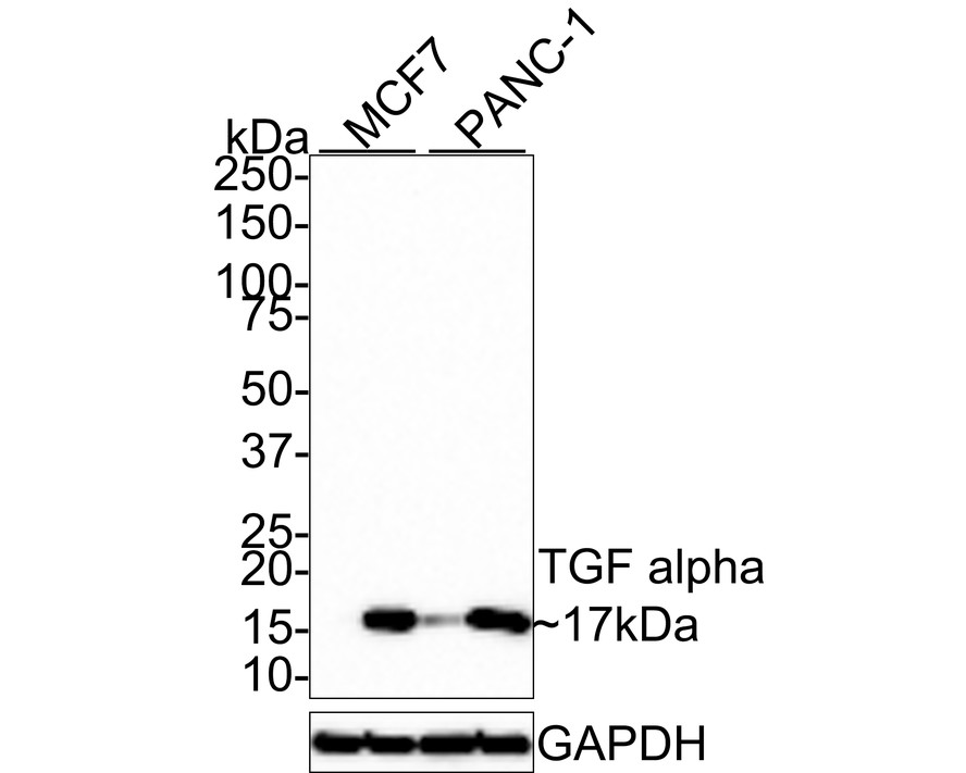
Western blot analysis of TGF alpha on different lysates with Rabbit anti-TGF alpha antibody at 1/500 dilution. Lane 1: MCF7 cell lysate (RIPA lysis), Lane 2: MCF7 cell lysate (hot lysis), Lane 3: PANC-1 cell lysate (RIPA lysis), Lane 4: PANC-1 cell lysate (hot lysis), Lysates/proteins at 20 µg/Lane. Exposure time: 2 minutes; 4-20% SDS-PAGE gel. Proteins were transferred to a PVDF membrane and blocked with 5% NFDM/TBST for 1 hour at room temperature. The primary antibody at 1/500 dilution was used in 5% NFDM/TBST at room temperature for 2 hours. Goat Anti-Rabbit IgG - HRP Secondary Antibody at 1:100,000 dilution was used for 1 hour at room temperature.IHC
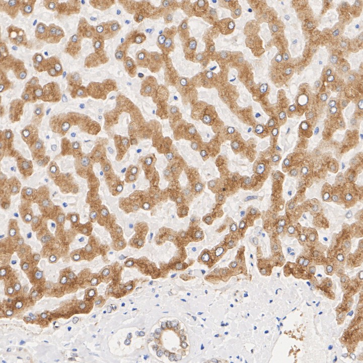
Immunohistochemical analysis of paraffin-embedded human liver tissue with Rabbit anti-TGF alpha antibody at 1/1,000 dilution. The section was pre-treated using heat mediated antigen retrieval with Tris-EDTA buffer (pH 9.0) for 20 minutes. The tissues were blocked in 1% BSA for 20 minutes at room temperature, washed with ddH2O and PBS, and then probed with the primary antibody at 1/1,000 dilution for 1 hour at room temperature. The detection was performed using an HRP conjugated compact polymer system. DAB was used as the chromogen. Tissues were counterstained with hematoxylin and mounted with DPX.IHC
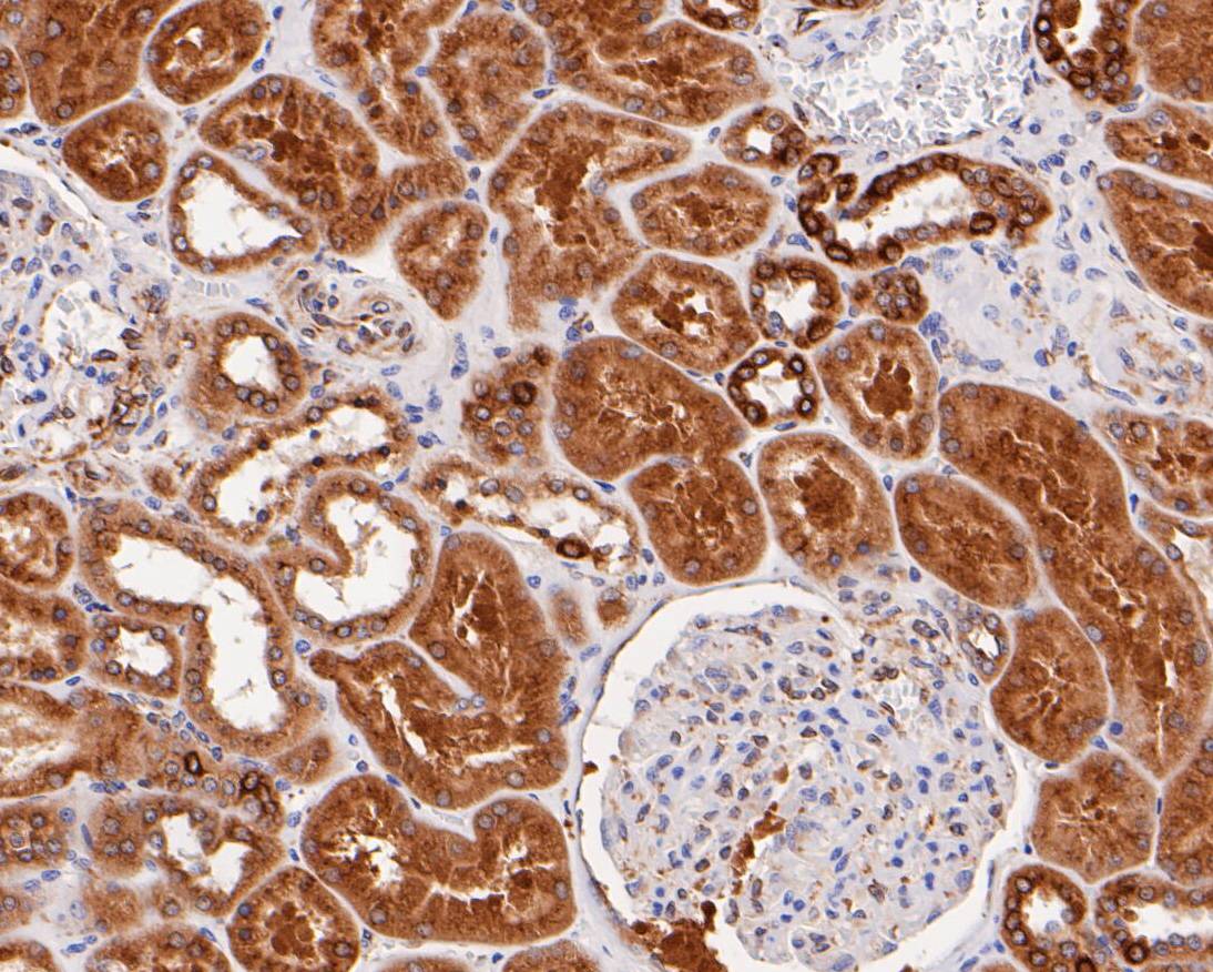
Immunohistochemical analysis of paraffin-embedded human kidney tissue using anti-TGF alpha antibody. Counter stained with hematoxylin.IHC
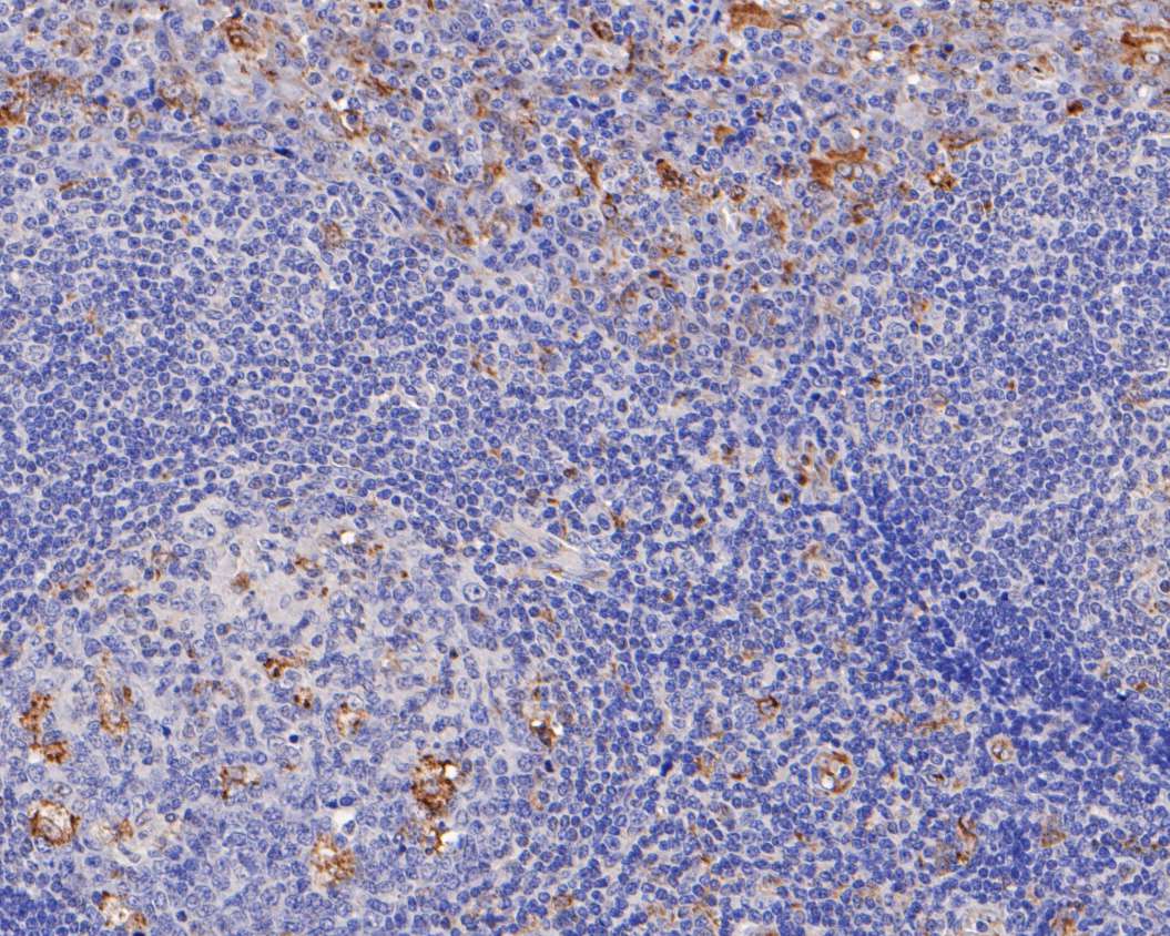
Immunohistochemical analysis of paraffin-embedded human tonsil tissue using anti-TGF alpha antibody. Counter stained with hematoxylin.IHC
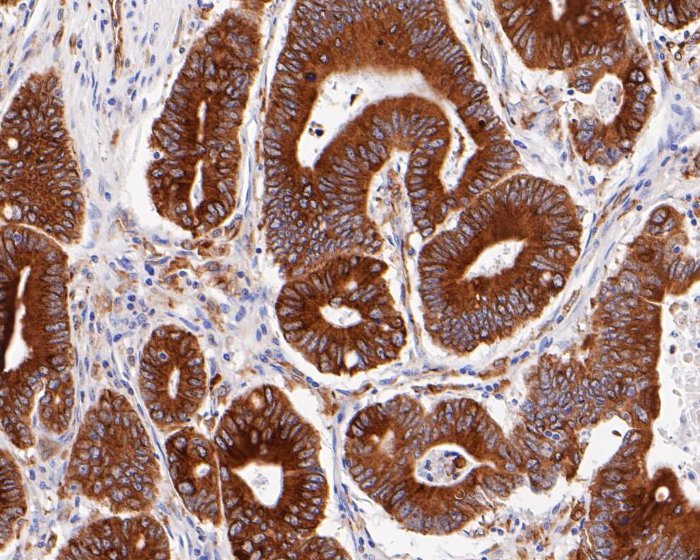
Immunohistochemical analysis of paraffin-embedded human colon cancer tissue using anti-TGF alpha antibody. Counter stained with hematoxylin.| Product Name | TGF alpha Recombinant Rabbit Monoclonal Antibody |
|---|---|
| Antibody Type | Primary Antibodies |
| Product description | Transforming growth factor-α (TGFα) is an acid- and heat-stable 50 amino acid protein originally found in rodents and humans. TGFα is 33% homologous at the amino acid level to epidermal growth factor (EGF). TGFα binds to the EGF receptor, mediates tyrosine phosphorylation of the receptor and promotes anchorage-independent growth of normal rat fibroblasts in soft agar in the presence of transforming growth factor-β. TGFα is secreted by a variety of transformed cells and tumors, embryonic cells and some normal adult cells. TGFα bioactivity has been found in the urine of cancer patients. It has been suggested that it may act as an autocrine growth factor for the induction or maintenance of malignancy. |
| Immunogen | Recombinant protein within Human TGF alpha aa 1-160 / 160. |
| Clonality | Monoclonal |
|---|---|
| Isotype | IgG |
| Host Species | Recombinant rabbit |
| Tested Applications | IHCWB |
| WB:1:500 IHC:1:200-1:1000 |
|
| Species Reactivity | Human |
| Concentration | 1mg/ml |
| Purification | Protein A |
| Alternative Names | EGF like TGF antibody EGF-like TGF antibody ETGF antibody TFGA antibody TGF A antibody TGF type 1 antibody TGF-alpha antibody Tgfa antibody TGFA_HUMAN antibody Transforming growth factor alpha antibody Transforming growth factor alpha precursor antibody Wa1 antibody Waved 1 antibody |
|---|---|
| Molecular Weight(MW) | 17 kDa |
| Cellular Localization | Plasma membrane. Extracellular region. Secreted. |
| SwissProt ID | P01135 |
|---|
WB
Western blot analysis of TGF alpha on different lysates with Rabbit anti-TGF alpha antibody at 1/500 dilution. Lane 1: MCF7 cell lysate (RIPA lysis), Lane 2: MCF7 cell lysate (hot lysis), Lane 3: PANC-1 cell lysate (RIPA lysis), Lane 4: PANC-1 cell lysate (hot lysis), Lysates/proteins at 20 µg/Lane. Exposure time: 2 minutes; 4-20% SDS-PAGE gel. Proteins were transferred to a PVDF membrane and blocked with 5% NFDM/TBST for 1 hour at room temperature. The primary antibody at 1/500 dilution was used in 5% NFDM/TBST at room temperature for 2 hours. Goat Anti-Rabbit IgG - HRP Secondary Antibody at 1:100,000 dilution was used for 1 hour at room temperature.
IHC
Immunohistochemical analysis of paraffin-embedded human liver tissue with Rabbit anti-TGF alpha antibody at 1/1,000 dilution. The section was pre-treated using heat mediated antigen retrieval with Tris-EDTA buffer (pH 9.0) for 20 minutes. The tissues were blocked in 1% BSA for 20 minutes at room temperature, washed with ddH2O and PBS, and then probed with the primary antibody at 1/1,000 dilution for 1 hour at room temperature. The detection was performed using an HRP conjugated compact polymer system. DAB was used as the chromogen. Tissues were counterstained with hematoxylin and mounted with DPX.
IHC
Immunohistochemical analysis of paraffin-embedded human kidney tissue using anti-TGF alpha antibody. Counter stained with hematoxylin.
IHC
Immunohistochemical analysis of paraffin-embedded human tonsil tissue using anti-TGF alpha antibody. Counter stained with hematoxylin.
IHC
Immunohistochemical analysis of paraffin-embedded human colon cancer tissue using anti-TGF alpha antibody. Counter stained with hematoxylin.| Application Notes | WB:1:500 IHC:1:200-1:1000 |
|---|
| Form | Liquid |
|---|---|
| Storage Instructions | Store at +4℃ after thawing. Aliquot store at -20℃ or -80℃. Avoid repeated freeze / thaw cycles. |
| Storage Buffer | 1*TBS (pH7.4), 1%BSA, 40%Glycerol. Preservative: 0.05% Sodium Azide. |


