Application
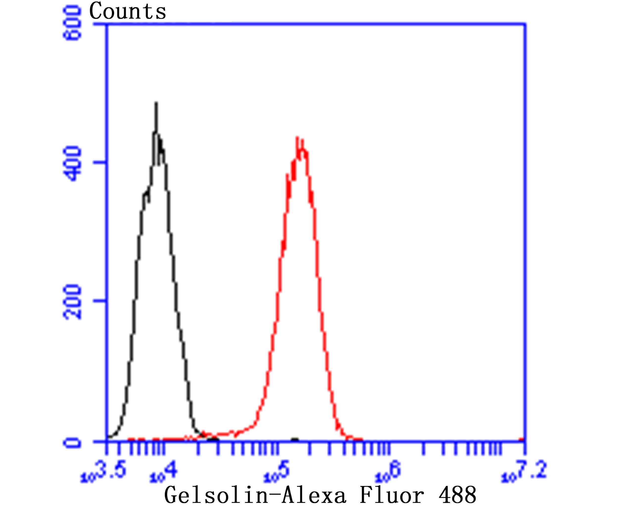
Fig7: Flow cytometric analysis of THP-1 cells with Gelsolin antibody at 1/100 dilution (red) compared with an unlabelled control (cells without incubation with primary antibody; black). Alexa Fluor 488-conjugated goat anti-rabbit IgG was used as the secondary antibody.Application
Fig7: Flow cytometric analysis of THP-1 cells with Gelsolin antibody at 1/100 dilution (red) compared with an unlabelled control (cells without incubation with primary antibody; black). Alexa Fluor 488-conjugated goat anti-rabbit IgG was used as the secondary antibody.Application
Fig7: Flow cytometric analysis of THP-1 cells with Gelsolin antibody at 1/100 dilution (red) compared with an unlabelled control (cells without incubation with primary antibody; black). Alexa Fluor 488-conjugated goat anti-rabbit IgG was used as the secondary antibody.Application
Fig7: Flow cytometric analysis of THP-1 cells with Gelsolin antibody at 1/100 dilution (red) compared with an unlabelled control (cells without incubation with primary antibody; black). Alexa Fluor 488-conjugated goat anti-rabbit IgG was used as the secondary antibody.Application
Fig7: Flow cytometric analysis of THP-1 cells with Gelsolin antibody at 1/100 dilution (red) compared with an unlabelled control (cells without incubation with primary antibody; black). Alexa Fluor 488-conjugated goat anti-rabbit IgG was used as the secondary antibody.Application
Fig7: Flow cytometric analysis of THP-1 cells with Gelsolin antibody at 1/100 dilution (red) compared with an unlabelled control (cells without incubation with primary antibody; black). Alexa Fluor 488-conjugated goat anti-rabbit IgG was used as the secondary antibody.Application
Fig7: Flow cytometric analysis of THP-1 cells with Gelsolin antibody at 1/100 dilution (red) compared with an unlabelled control (cells without incubation with primary antibody; black). Alexa Fluor 488-conjugated goat anti-rabbit IgG was used as the secondary antibody.| Product Name | Gelsolin [JB36-68] |
|---|---|
| Antibody Type | Primary Antibodies |
| Antigen Alias | Actin depolymerizing factor antibody Actin-depolymerizing factor antibody ADF antibody AGEL antibody Brevin antibody DKFZp313L0718 antibody GELS_HUMAN antibody Gelsolin antibody Gsn antibody |
| Product description | Gelsolin (also known as brevin, Actin-depolymerizing factor or ADF), a protein of leukocytes, platelets and other cells, severs Actin filaments in the presence of submicromolar calcium, thereby isolating cytoplasmic Actin gels. A calcium-independent mechanism reverses the process. A Gelsolin variant with 23 more amino-terminal amino acids is a plasma component probably involved in the clearance of Actin, the most abundant human protein, from the circulation. It has been suggested that a single gene encodes both cell and plasma gelsolins. Gelsolin may be unique in that it is made for both secretion and intracytoplasmic location. Amino acid homology was identified between Gelsolin and the amyloid of the Finnish variety of amyloidosis. The amyloid in this disorder is antigenically and structurally related to Gelsolin. Gelsolin is the principal intracellular and extracellular Actin-severing protein. Gelsolin and Gc protein together constitute the extracellular Actin-scavenger system which prevents the toxic effects of Actin release into the extracellular space under circumstances of cell necrosis. |
| Immunogen | Recombinant protein |
| Clonality | Monoclonal |
|---|---|
| Isotype | IgG |
| Host Species | Recombinant rabbit |
| Tested Applications | WBICCIFIHCFC |
| WB:1:500-1:2,000 ICC:1:50-1:200 IHC:1:50-1:200 FC:1:50-1:100 |
|
| Species Reactivity | HumanMouse |
| Concentration | 1mg/ml |
| Alternative Names | Actin depolymerizing factor antibody Actin-depolymerizing factor antibody ADF antibody AGEL antibody Brevin antibody DKFZp313L0718 antibody GELS_HUMAN antibody Gelsolin antibody Gsn antibody |
|---|---|
| Molecular Weight(MW) | 86 kDa |
| Cellular Localization | Cytoskeleton. Secreted. |
Application
Fig7: Flow cytometric analysis of THP-1 cells with Gelsolin antibody at 1/100 dilution (red) compared with an unlabelled control (cells without incubation with primary antibody; black). Alexa Fluor 488-conjugated goat anti-rabbit IgG was used as the secondary antibody.
Application
Fig7: Flow cytometric analysis of THP-1 cells with Gelsolin antibody at 1/100 dilution (red) compared with an unlabelled control (cells without incubation with primary antibody; black). Alexa Fluor 488-conjugated goat anti-rabbit IgG was used as the secondary antibody.
Application
Fig7: Flow cytometric analysis of THP-1 cells with Gelsolin antibody at 1/100 dilution (red) compared with an unlabelled control (cells without incubation with primary antibody; black). Alexa Fluor 488-conjugated goat anti-rabbit IgG was used as the secondary antibody.
Application
Fig7: Flow cytometric analysis of THP-1 cells with Gelsolin antibody at 1/100 dilution (red) compared with an unlabelled control (cells without incubation with primary antibody; black). Alexa Fluor 488-conjugated goat anti-rabbit IgG was used as the secondary antibody.
Application
Fig7: Flow cytometric analysis of THP-1 cells with Gelsolin antibody at 1/100 dilution (red) compared with an unlabelled control (cells without incubation with primary antibody; black). Alexa Fluor 488-conjugated goat anti-rabbit IgG was used as the secondary antibody.
Application
Fig7: Flow cytometric analysis of THP-1 cells with Gelsolin antibody at 1/100 dilution (red) compared with an unlabelled control (cells without incubation with primary antibody; black). Alexa Fluor 488-conjugated goat anti-rabbit IgG was used as the secondary antibody.
Application
Fig7: Flow cytometric analysis of THP-1 cells with Gelsolin antibody at 1/100 dilution (red) compared with an unlabelled control (cells without incubation with primary antibody; black). Alexa Fluor 488-conjugated goat anti-rabbit IgG was used as the secondary antibody.| Positive Control | Human spleen tissue, human kidney tissue, mouse lung tissue, NIH-3T3, SiHa, mouse thymus tissue lysate, mouse lung tissue lysate, THP-1, HepG2, THP-1. |
|---|---|
| Application Notes | WB:1:500-1:2,000 ICC:1:50-1:200 IHC:1:50-1:200 FC:1:50-1:100 |
| Form | Liquid |
|---|---|
| Storage Instructions | Store at +4℃ after thawing. Aliquot store at -20℃ or -80℃. Avoid repeated freeze / thaw cycles. |
| Storage Buffer | 1*TBS (pH7.4), 1%BSA, 40%Glycerol. Preservative: 0.05% Sodium Azide. |


