Application
Fig1: Western blot analysis of Galectin 8 on mouse testis and rat testis tissue lysates using anti-Galectin 8 antibody at 1/500 dilution.Application
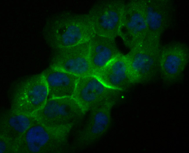
Fig2: ICC staining Galectin 8 in A431 cells (green). The nuclear counter stain is DAPI (blue). Cells were fixed in paraformaldehyde, permeabilised with 0.25% Triton X100/PBS.Application
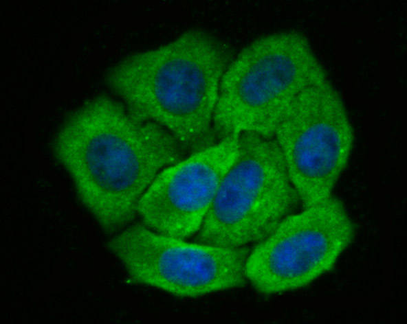
Fig3: ICC staining Galectin 8 in HepG2 cells (green). The nuclear counter stain is DAPI (blue). Cells were fixed in paraformaldehyde, permeabilised with 0.25% Triton X100/PBS.Application
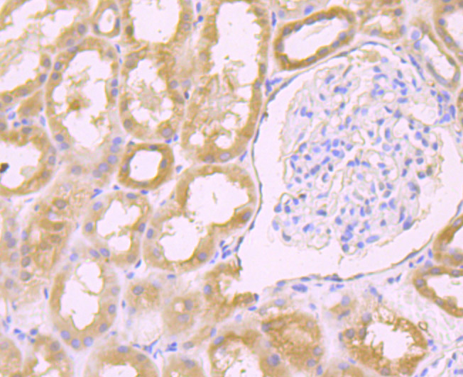
Fig4: Immunohistochemical analysis of paraffin-embedded human kidney tissue using anti-Galectin 8 antibody. Counter stained with hematoxylin.Application
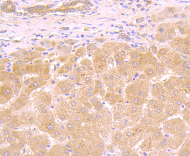
Fig5: Immunohistochemical analysis of paraffin-embedded human liver tissue using anti-Galectin 8 antibody. Counter stained with hematoxylin.Application
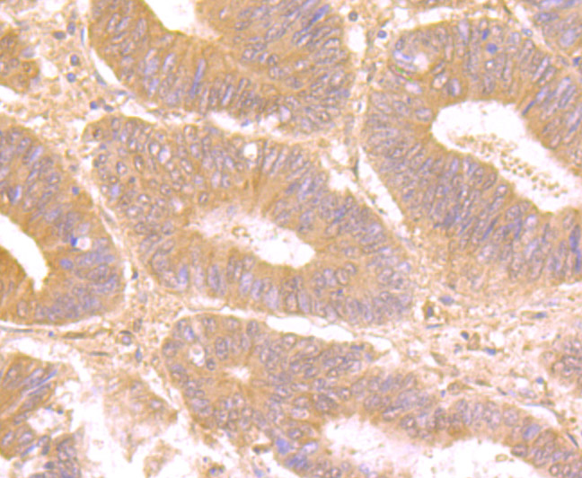
Fig6: Immunohistochemical analysis of paraffin-embedded human colon cancer tissue using anti-Galectin 8 antibody. Counter stained with hematoxylin.Application
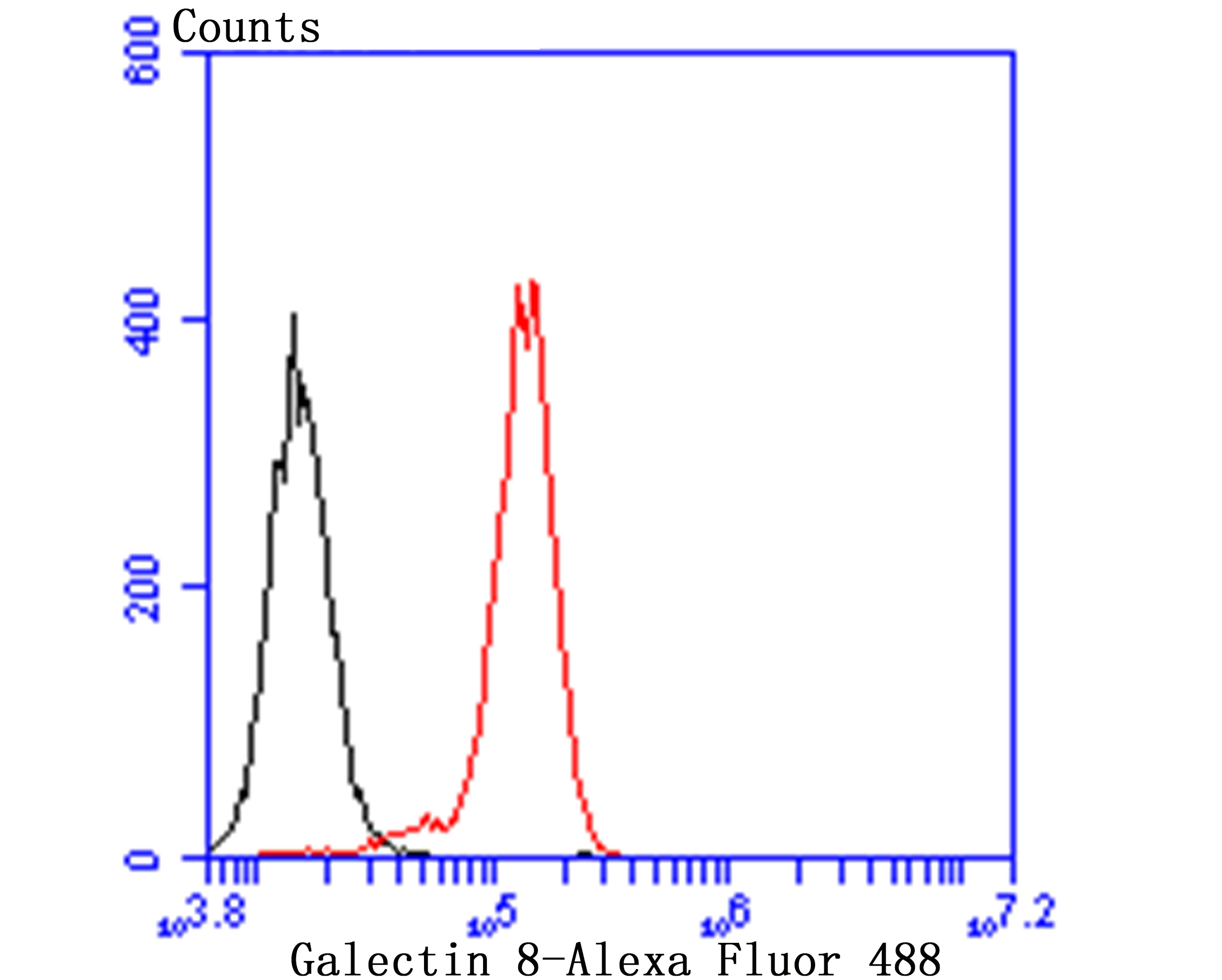
Fig7: Flow cytometric analysis of PC-3M cells with Galectin 8 antibody at 1/100 dilution (red) compared with an unlabelled control (cells without incubation with primary antibody; black). Alexa Fluor 488-conjugated goat anti-rabbit IgG was used as the secondary antibody.| Product Name | Galectin 8 [JB85-35] |
|---|---|
| Antibody Type | Primary Antibodies |
| Antigen Alias | Gal 8 antibody Gal-8 antibody Gal8 antibody Galectin-8 antibody galectin-8g antibody Lectin galactoside binding soluble 8 antibody LEG8_HUMAN antibody LGAL S8 antibody Lgals8 antibody PCTA 1 antibody PCTA-1 antibody PCTA1 antibody Po66 carbohydrate binding protein antibody Po66 carbohydrate-binding protein antibody Po66 CBP antibody Po66-CBP antibody Prostate carcinoma tumor antigen 1 antibody |
| Product description | Several proteins have been identified as specific markers of prostate cancer, and they may be useful as diagnostic indicators. PSA, prostate specific antigen, is the classical indicator for transformed prostate tissue; however, in addition to being up-regulated in prostate cancer, PSA is also up-regulated in non-malignant conditions, such as benign prostatic hyperplasia prostate. Galectin-8, also known as prostate-specific membrane antigen (PCTA-1), is an additional prostate-specific antigen that is overexpressed only in malignant tumors and therefore is a more specific identifier of malignancies. It is a member of the galectin gene family which mediates bothe cell-cell and cell-matrix interactions in a manner similar to the selectin subgroup of C-type lectins. |
| Immunogen | Recombinant protein |
| Clonality | Monoclonal |
|---|---|
| Isotype | IgG |
| Host Species | Recombinant rabbit |
| Tested Applications | WBICCIFIHCFCIP |
| WB:1:500-1:1,000 ICC/IF:1:400-1:800 IHC:1:50-1:200 FC:1:50-1:100 |
|
| Species Reactivity | HumanMouseRat |
| Concentration | 1mg/ml |
| Alternative Names | Gal 8 antibody Gal-8 antibody Gal8 antibody Galectin-8 antibody galectin-8g antibody Lectin galactoside binding soluble 8 antibody LEG8_HUMAN antibody LGAL S8 antibody Lgals8 antibody PCTA 1 antibody PCTA-1 antibody PCTA1 antibody Po66 carbohydrate binding protein antibody Po66 carbohydrate-binding protein antibody Po66 CBP antibody Po66-CBP antibody Prostate carcinoma tumor antigen 1 antibody |
|---|---|
| Molecular Weight(MW) | 40 kDa |
| Cellular Localization | Cytosol. |
| SwissProt ID | O00214 |
|---|
Application
Fig1: Western blot analysis of Galectin 8 on mouse testis and rat testis tissue lysates using anti-Galectin 8 antibody at 1/500 dilution.
Application
Fig2: ICC staining Galectin 8 in A431 cells (green). The nuclear counter stain is DAPI (blue). Cells were fixed in paraformaldehyde, permeabilised with 0.25% Triton X100/PBS.
Application
Fig3: ICC staining Galectin 8 in HepG2 cells (green). The nuclear counter stain is DAPI (blue). Cells were fixed in paraformaldehyde, permeabilised with 0.25% Triton X100/PBS.
Application
Fig4: Immunohistochemical analysis of paraffin-embedded human kidney tissue using anti-Galectin 8 antibody. Counter stained with hematoxylin.
Application
Fig5: Immunohistochemical analysis of paraffin-embedded human liver tissue using anti-Galectin 8 antibody. Counter stained with hematoxylin.
Application
Fig6: Immunohistochemical analysis of paraffin-embedded human colon cancer tissue using anti-Galectin 8 antibody. Counter stained with hematoxylin.
Application
Fig7: Flow cytometric analysis of PC-3M cells with Galectin 8 antibody at 1/100 dilution (red) compared with an unlabelled control (cells without incubation with primary antibody; black). Alexa Fluor 488-conjugated goat anti-rabbit IgG was used as the secondary antibody.| Positive Control | Mouse testis and rat testis tissue lysates, Human liver tissue, human colon cancer tissue, human kidney tissue, HepG2, A431, PC-3M. |
|---|---|
| Application Notes | WB:1:500-1:1,000 ICC/IF:1:400-1:800 IHC:1:50-1:200 FC:1:50-1:100 |
| Form | Liquid |
|---|---|
| Storage Instructions | Store at +4℃ after thawing. Aliquot store at -20℃ or -80℃. Avoid repeated freeze / thaw cycles. |
| Storage Buffer | 1*TBS (pH7.4), 1%BSA, 40%Glycerol. Preservative: 0.05% Sodium Azide. |


