WB
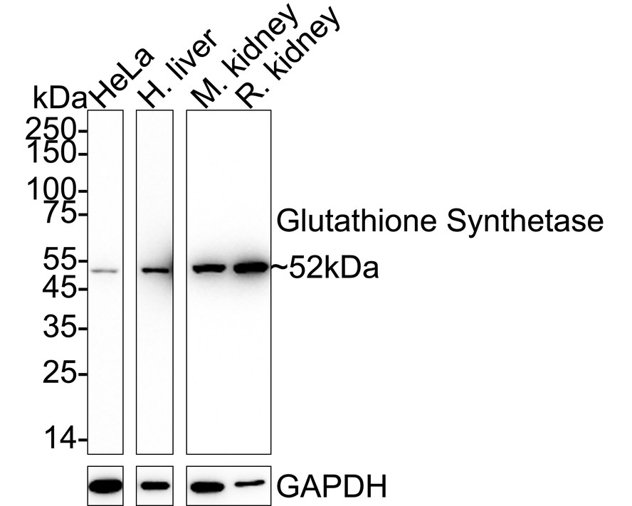
Western blot analysis of Glutathione Synthetase on different lysates with Rabbit anti-Glutathione Synthetase antibody at 1/1,000 dilution. Lane 1: HeLa cell lysate, Lane 2: Human liver tissue lysate, Lane 3: Mouse kidney tissue lysate, Lane 4: Rat kidney tissue lysate, Lysates/proteins at 20 µg/Lane. Exposure time: Lane 1-2: 3 minutes; Lane 3-4: 30 seconds; 4-20% SDS-PAGE gel. Proteins were transferred to a PVDF membrane and blocked with 5% NFDM/TBST for 1 hour at room temperature. The primary antibody at 1/1,000 dilution was used in 5% NFDM/TBST at 4℃ overnight. Goat Anti-Rabbit IgG - HRP Secondary Antibody at 1/50,000 dilution was used for 1 hour at room temperature.IHC
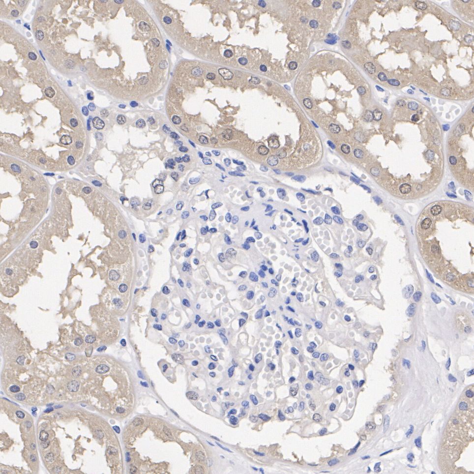
Immunohistochemical analysis of paraffin-embedded human kidney tissue with Rabbit anti-Glutathione Synthetase antibody at 1/1,000 dilution. The section was pre-treated using heat mediated antigen retrieval with Tris-EDTA buffer (pH 9.0) for 20 minutes. The tissues were blocked in 1% BSA for 20 minutes at room temperature, washed with ddH2O and PBS, and then probed with the primary antibody at 1/1,000 dilution for 1 hour at room temperature. The detection was performed using an HRP conjugated compact polymer system. DAB was used as the chromogen. Tissues were counterstained with hematoxylin and mounted with DPX.IHC
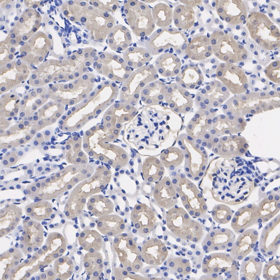
Immunohistochemical analysis of paraffin-embedded mouse kidney tissue with Rabbit anti-Glutathione Synthetase antibody at 1/1,000 dilution. The section was pre-treated using heat mediated antigen retrieval with Tris-EDTA buffer (pH 9.0) for 20 minutes. The tissues were blocked in 1% BSA for 20 minutes at room temperature, washed with ddH2O and PBS, and then probed with the primary antibody at 1/1,000 dilution for 1 hour at room temperature. The detection was performed using an HRP conjugated compact polymer system. DAB was used as the chromogen. Tissues were counterstained with hematoxylin and mounted with DPX.IHC
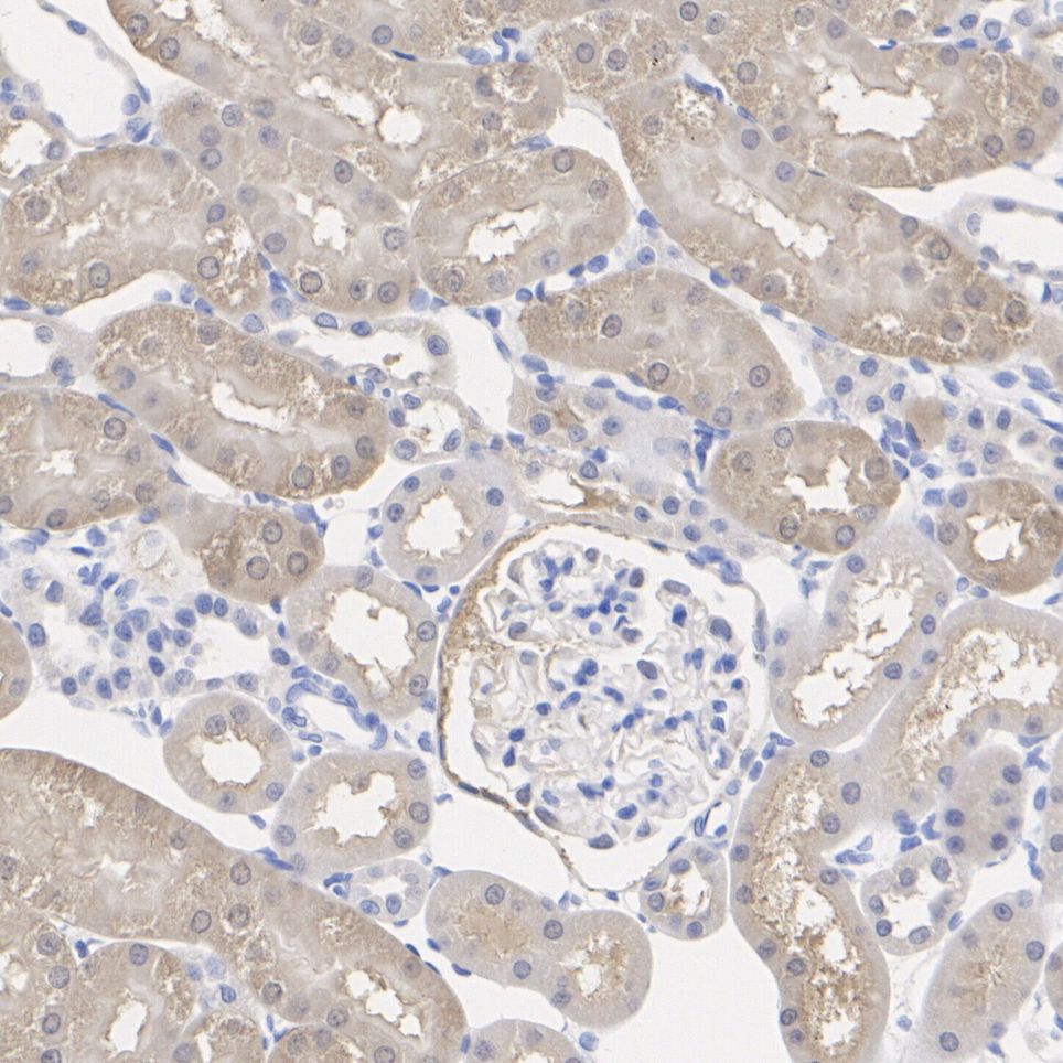
Immunohistochemical analysis of paraffin-embedded rat kidney tissue with Rabbit anti-Glutathione Synthetase antibody at 1/1,000 dilution. The section was pre-treated using heat mediated antigen retrieval with Tris-EDTA buffer (pH 9.0) for 20 minutes. The tissues were blocked in 1% BSA for 20 minutes at room temperature, washed with ddH2O and PBS, and then probed with the primary antibody at 1/1,000 dilution for 1 hour at room temperature. The detection was performed using an HRP conjugated compact polymer system. DAB was used as the chromogen. Tissues were counterstained with hematoxylin and mounted with DPX.| Product Name | Glutathione Synthetase Recombinant Rabbit Monoclonal Antibody |
|---|---|
| Antibody Type | Primary Antibodies |
| Product description | GSS (Glutathione synthetase) is a 474 amino acid protein encoded by the gene located at human chromosome 20q11.2. GSS consists of three loops projecting from an antiparallel β-sheet, a parallel β-sheet and a lid of anti-parallel sheets, which provide access to the ATP-binding site. Although Southern blot and gene analysis suggest that GSS may be the only member of a unique family, the crystal structure indicates that GSS belongs to the ATP-GRASP superfamily. GSS is expressed in hemocytes and nucleated cells, including the brain. GSS occurs as a homodimer. There are two steps in the production of Glutathione, begining with γ-GCS and ending with GSS. In an ATP-dependent reaction, GSS produces Glutathione from γ-glutamylcysteine and glycine precursors. Partial hepatectomy, diethyl maleate, buthionine sulfoximine, tert-butylhaydroquinone and thioacetamide increase the ex-pression of GSS, which causes an increase in Glutathione levels. An inherited autosomal recessive disorder, 5-oxoprolinuria (pyroglutamic aciduria), is caused by GSS deficiencies, which leads to central nervous system damage, hemolytic anemia, metabolic acidosis and urinary excretion of 5-oxoproline. |
| Immunogen | Recombinant protein within Human Glutathione Synthetase aa 301-420 / 474. |
| Clonality | Monoclonal |
|---|---|
| Isotype | IgG |
| Host Species | Recombinant rabbit |
| Tested Applications | IHCWB |
| WB:1:1000 IHC:1:200-1:1000 |
|
| Species Reactivity | HumanMouseRat |
| Concentration | 1mg/ml |
| Purification | Protein A |
| Alternative Names | epididymis secretory sperm binding protein Li 64p antibody epididymis secretory sperm binding protein Li 88n antibody Glutathione synthase antibody Glutathione synthetase antibody GSH S antibody GSH synthetase antibody GSH-S antibody GSHB_HUMAN antibody GSHS antibody GSS antibody HEL-S-64p antibody HEL-S-88n antibody MGC14098 antibody OTTHUMP00000030711 antibody |
|---|---|
| Molecular Weight(MW) | 52 kDa |
| Cellular Localization | Cytosol. Extracellular region or secreted. |
WB
Western blot analysis of Glutathione Synthetase on different lysates with Rabbit anti-Glutathione Synthetase antibody at 1/1,000 dilution. Lane 1: HeLa cell lysate, Lane 2: Human liver tissue lysate, Lane 3: Mouse kidney tissue lysate, Lane 4: Rat kidney tissue lysate, Lysates/proteins at 20 µg/Lane. Exposure time: Lane 1-2: 3 minutes; Lane 3-4: 30 seconds; 4-20% SDS-PAGE gel. Proteins were transferred to a PVDF membrane and blocked with 5% NFDM/TBST for 1 hour at room temperature. The primary antibody at 1/1,000 dilution was used in 5% NFDM/TBST at 4℃ overnight. Goat Anti-Rabbit IgG - HRP Secondary Antibody at 1/50,000 dilution was used for 1 hour at room temperature.
IHC
Immunohistochemical analysis of paraffin-embedded human kidney tissue with Rabbit anti-Glutathione Synthetase antibody at 1/1,000 dilution. The section was pre-treated using heat mediated antigen retrieval with Tris-EDTA buffer (pH 9.0) for 20 minutes. The tissues were blocked in 1% BSA for 20 minutes at room temperature, washed with ddH2O and PBS, and then probed with the primary antibody at 1/1,000 dilution for 1 hour at room temperature. The detection was performed using an HRP conjugated compact polymer system. DAB was used as the chromogen. Tissues were counterstained with hematoxylin and mounted with DPX.
IHC
Immunohistochemical analysis of paraffin-embedded mouse kidney tissue with Rabbit anti-Glutathione Synthetase antibody at 1/1,000 dilution. The section was pre-treated using heat mediated antigen retrieval with Tris-EDTA buffer (pH 9.0) for 20 minutes. The tissues were blocked in 1% BSA for 20 minutes at room temperature, washed with ddH2O and PBS, and then probed with the primary antibody at 1/1,000 dilution for 1 hour at room temperature. The detection was performed using an HRP conjugated compact polymer system. DAB was used as the chromogen. Tissues were counterstained with hematoxylin and mounted with DPX.
IHC
Immunohistochemical analysis of paraffin-embedded rat kidney tissue with Rabbit anti-Glutathione Synthetase antibody at 1/1,000 dilution. The section was pre-treated using heat mediated antigen retrieval with Tris-EDTA buffer (pH 9.0) for 20 minutes. The tissues were blocked in 1% BSA for 20 minutes at room temperature, washed with ddH2O and PBS, and then probed with the primary antibody at 1/1,000 dilution for 1 hour at room temperature. The detection was performed using an HRP conjugated compact polymer system. DAB was used as the chromogen. Tissues were counterstained with hematoxylin and mounted with DPX.| Application Notes | WB:1:1000 IHC:1:200-1:1000 |
|---|
| Form | Liquid |
|---|---|
| Storage Instructions | Store at +4℃ after thawing. Aliquot store at -20℃ or -80℃. Avoid repeated freeze / thaw cycles. |
| Storage Buffer | 1*TBS (pH7.4), 1%BSA, 40%Glycerol. Preservative: 0.05% Sodium Azide. |


