Application
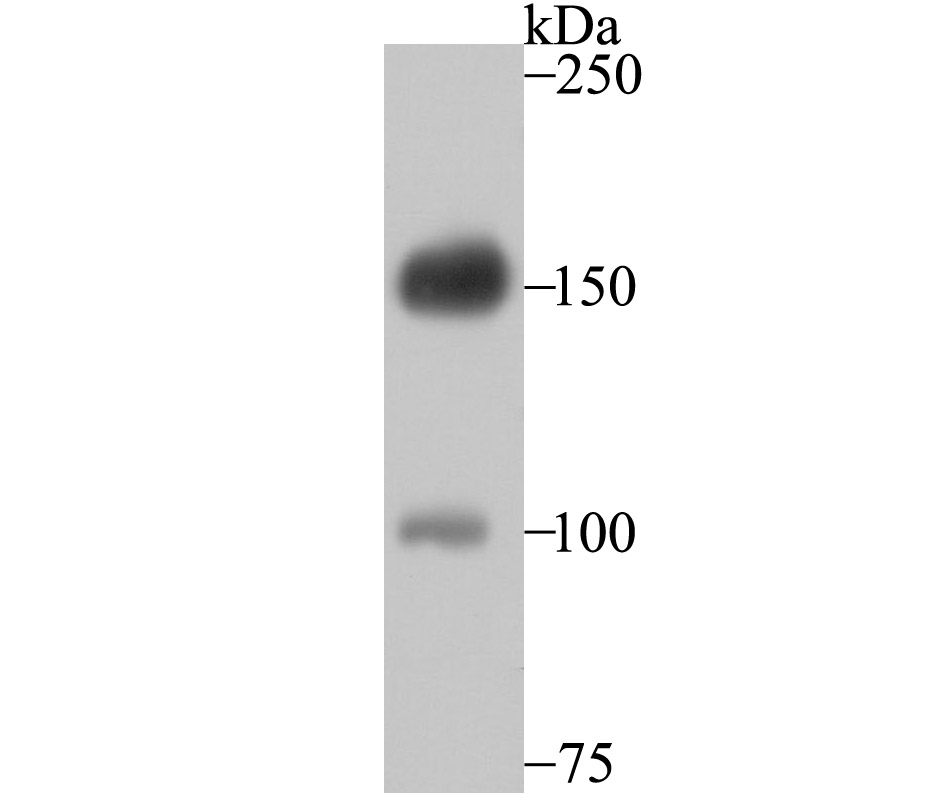
Fig1: Western blot analysis of Desmoglein 2 on A431 cell using anti-Desmoglein 2 antibody at 1/500 dilution.Application
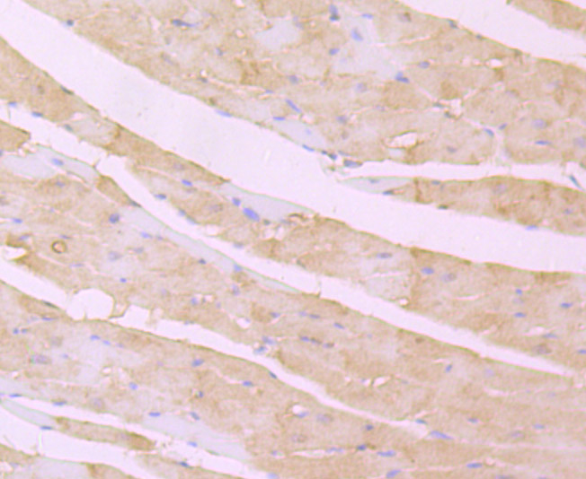
Fig2: Immunohistochemical analysis of paraffin-embedded rat heart tissue using anti-Desmoglein 2 antibody. Counter stained with hematoxylin.Application
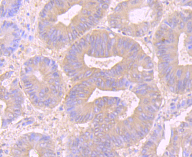
Fig3: Immunohistochemical analysis of paraffin-embedded human colon cancer tissue using anti-Desmoglein 2 antibody. Counter stained with hematoxylin.Application
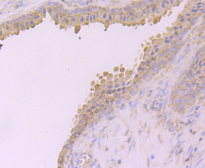
Fig4: Immunohistochemical analysis of paraffin-embedded human prostate tissue using anti-Desmoglein 2 antibody. Counter stained with hematoxylin.Application
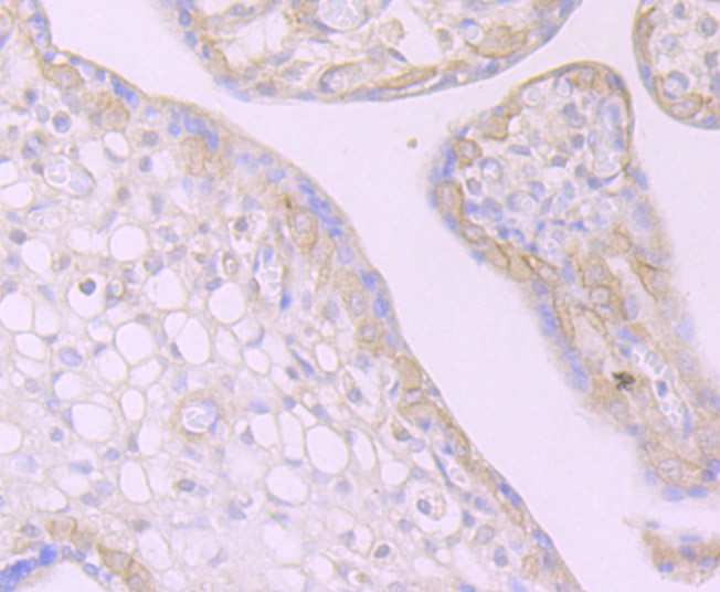
Fig5: Immunohistochemical analysis of paraffin-embedded human placenta tissue using anti-Desmoglein 2 antibody. Counter stained with hematoxylin.Application
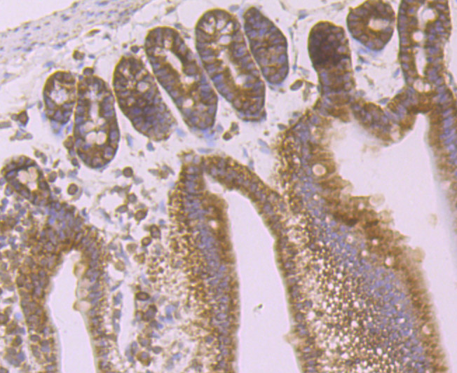
Fig6: Immunohistochemical analysis of paraffin-embedded mouse small intestine tissue using anti-Desmoglein 2 antibody. Counter stained with hematoxylin.Application
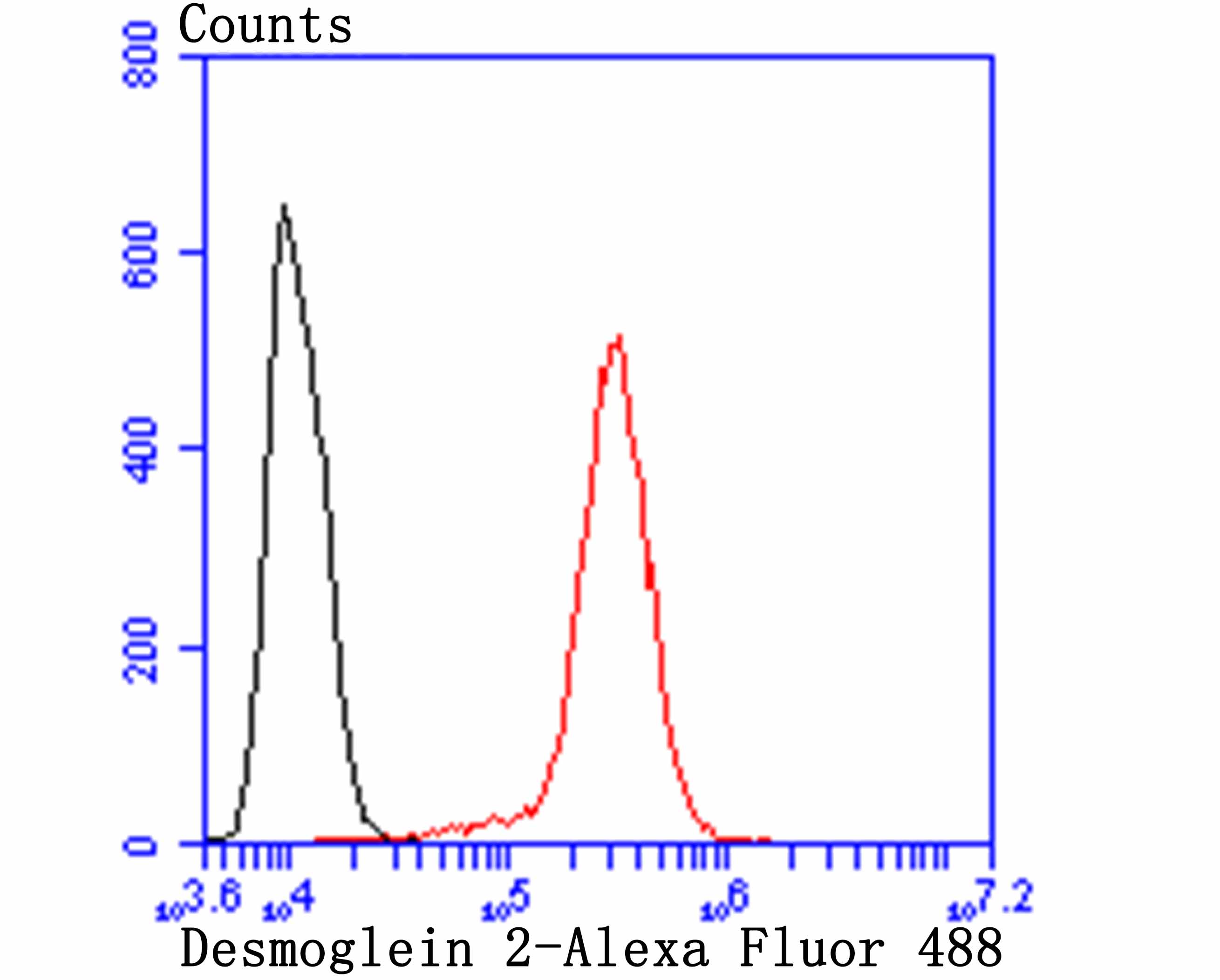
Fig7: Flow cytometric analysis of A431 cells with Desmoglein 2 antibody at 1/100 dilution (red) compared with an unlabelled control (cells without incubation with primary antibody; black). Alexa Fluor 488-conjugated goat anti-rabbit IgG was used as the secondary antibody.| Product Name | Desmoglein 2 [JB96-37] |
|---|---|
| Antibody Type | Primary Antibodies |
| Antigen Alias | ARVC 10 antibody ARVC10 antibody ARVD 10 antibody ARVD10 antibody Cadherin family member 5 antibody CDHF 5 antibody CDHF5 antibody CMD1BB antibody Desmoglein 2 antibody Desmoglein-2 antibody Desmoglein2 antibody DSG 2 antibody DSG2 antibody DSG2_HUMAN antibody HDGC antibody HDGC included antibody Human Desmoglein colon antibody MGC117034 antibody MGC117036 antibody MGC117037 antibody |
| Product description | Pemphigus is an autoimmune disease of skin adhesion associated with auto-antibodies against a number of keratinocyte antigens, such as the adhesion molecules desmoglein (dsg) 1 and 3 and acetylcholine receptors. Desmogleins, Type I membrane proteins, are important for cell adhesion and are expressed in great abundance at the desmosomes, which are adhesive cell junctions. Desmogleins belong to the cadherin family and consist of dsg1, dsg2 and dsg3. Calcium binds to the putative calcium binding sites at the extracellular N-terminal domain, which has cadherin-like repeats. Unlike normal human keratinocytes, the squamous cell carcinoma cells exhibit diminished or un-usual expression of dsg3 and dsg1, which bear pemphigus vulgaris and pemphigus foliaceus antigens, respectively. Several carcinoma cell lines constantly express dsg2 and dsg3 mRNA, whereas cultured normal human keratinocytes always express dsg1 and dsg3 mRNA, with or without dsg2 mRNA. This expression pattern indicates that desmoglein isoforms exhibit abnormal expression and may be related to tumor cell kinetics, such as cell invasion and metastasis. dsg2 is the fundamental dsg common to all desmosome-possessing tissues and is the largest desmoglein in the family. |
| Immunogen | Recombinant protein |
| Clonality | Monoclonal |
|---|---|
| Isotype | IgG |
| Host Species | Recombinant rabbit |
| Tested Applications | WBIHCFC |
| WB:1:500-1:1,000 IHC:1:50-1:200 FC:1:50-1:100 |
|
| Species Reactivity | HumanMouseRat |
| Concentration | 1mg/ml |
| Alternative Names | ARVC 10 antibody ARVC10 antibody ARVD 10 antibody ARVD10 antibody Cadherin family member 5 antibody CDHF 5 antibody CDHF5 antibody CMD1BB antibody Desmoglein 2 antibody Desmoglein-2 antibody Desmoglein2 antibody DSG 2 antibody DSG2 antibody DSG2_HUMAN antibody HDGC antibody HDGC included antibody Human Desmoglein colon antibody MGC117034 antibody MGC117036 antibody MGC117037 antibody |
|---|---|
| Molecular Weight(MW) | 150 kDa |
| Cellular Localization | Cell membrane. |
Application
Fig1: Western blot analysis of Desmoglein 2 on A431 cell using anti-Desmoglein 2 antibody at 1/500 dilution.
Application
Fig2: Immunohistochemical analysis of paraffin-embedded rat heart tissue using anti-Desmoglein 2 antibody. Counter stained with hematoxylin.
Application
Fig3: Immunohistochemical analysis of paraffin-embedded human colon cancer tissue using anti-Desmoglein 2 antibody. Counter stained with hematoxylin.
Application
Fig4: Immunohistochemical analysis of paraffin-embedded human prostate tissue using anti-Desmoglein 2 antibody. Counter stained with hematoxylin.
Application
Fig5: Immunohistochemical analysis of paraffin-embedded human placenta tissue using anti-Desmoglein 2 antibody. Counter stained with hematoxylin.
Application
Fig6: Immunohistochemical analysis of paraffin-embedded mouse small intestine tissue using anti-Desmoglein 2 antibody. Counter stained with hematoxylin.
Application
Fig7: Flow cytometric analysis of A431 cells with Desmoglein 2 antibody at 1/100 dilution (red) compared with an unlabelled control (cells without incubation with primary antibody; black). Alexa Fluor 488-conjugated goat anti-rabbit IgG was used as the secondary antibody.| Positive Control | A431, rat heart tissue, human colon cancer tissue, human prostate tissue, human placenta tissue, mouse small intestine tissue. |
|---|---|
| Application Notes | WB:1:500-1:1,000 IHC:1:50-1:200 FC:1:50-1:100 |
| Form | Liquid |
|---|---|
| Storage Instructions | Store at +4℃ after thawing. Aliquot store at -20℃ or -80℃. Avoid repeated freeze / thaw cycles. |
| Storage Buffer | 1*TBS (pH7.4), 1%BSA, 40%Glycerol. Preservative: 0.05% Sodium Azide. |


