Application
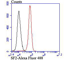
Fig8: Flow cytometric analysis of K562 cells with SF2 antibody at 1/100 dilution (red) compared with an unlabelled control (cells without incubation with primary antibody; black). Alexa Fluor 488-conjugated goat anti rabbit IgG was used as the secondary antibody.Application
Fig8: Flow cytometric analysis of K562 cells with SF2 antibody at 1/100 dilution (red) compared with an unlabelled control (cells without incubation with primary antibody; black). Alexa Fluor 488-conjugated goat anti rabbit IgG was used as the secondary antibody.Application
Fig8: Flow cytometric analysis of K562 cells with SF2 antibody at 1/100 dilution (red) compared with an unlabelled control (cells without incubation with primary antibody; black). Alexa Fluor 488-conjugated goat anti rabbit IgG was used as the secondary antibody.Application
Fig8: Flow cytometric analysis of K562 cells with SF2 antibody at 1/100 dilution (red) compared with an unlabelled control (cells without incubation with primary antibody; black). Alexa Fluor 488-conjugated goat anti rabbit IgG was used as the secondary antibody.Application
Fig8: Flow cytometric analysis of K562 cells with SF2 antibody at 1/100 dilution (red) compared with an unlabelled control (cells without incubation with primary antibody; black). Alexa Fluor 488-conjugated goat anti rabbit IgG was used as the secondary antibody.Application
Fig8: Flow cytometric analysis of K562 cells with SF2 antibody at 1/100 dilution (red) compared with an unlabelled control (cells without incubation with primary antibody; black). Alexa Fluor 488-conjugated goat anti rabbit IgG was used as the secondary antibody.Application
Fig8: Flow cytometric analysis of K562 cells with SF2 antibody at 1/100 dilution (red) compared with an unlabelled control (cells without incubation with primary antibody; black). Alexa Fluor 488-conjugated goat anti rabbit IgG was used as the secondary antibody.Application
Fig8: Flow cytometric analysis of K562 cells with SF2 antibody at 1/100 dilution (red) compared with an unlabelled control (cells without incubation with primary antibody; black). Alexa Fluor 488-conjugated goat anti rabbit IgG was used as the secondary antibody.| Product Name | SF2 [JB40-76] |
|---|---|
| Antibody Type | Primary Antibodies |
| Antigen Alias | Alternative splicing factor 1 antibody Alternative-splicing factor 1 antibody arginine/serine-rich 1 antibody ASF 1 antibody ASF antibody ASF-1 antibody ASF1 antibody FLJ53078 antibody MGC5228 antibody P33 subunit antibody Pre mRNA splicing factor SF2 P33 subunit antibody pre-mRNA-splicing factor SF2 antibody Serine/arginine-rich splicing factor 1 antibody SF2 antibody SF2P33 antibody SFRS1 antibody Splicing factor 2 alternate splicing factor antibody Splicing factor 2 antibody Splicing factor antibody Splicing factor arginine/serine rich 1 antibody SR Splicing factor 1 antibody SRp30a antibody srsf1 antibody SRSF1_HUMAN antibody |
| Product description | Pre-mRNA splicing enhancer elements are short RNA sequences capable of activating weak splice sites in nearby introns that are required for accurate splice site recognition and the control of alternative splicing. Splicing enhancer elements contain specific binding sites for serine/arginine (SR)-rich splicing factors, which include SC35, 9G8, SRp20, and SF2/ASF. The family of SR factors all contain one or more RNA recognition motifs (RRM) and an arginine/ serine (RS)-rich domain. They are not only essential for constitutive splicing but also regulate splicing in a concentration-dependent manner by influencing the selection of alternative splice sites. The majority of SR proteins, including SC35 and SRp40, are confined to the nucleus, while SF2/ASF, SRp20, and 9G8 are continuously shuttled between the nucleus and the cytoplasm and contribute to mRNA transport. The activity of SR proteins in regulated splicing is antagonized by members of the hnRNP A/B family of proteins, which induce drastic shifts in the selection of splicing sites. |
| Immunogen | Recombinant protein |
| Clonality | Monoclonal |
|---|---|
| Isotype | IgG |
| Host Species | Recombinant rabbit |
| Tested Applications | WBICCIFIHCFC |
| WB:1:500-1:1,000 ICC:1:50 IHC:1:50-1:200 FC:1:50-1:100 |
|
| Species Reactivity | HumanMouseRat |
| Concentration | 1mg/ml |
| Alternative Names | Alternative splicing factor 1 antibody Alternative-splicing factor 1 antibody arginine/serine-rich 1 antibody ASF 1 antibody ASF antibody ASF-1 antibody ASF1 antibody FLJ53078 antibody MGC5228 antibody P33 subunit antibody Pre mRNA splicing factor SF2 P33 subunit antibody pre-mRNA-splicing factor SF2 antibody Serine/arginine-rich splicing factor 1 antibody SF2 antibody SF2P33 antibody SFRS1 antibody Splicing factor 2 alternate splicing factor antibody Splicing factor 2 antibody Splicing factor antibody Splicing factor arginine/serine rich 1 antibody SR Splicing factor 1 antibody SRp30a antibody srsf1 antibody SRSF1_HUMAN antibody |
|---|---|
| Cellular Localization | Nucleus. Cytoplasm. |
Application
Fig8: Flow cytometric analysis of K562 cells with SF2 antibody at 1/100 dilution (red) compared with an unlabelled control (cells without incubation with primary antibody; black). Alexa Fluor 488-conjugated goat anti rabbit IgG was used as the secondary antibody.
Application
Fig8: Flow cytometric analysis of K562 cells with SF2 antibody at 1/100 dilution (red) compared with an unlabelled control (cells without incubation with primary antibody; black). Alexa Fluor 488-conjugated goat anti rabbit IgG was used as the secondary antibody.
Application
Fig8: Flow cytometric analysis of K562 cells with SF2 antibody at 1/100 dilution (red) compared with an unlabelled control (cells without incubation with primary antibody; black). Alexa Fluor 488-conjugated goat anti rabbit IgG was used as the secondary antibody.
Application
Fig8: Flow cytometric analysis of K562 cells with SF2 antibody at 1/100 dilution (red) compared with an unlabelled control (cells without incubation with primary antibody; black). Alexa Fluor 488-conjugated goat anti rabbit IgG was used as the secondary antibody.
Application
Fig8: Flow cytometric analysis of K562 cells with SF2 antibody at 1/100 dilution (red) compared with an unlabelled control (cells without incubation with primary antibody; black). Alexa Fluor 488-conjugated goat anti rabbit IgG was used as the secondary antibody.
Application
Fig8: Flow cytometric analysis of K562 cells with SF2 antibody at 1/100 dilution (red) compared with an unlabelled control (cells without incubation with primary antibody; black). Alexa Fluor 488-conjugated goat anti rabbit IgG was used as the secondary antibody.
Application
Fig8: Flow cytometric analysis of K562 cells with SF2 antibody at 1/100 dilution (red) compared with an unlabelled control (cells without incubation with primary antibody; black). Alexa Fluor 488-conjugated goat anti rabbit IgG was used as the secondary antibody.
Application
Fig8: Flow cytometric analysis of K562 cells with SF2 antibody at 1/100 dilution (red) compared with an unlabelled control (cells without incubation with primary antibody; black). Alexa Fluor 488-conjugated goat anti rabbit IgG was used as the secondary antibody.| Positive Control | Mouse heart tissue lysate, mouse liver tissue lysate, K562, A431, human lung cancer tissue, human tonsil tissue, human breast tissue, mouse colon tissue, rat epididymis tissue. |
|---|---|
| Application Notes | WB:1:500-1:1,000 ICC:1:50 IHC:1:50-1:200 FC:1:50-1:100 |
| Form | Liquid |
|---|---|
| Storage Instructions | Store at +4℃ after thawing. Aliquot store at -20℃ or -80℃. Avoid repeated freeze / thaw cycles. |
| Storage Buffer | 1*TBS (pH7.4), 1%BSA, 40%Glycerol. Preservative: 0.05% Sodium Azide. |


