WB
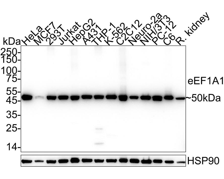
Western blot analysis of eEF1A1 on different lysates with Rabbit anti-eEF1A1 antibody at 1/2,000 dilution. Lane 1: HeLa cell lysate (10 µg/Lane), Lane 2: MCF7 cell lysate (10 µg/Lane), Lane 3: 293T cell lysate (10 µg/Lane), Lane 4: Jurkat cell lysate (10 µg/Lane), Lane 5: HepG2 cell lysate (10 µg/Lane), Lane 6: A431 cell lysate (10 µg/Lane), Lane 7: THP-1 cell lysate (10 µg/Lane), Lane 8: K-562 cell lysate (10 µg/Lane), Lane 9: C2C12 cell lysate (10 µg/Lane), Lane 10: Neuro-2a cell lysate (10 µg/Lane), Lane 11: NIH/3T3 cell lysate (10 µg/Lane), Lane 12: PC-12 cell lysate (10 µg/Lane), Lane 13: C6 cell lysate (10 µg/Lane), Lane 14: Rat kidney tissue lysate (40 µg/Lane), Exposure time: 6 seconds; 4-20% SDS-PAGE gel. Proteins were transferred to a PVDF membrane and blocked with 5% NFDM/TBST for 1 hour at room temperature. The primary antibody at 1/2,000 dilution was used in 5% NFDM/TBST at 4℃ overnight. Goat Anti-Rabbit IgG - HRP Secondary Antibody at 1/50,000 dilution was used for 1 hour at room temperature.IHC
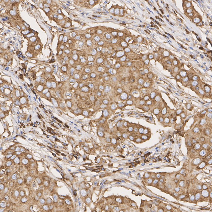
Immunohistochemical analysis of paraffin-embedded human breast cancer tissue with Rabbit anti-eEF1A1 antibody at 1/200 dilution. The section was pre-treated using heat mediated antigen retrieval with Tris-EDTA buffer (pH 9.0) for 20 minutes. The tissues were blocked in 1% BSA for 20 minutes at room temperature, washed with ddH2O and PBS, and then probed with the primary antibody at 1/200 dilution for 1 hour at room temperature. The detection was performed using an HRP conjugated compact polymer system. DAB was used as the chromogen. Tissues were counterstained with hematoxylin and mounted with DPX.IHC
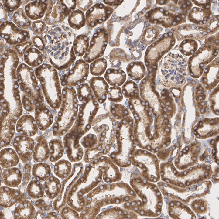
Immunohistochemical analysis of paraffin-embedded mouse kidney tissue with Rabbit anti-eEF1A1 antibody at 1/200 dilution. The section was pre-treated using heat mediated antigen retrieval with Tris-EDTA buffer (pH 9.0) for 20 minutes. The tissues were blocked in 1% BSA for 20 minutes at room temperature, washed with ddH2O and PBS, and then probed with the primary antibody at 1/200 dilution for 1 hour at room temperature. The detection was performed using an HRP conjugated compact polymer system. DAB was used as the chromogen. Tissues were counterstained with hematoxylin and mounted with DPX.IHC
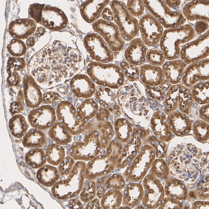
Immunohistochemical analysis of paraffin-embedded rat kidney tissue with Rabbit anti-eEF1A1 antibody at 1/200 dilution. The section was pre-treated using heat mediated antigen retrieval with Tris-EDTA buffer (pH 9.0) for 20 minutes. The tissues were blocked in 1% BSA for 20 minutes at room temperature, washed with ddH2O and PBS, and then probed with the primary antibody at 1/200 dilution for 1 hour at room temperature. The detection was performed using an HRP conjugated compact polymer system. DAB was used as the chromogen. Tissues were counterstained with hematoxylin and mounted with DPX.ICC/IF
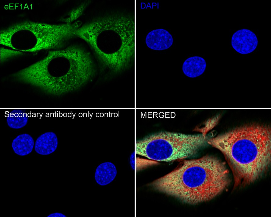
Immunocytochemistry analysis of C2C12 cells labeling eEF1A1 with Rabbit anti-eEF1A1 antibody at 1/250 dilution. Cells were fixed in 4% paraformaldehyde for 20 minutes at room temperature, permeabilized with 0.1% Triton X-100 in PBS for 5 minutes at room temperature, then blocked with 1% BSA in 10% negative goat serum for 1 hour at room temperature. Cells were then incubated with Rabbit anti-eEF1A1 antibody at 1/250 dilution in 1% BSA in PBST overnight at 4 ℃. Goat Anti-Rabbit IgG H&L (488) was used as the secondary antibody at 1/1,000 dilution. PBS instead of the primary antibody was used as the secondary antibody only control. Nuclear DNA was labelled in blue with DAPI. Beta tubulin (red) was stained at 1/100 dilution overnight at +4℃. Goat Anti-Mouse IgG H&L (594) was used as the secondary antibody at 1/1,000 dilution.FC
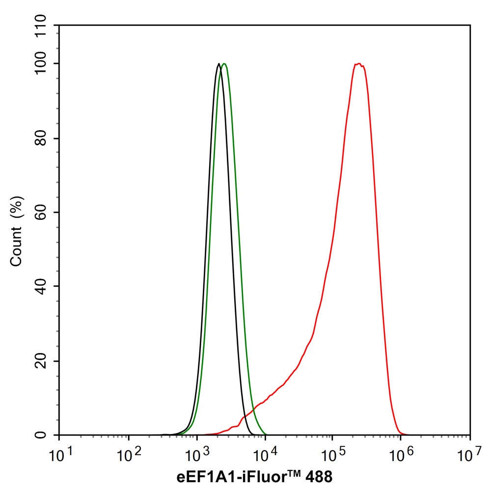
Flow cytometric analysis of PC-12 cells labeling eEF1A1. Cells were fixed and permeabilized. Then stained with the primary antibody (1/1,000) (red) compared with Rabbit IgG Isotype Control (green). After incubation of the primary antibody at +4℃ for an hour, the cells were stained with a 488 conjugate-Goat anti-Rabbit IgG Secondary antibody at 1/1,000 dilution for 30 minutes at +4℃. Unlabelled sample was used as a control (cells without incubation with primary antibody; black).| Product Name | eEF1A1 Recombinant Rabbit Monoclonal Antibody |
|---|---|
| Antibody Type | Primary Antibodies |
| Product description | The elongation factor-1 complex is composed of two subunits, EF-1 α1 (elongation factor 1-alpha 1) and EF-1 α2 (elongation factor 1-alpha 2), and is responsible for the delivery of aminoacyl tRNAs to the ribosome. EF-1 α1 is expressed predominately in brain, placenta, lung, liver, kidney and pancreas, while EF-1 α2 is highly expressed in heart, brain and skeletal muscle. Both EF-1 α1 and α2 localize to the nucleus and belong to the GTP-binding elongation factor family. The gene encoding EF-1 α2, which maps to human chromosome 20q13.3, may play a role in the development of ovarian cancer, while the EF-1 α1 gene, mapping to chromosome 6Q14.1, is commonly present as an autoantigen in patients with Felty syndrome. Felty syndrome is a disorder characterized by rheumatoid arthritis, a swollen spleen, decreased white blood cell count, and increased susceptibility to infection. |
| Immunogen | Synthetic peptide within Human eEF1A1 aa 413-462 / 462. |
| Clonality | Monoclonal |
|---|---|
| Isotype | IgG |
| Host Species | Recombinant rabbit |
| Tested Applications | FCICC/IFIHCWB |
| WB:1:2000 IHC:1:200-1:1000 ICC/IF:1:100-1:250 FC:1:1000 |
|
| Species Reactivity | HumanMouseRat |
| Concentration | 1mg/ml |
| Purification | Protein A |
| Alternative Names | CCS 3 antibody CCS3 antibody Cervical cancer suppressor 3 antibody chunp6927 antibody CTCL tumor antigen antibody EE1A1 antibody EEF 1 antibody EEF1A antibody eEF1A-1 antibody EEF1A1 antibody EF-1-alpha-1 antibody EF-Tu antibody EF1A antibody EF1a like protein antibody EF1A1_HUMAN antibody Elongation factor 1 alpha subunit antibody Elongation factor 1-alpha 1 antibody Elongation factor Tu antibody Eukaryotic elongation factor 1 A-1 antibody Eukaryotic translation elongation factor 1 alpha 1 antibody Eukaryotic translation elongation factor 1 alpha 1 like 14 antibody Glucocorticoid receptor AF 1 specific elongation factor antibody GRAF 1EF antibody HNGC:16303 antibody ik:tdsubc_2a3 antibody ik:tdsubc_2b3 antibody LENG7 antibody Leukocyte receptor cluster (LRC) member 7 antibody Leukocyte receptor cluster member 7 antibody Prostate tumor inducing protein 1 antibody PTI1 antibody tdsubc_2a3 antibody Translation elongation factor 1 alpha 1 like 14 antibody wu:fa91c07 antibody wu:fa94b03 antibody wu:fi13b09 antibody xx:tdsubc_2a3 antibody xx:tdsubc_2b3 antibody |
|---|---|
| Molecular Weight(MW) | 50 kDa |
| Cellular Localization | Cell membrane, Cytoplasm, Membrane, Nucleus. |
WB
Western blot analysis of eEF1A1 on different lysates with Rabbit anti-eEF1A1 antibody at 1/2,000 dilution. Lane 1: HeLa cell lysate (10 µg/Lane), Lane 2: MCF7 cell lysate (10 µg/Lane), Lane 3: 293T cell lysate (10 µg/Lane), Lane 4: Jurkat cell lysate (10 µg/Lane), Lane 5: HepG2 cell lysate (10 µg/Lane), Lane 6: A431 cell lysate (10 µg/Lane), Lane 7: THP-1 cell lysate (10 µg/Lane), Lane 8: K-562 cell lysate (10 µg/Lane), Lane 9: C2C12 cell lysate (10 µg/Lane), Lane 10: Neuro-2a cell lysate (10 µg/Lane), Lane 11: NIH/3T3 cell lysate (10 µg/Lane), Lane 12: PC-12 cell lysate (10 µg/Lane), Lane 13: C6 cell lysate (10 µg/Lane), Lane 14: Rat kidney tissue lysate (40 µg/Lane), Exposure time: 6 seconds; 4-20% SDS-PAGE gel. Proteins were transferred to a PVDF membrane and blocked with 5% NFDM/TBST for 1 hour at room temperature. The primary antibody at 1/2,000 dilution was used in 5% NFDM/TBST at 4℃ overnight. Goat Anti-Rabbit IgG - HRP Secondary Antibody at 1/50,000 dilution was used for 1 hour at room temperature.
IHC
Immunohistochemical analysis of paraffin-embedded human breast cancer tissue with Rabbit anti-eEF1A1 antibody at 1/200 dilution. The section was pre-treated using heat mediated antigen retrieval with Tris-EDTA buffer (pH 9.0) for 20 minutes. The tissues were blocked in 1% BSA for 20 minutes at room temperature, washed with ddH2O and PBS, and then probed with the primary antibody at 1/200 dilution for 1 hour at room temperature. The detection was performed using an HRP conjugated compact polymer system. DAB was used as the chromogen. Tissues were counterstained with hematoxylin and mounted with DPX.
IHC
Immunohistochemical analysis of paraffin-embedded mouse kidney tissue with Rabbit anti-eEF1A1 antibody at 1/200 dilution. The section was pre-treated using heat mediated antigen retrieval with Tris-EDTA buffer (pH 9.0) for 20 minutes. The tissues were blocked in 1% BSA for 20 minutes at room temperature, washed with ddH2O and PBS, and then probed with the primary antibody at 1/200 dilution for 1 hour at room temperature. The detection was performed using an HRP conjugated compact polymer system. DAB was used as the chromogen. Tissues were counterstained with hematoxylin and mounted with DPX.
IHC
Immunohistochemical analysis of paraffin-embedded rat kidney tissue with Rabbit anti-eEF1A1 antibody at 1/200 dilution. The section was pre-treated using heat mediated antigen retrieval with Tris-EDTA buffer (pH 9.0) for 20 minutes. The tissues were blocked in 1% BSA for 20 minutes at room temperature, washed with ddH2O and PBS, and then probed with the primary antibody at 1/200 dilution for 1 hour at room temperature. The detection was performed using an HRP conjugated compact polymer system. DAB was used as the chromogen. Tissues were counterstained with hematoxylin and mounted with DPX.
ICC/IF
Immunocytochemistry analysis of C2C12 cells labeling eEF1A1 with Rabbit anti-eEF1A1 antibody at 1/250 dilution. Cells were fixed in 4% paraformaldehyde for 20 minutes at room temperature, permeabilized with 0.1% Triton X-100 in PBS for 5 minutes at room temperature, then blocked with 1% BSA in 10% negative goat serum for 1 hour at room temperature. Cells were then incubated with Rabbit anti-eEF1A1 antibody at 1/250 dilution in 1% BSA in PBST overnight at 4 ℃. Goat Anti-Rabbit IgG H&L (488) was used as the secondary antibody at 1/1,000 dilution. PBS instead of the primary antibody was used as the secondary antibody only control. Nuclear DNA was labelled in blue with DAPI. Beta tubulin (red) was stained at 1/100 dilution overnight at +4℃. Goat Anti-Mouse IgG H&L (594) was used as the secondary antibody at 1/1,000 dilution.
FC
Flow cytometric analysis of PC-12 cells labeling eEF1A1. Cells were fixed and permeabilized. Then stained with the primary antibody (1/1,000) (red) compared with Rabbit IgG Isotype Control (green). After incubation of the primary antibody at +4℃ for an hour, the cells were stained with a 488 conjugate-Goat anti-Rabbit IgG Secondary antibody at 1/1,000 dilution for 30 minutes at +4℃. Unlabelled sample was used as a control (cells without incubation with primary antibody; black).| Application Notes | WB:1:2000 IHC:1:200-1:1000 ICC/IF:1:100-1:250 FC:1:1000 |
|---|
| Form | Liquid |
|---|---|
| Storage Instructions | Store at +4℃ after thawing. Aliquot store at -20℃ or -80℃. Avoid repeated freeze / thaw cycles. |
| Storage Buffer | 1*TBS (pH7.4), 1%BSA, 40%Glycerol. Preservative: 0.05% Sodium Azide. |


