WB
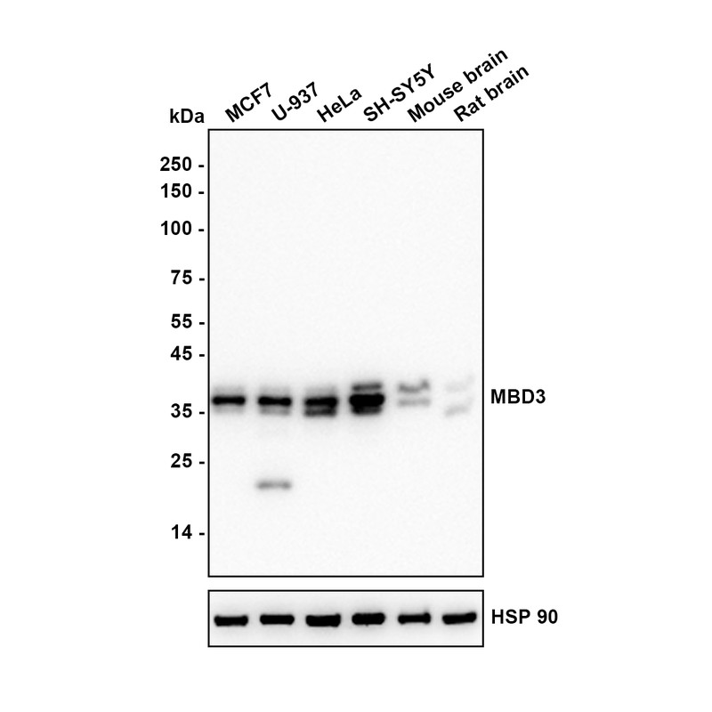
Western blot analysis of MBD3 on different lysates with Rabbit anti-MBD3 antibody at 1/5,000 dilution. Lane 1: MCF7 (Human breast cancer cell) cell lysate (15 µg/Lane), Lane 2: U-937 (Human acute monocytic leukemia cell) cell lysate (15 µg/Lane), Lane 3: HeLa (Human cervical adenocarcinoma cell) cell lysate (15 µg/Lane), Lane 4: SH-SY5Y (Human neuroblastoma cell) cell lysate (15 µg/Lane), Lane 5: Mouse brain tissue lysate (30 µg/Lane) Lane 6: Rat brainc tissue lysate (30 µg/Lane), Exposure time: 25 seconds; Blocking: 5% NFDM/TBST, 1 hour at room temperature Primary antibody: 1/5,000 in primary antibody dilution buffer, overnight at 4 ℃ Secondary antibody: Goat anti-Rabbit IgG-HRP, 1/50,000 in 5% NFDM/TBST, 1 hour at room temperature.ICC/IF
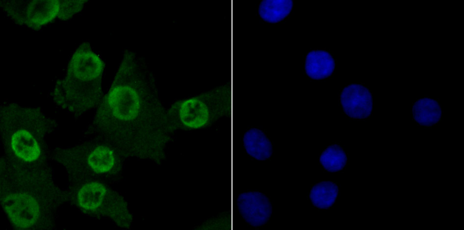
ICC staining MBD3 in A549 cells (green). The nuclear counter stain is DAPI (blue). Cells were fixed in paraformaldehyde, permeabilised with 0.25% Triton X100/PBS.ICC/IF
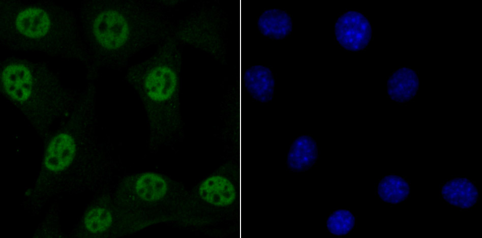
ICC staining MBD3 in NIH-3T3 cells (green). The nuclear counter stain is DAPI (blue). Cells were fixed in paraformaldehyde, permeabilised with 0.25% Triton X100/PBS.ICC/IF
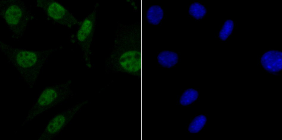
ICC staining MBD3 in SH-SY-5Y cells (green). The nuclear counter stain is DAPI (blue). Cells were fixed in paraformaldehyde, permeabilised with 0.25% Triton X100/PBS.IHC
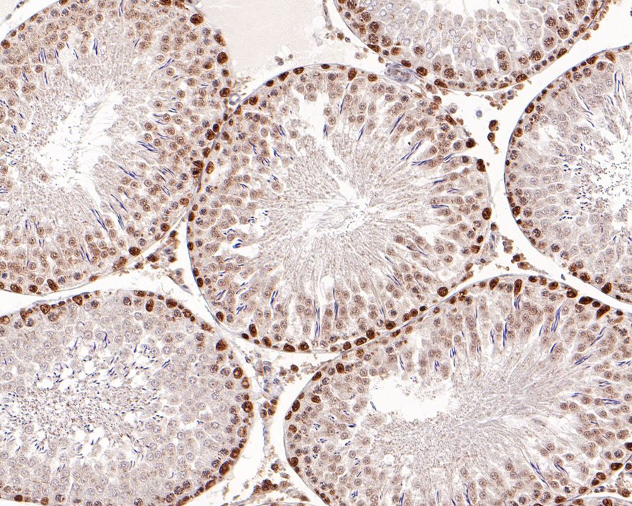
Immunohistochemical analysis of paraffin-embedded rat testis tissue with Rabbit anti-MBD3 antibody at 1/500 dilution. The section was pre-treated using heat mediated antigen retrieval with sodium citrate buffer (pH 6.0) (high pressure) for 2 minutes. The tissues were blocked in 1% BSA for 20 minutes at room temperature, washed with ddH2O and PBS, and then probed with the primary antibody at 1/500 dilution for 1 hour at room temperature. The detection was performed using an HRP conjugated compact polymer system. DAB was used as the chromogen. Tissues were counterstained with hematoxylin and mounted with DPX.IHC
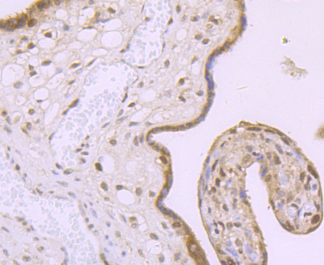
Immunohistochemical analysis of paraffin-embedded human placenta tissue using anti-MBD3 antibody. Counter stained with hematoxylin.IHC
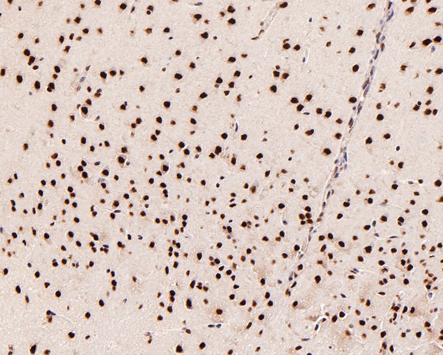
Immunohistochemical analysis of paraffin-embedded mouse brain tissue with Rabbit anti-MBD3 antibody at 1/500 dilution. The section was pre-treated using heat mediated antigen retrieval with sodium citrate buffer (pH 6.0) (high pressure) for 2 minutes. The tissues were blocked in 1% BSA for 20 minutes at room temperature, washed with ddH2O and PBS, and then probed with the primary antibody at 1/500 dilution for 1 hour at room temperature. The detection was performed using an HRP conjugated compact polymer system. DAB was used as the chromogen. Tissues were counterstained with hematoxylin and mounted with DPX.| Product Name | MBD3 Recombinant Rabbit Monoclonal Antibody |
|---|---|
| Antibody Type | Primary Antibodies |
| Product description | Methylation of DNA contributes to the regulation of gene transcription in both mammalian and invertebrate systems. DNA methylation predominates on cytosine residues that are present in dinucleotide motifs consisting of a 5′ cytosine followed by guanosine (CpG), and it requires the enzymatic activity of DNA methyltransferase, which results in transcriptional repression of the methylated gene. Several proteins have been identified that associate with the methyl-CpG sites, and they include methyl-CpG binding protein-1 (MBD1), MBD2, MBD3, MBD4 and MeCP2. Expression of the MBD proteins is highest in somatic tissues. |
| Immunogen | Recombinant protein within Human MBD3 aa 160-291 / 291. |
| Clonality | Monoclonal |
|---|---|
| Isotype | IgG |
| Host Species | Recombinant rabbit |
| Tested Applications | ICC/IFIHCWB |
| WB:1:5000 IHC:1:50-1:500 ICC/IF:1:50-1:200 |
|
| Species Reactivity | HumanMouseRat |
| Concentration | 1mg/ml |
| Purification | Protein A |
| Alternative Names | AI181826 antibody AU019209 antibody MBD 3 antibody Mbd3 antibody MBD3: methyl CpG binding domain protein 3 antibody MBD3_HUMAN antibody Methyl CpG binding domain protein 3 antibody Methyl CpG binding protein MBD3 antibody Methyl-CpG-binding domain protein 3 antibody Methyl-CpG-binding protein MBD3 antibody |
|---|---|
| Molecular Weight(MW) | 33 kDa(Observed band size: 35 kDa) |
| Cellular Localization | Nucleus. |
WB
Western blot analysis of MBD3 on different lysates with Rabbit anti-MBD3 antibody at 1/5,000 dilution. Lane 1: MCF7 (Human breast cancer cell) cell lysate (15 µg/Lane), Lane 2: U-937 (Human acute monocytic leukemia cell) cell lysate (15 µg/Lane), Lane 3: HeLa (Human cervical adenocarcinoma cell) cell lysate (15 µg/Lane), Lane 4: SH-SY5Y (Human neuroblastoma cell) cell lysate (15 µg/Lane), Lane 5: Mouse brain tissue lysate (30 µg/Lane) Lane 6: Rat brainc tissue lysate (30 µg/Lane), Exposure time: 25 seconds; Blocking: 5% NFDM/TBST, 1 hour at room temperature Primary antibody: 1/5,000 in primary antibody dilution buffer, overnight at 4 ℃ Secondary antibody: Goat anti-Rabbit IgG-HRP, 1/50,000 in 5% NFDM/TBST, 1 hour at room temperature.
ICC/IF
ICC staining MBD3 in A549 cells (green). The nuclear counter stain is DAPI (blue). Cells were fixed in paraformaldehyde, permeabilised with 0.25% Triton X100/PBS.
ICC/IF
ICC staining MBD3 in NIH-3T3 cells (green). The nuclear counter stain is DAPI (blue). Cells were fixed in paraformaldehyde, permeabilised with 0.25% Triton X100/PBS.
ICC/IF
ICC staining MBD3 in SH-SY-5Y cells (green). The nuclear counter stain is DAPI (blue). Cells were fixed in paraformaldehyde, permeabilised with 0.25% Triton X100/PBS.
IHC
Immunohistochemical analysis of paraffin-embedded rat testis tissue with Rabbit anti-MBD3 antibody at 1/500 dilution. The section was pre-treated using heat mediated antigen retrieval with sodium citrate buffer (pH 6.0) (high pressure) for 2 minutes. The tissues were blocked in 1% BSA for 20 minutes at room temperature, washed with ddH2O and PBS, and then probed with the primary antibody at 1/500 dilution for 1 hour at room temperature. The detection was performed using an HRP conjugated compact polymer system. DAB was used as the chromogen. Tissues were counterstained with hematoxylin and mounted with DPX.
IHC
Immunohistochemical analysis of paraffin-embedded human placenta tissue using anti-MBD3 antibody. Counter stained with hematoxylin.
IHC
Immunohistochemical analysis of paraffin-embedded mouse brain tissue with Rabbit anti-MBD3 antibody at 1/500 dilution. The section was pre-treated using heat mediated antigen retrieval with sodium citrate buffer (pH 6.0) (high pressure) for 2 minutes. The tissues were blocked in 1% BSA for 20 minutes at room temperature, washed with ddH2O and PBS, and then probed with the primary antibody at 1/500 dilution for 1 hour at room temperature. The detection was performed using an HRP conjugated compact polymer system. DAB was used as the chromogen. Tissues were counterstained with hematoxylin and mounted with DPX.| Application Notes | WB:1:5000 IHC:1:50-1:500 ICC/IF:1:50-1:200 |
|---|
| Form | Liquid |
|---|---|
| Storage Instructions | Store at +4℃ after thawing. Aliquot store at -20℃ or -80℃. Avoid repeated freeze / thaw cycles. |
| Storage Buffer | 1*TBS (pH7.4), 1%BSA, 40%Glycerol. Preservative: 0.05% Sodium Azide. |


