Application
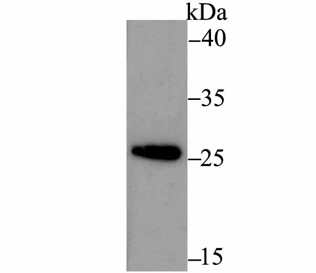
Fig1: Western blot analysis of ERp29 on THP-1 cell using anti-ERp29 antibody at 1/500 dilution.Application
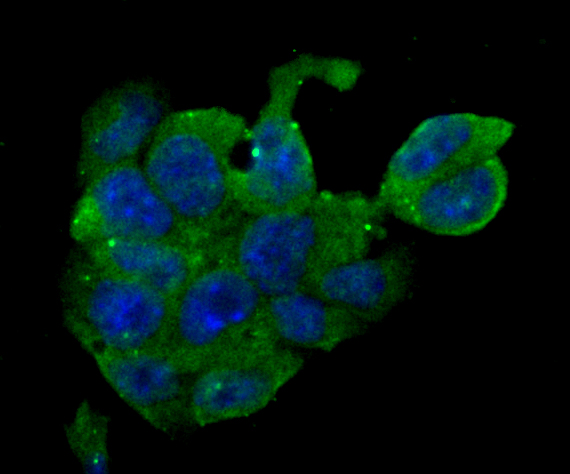
Fig2: ICC staining ERp29 in F9 cells (green). The nuclear counter stain is DAPI (blue). Cells were fixed in paraformaldehyde, permeabilised with 0.25% Triton X100/PBS.Application
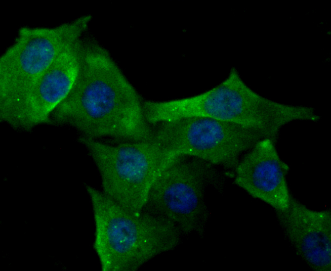
Fig3: ICC staining ERp29 in PMVEC cells (green). The nuclear counter stain is DAPI (blue). Cells were fixed in paraformaldehyde, permeabilised with 0.25% Triton X100/PBS.Application
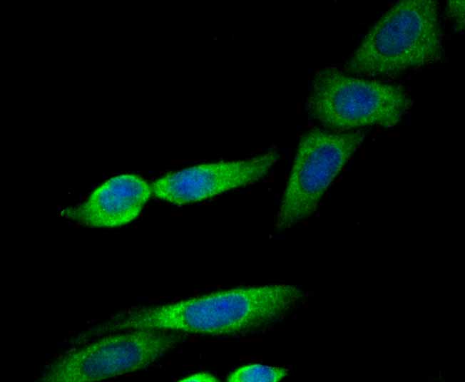
Fig4: ICC staining ERp29 in SiHa cells (green). The nuclear counter stain is DAPI (blue). Cells were fixed in paraformaldehyde, permeabilised with 0.25% Triton X100/PBS.Application
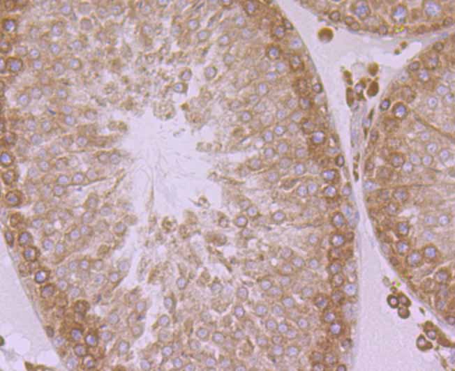
Fig5: Immunohistochemical analysis of paraffin-embedded rat testis tissue using anti-ERp29 antibody. Counter stained with hematoxylin.Application
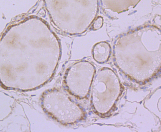
Fig6: Immunohistochemical analysis of paraffin-embedded human thyroid gland tissue using anti-ERp29 antibody. Counter stained with hematoxylin.Application
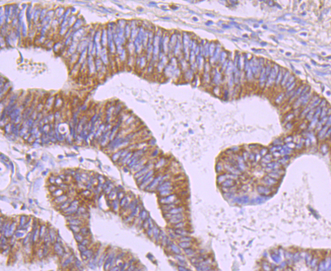
Fig7: Immunohistochemical analysis of paraffin-embedded human colon cancer tissue using anti-ERp29 antibody. Counter stained with hematoxylin.| Product Name | ERp29 [JG34-42] |
|---|---|
| Antibody Type | Primary Antibodies |
| Product description | ERp29 is a reticuloplasmin, a protein which resides in the lumen of the endoplasmic reticulum (ER). The protein shows sequence similarity to the protein disulfide-isomerase family. However, it lacks the thioredoxin motif characteristic of this family, suggesting that this protein does not function as a disulfide isomerase. The protein dimerizes and is thought to play a role in the processing of secretory proteins within the ER. Alternative splicing results in multiple transcript variants encoding different isoforms. |
| Immunogen | Recombinant protein within human ERp29 aa 1-150. |
| Clonality | Monoclonal |
|---|---|
| Isotype | IgG |
| Host Species | Recombinant rabbit |
| Tested Applications | WB,ICC,IF,IHC |
| WB:1:500-1:1,000 ICC:1:50-1:200 IHC:1:50-1:200 |
|
| Species Reactivity | Human,Mouse,Rat |
| Concentration | 1mg/ml |
| Purification | Unpurified |
| Alternative Names | C12orf8 antibody Chromosome 12 open reading frame 8 antibody Endoplasmic reticulum lumenal protein ERp28 antibody Endoplasmic reticulum protein 29 antibody Endoplasmic reticulum protein 29 isoform 1 antibody Endoplasmic reticulum protein ERp29 antibody Endoplasmic reticulum resident protein 28 antibody Endoplasmic reticulum resident protein 29 antibody Endoplasmic reticulum resident protein 31 antibody Epididymis secretory protein Li 107 antibody ERp28 antibody ERP29_HUMAN antibody ERp31 antibody HEL S 107 antibody PDI-DB antibody PDIA9 antibody Protein disulfide isomerase family A member 9 antibody |
|---|---|
| Molecular Weight(MW) | 29 kDa |
| Cellular Localization | Endoplasmic reticulum. |
Application
Fig1: Western blot analysis of ERp29 on THP-1 cell using anti-ERp29 antibody at 1/500 dilution.
Application
Fig2: ICC staining ERp29 in F9 cells (green). The nuclear counter stain is DAPI (blue). Cells were fixed in paraformaldehyde, permeabilised with 0.25% Triton X100/PBS.
Application
Fig3: ICC staining ERp29 in PMVEC cells (green). The nuclear counter stain is DAPI (blue). Cells were fixed in paraformaldehyde, permeabilised with 0.25% Triton X100/PBS.
Application
Fig4: ICC staining ERp29 in SiHa cells (green). The nuclear counter stain is DAPI (blue). Cells were fixed in paraformaldehyde, permeabilised with 0.25% Triton X100/PBS.
Application
Fig5: Immunohistochemical analysis of paraffin-embedded rat testis tissue using anti-ERp29 antibody. Counter stained with hematoxylin.
Application
Fig6: Immunohistochemical analysis of paraffin-embedded human thyroid gland tissue using anti-ERp29 antibody. Counter stained with hematoxylin.
Application
Fig7: Immunohistochemical analysis of paraffin-embedded human colon cancer tissue using anti-ERp29 antibody. Counter stained with hematoxylin.| Positive Control | THP-1, F9, PMVEC, SiHa, rat testis tissue, human thyroid gland tissue, human colon cancer tissue. |
|---|---|
| Application Notes | WB:1:500-1:1,000 ICC:1:50-1:200 IHC:1:50-1:200 |
| Form | Liquid |
|---|---|
| Storage Instructions | Store at +4℃ after thawing. Aliquot store at -20℃. Avoid repeated freeze / thaw cycles. |
| Storage Buffer | 1*TBS (pH7.4), 1%BSA, 40%Glycerol. Preservative: 0.05% Sodium Azide. |


