Application
Fig1: Western blot analysis of Nuf2 on different cell lysates using anti-Nuf2 at 1/1,000 dilution. Positive control: Line 1: K562 Line 2: PC-3M Line 3: THP-1Application
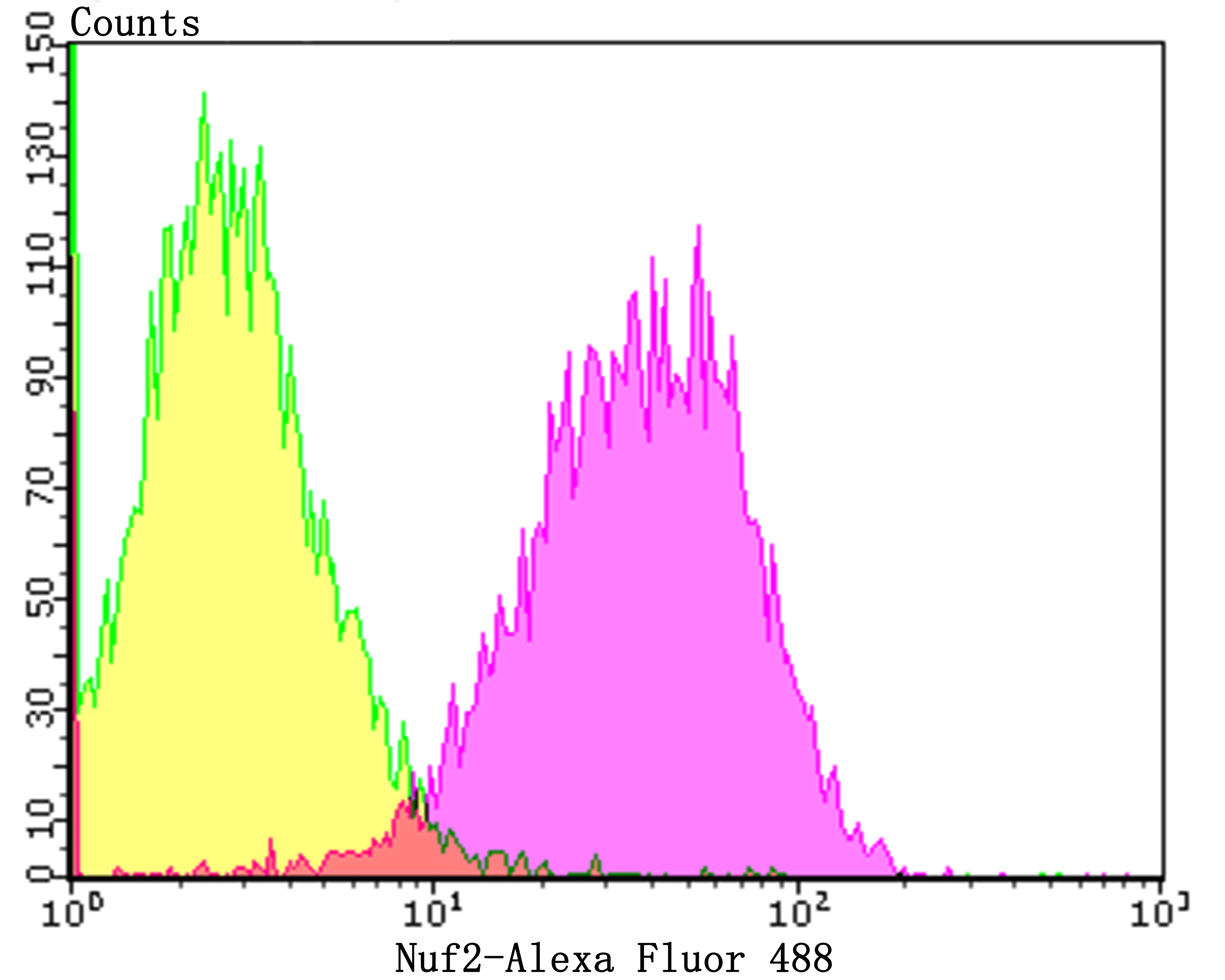
Fig2: Flow cytometric analysis of A431 cells with Nuf2 antibody at 1/50 dilution (purple) compared with an unlabelled control (cells without incubation with primary antibody; yellow). Alexa Fluor 488-conjugated goat anti-rabbit IgG was used as the secondary antibody.| Product Name | Nuf2 [JG34-64] |
|---|---|
| Antibody Type | Primary Antibodies |
| Antigen Alias | Cancer/testis antigen 106 antibody CDCA 1 antibody CDCA1 antibody Cell division cycle associated 1 antibody Cell division cycle associated protein 1 antibody Cell division cycle-associated protein 1 antibody CT106 antibody hNuf 2 antibody hNuf2 antibody hNuf2R antibody hsNuf2 antibody Kinetochore protein Nuf2 antibody Nuf 2 antibody NUF2 antibody NUF2 NDC80 kinetochore complex component antibody NUF2 NDC80 kinetochore complex component homolog antibody NUF2, S. cerevisiae, homolog of antibody NUF2_HUMAN antibody NUF2R antibody |
| Product description | This gene encodes a protein that is highly similar to yeast Nuf2, a component of a conserved protein complex associated with the centromere. Yeast Nuf2 disappears from the centromere during meiotic prophase when centromeres lose their connection to the spindle pole body, and plays a regulatory role in chromosome segregation. The encoded protein is found to be associated with centromeres of mitotic HeLa cells, which suggests that this protein is a functional homolog of yeast Nuf2. Acts as a component of the essential kinetochore-associated NDC80 complex, which is required for chromosome segregation and spindle checkpoint activity. Required for kinetochore integrity and the organization of stable microtubule binding sites in the outer plate of the kinetochore. The NDC80 complex synergistically enhances the affinity of the SKA1 complex for microtubules and may allow the NDC80 complex to track depolymerizing microtubules. |
| Immunogen | Recombinant protein within human Nuf2 aa 1-200. |
| Clonality | Monoclonal |
|---|---|
| Isotype | IgG |
| Host Species | Recombinant rabbit |
| Tested Applications | WBFC |
| WB:1:500-1:2,000 FC:1:50-1:100 |
|
| Species Reactivity | Human |
| Concentration | 1mg/ml |
| Alternative Names | Cancer/testis antigen 106 antibody
CDCA 1 antibody
CDCA1 antibody
Cell division cycle associated 1 antibody
Cell division cycle associated protein 1 antibody
Cell division cycle-associated protein 1 antibody
CT106 antibody
hNuf 2 antibody
hNuf2 antibody
hNuf2R antibody
hsNuf2 antibody
Kinetochore protein Nuf2 antibody
Nuf 2 antibody
NUF2 antibody
NUF2 NDC80 kinetochore complex component antibody
NUF2 NDC80 kinetochore complex component homolog antibody
NUF2 S. cerevisiae homolog of antibody NUF2_HUMAN antibody NUF2R antibody |
|---|---|
| Molecular Weight(MW) | 54 kDa |
| Cellular Localization | Nucleus. |
| SwissProt ID | Q9BZD4 |
|---|
Application
Fig1: Western blot analysis of Nuf2 on different cell lysates using anti-Nuf2 at 1/1,000 dilution. Positive control: Line 1: K562 Line 2: PC-3M Line 3: THP-1
Application
Fig2: Flow cytometric analysis of A431 cells with Nuf2 antibody at 1/50 dilution (purple) compared with an unlabelled control (cells without incubation with primary antibody; yellow). Alexa Fluor 488-conjugated goat anti-rabbit IgG was used as the secondary antibody.| Positive Control | K562, PC-3M, THP-1, A431. |
|---|---|
| Application Notes | WB:1:500-1:2,000 FC:1:50-1:100 |
| Form | Liquid |
|---|---|
| Storage Instructions | Store at +4℃ after thawing. Aliquot store at -20℃. Avoid repeated freeze / thaw cycles. |
| Storage Buffer | 1*TBS (pH7.4), 1%BSA, 40%Glycerol. Preservative: 0.05% Sodium Azide. |


