
Application
Fig1: Western blot analysis of eIF4A3 on SiHa cell lysates using anti-eIF4A3 at 1/500 dilution.Application
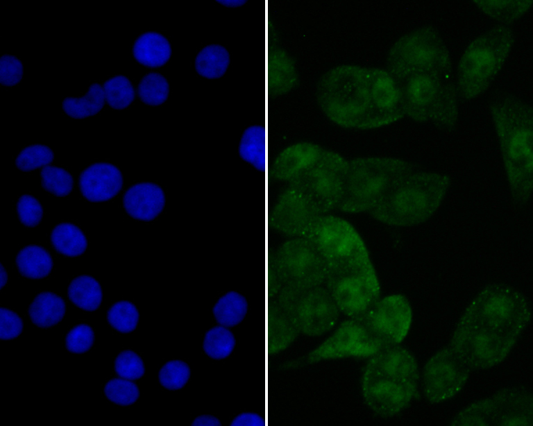
Fig2: ICC staining eIF4A3 in LOVO cells (green). The nuclear counter stain is DAPI (blue). Cells were fixed in paraformaldehyde, permeabilised with 0.25% Triton X100/PBS.Application
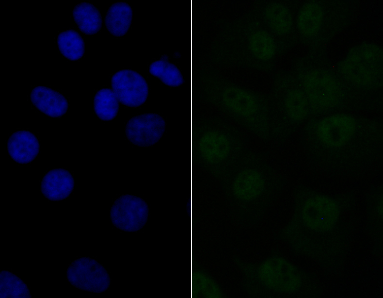
Fig3: ICC staining eIF4A3 in MCF-7 cells (green). The nuclear counter stain is DAPI (blue). Cells were fixed in paraformaldehyde, permeabilised with 0.25% Triton X100/PBS.Application
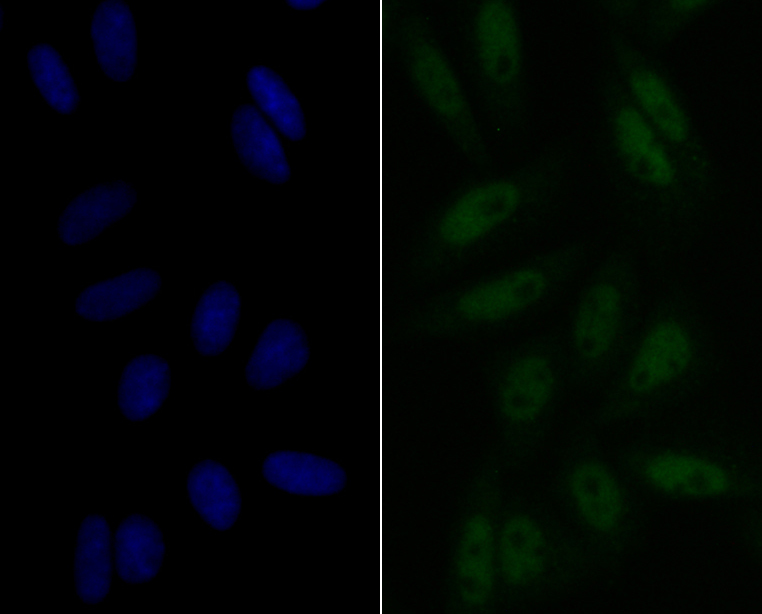
Fig4: ICC staining eIF4A3 in SiHa cells (green). The nuclear counter stain is DAPI (blue). Cells were fixed in paraformaldehyde, permeabilised with 0.25% Triton X100/PBS.Application
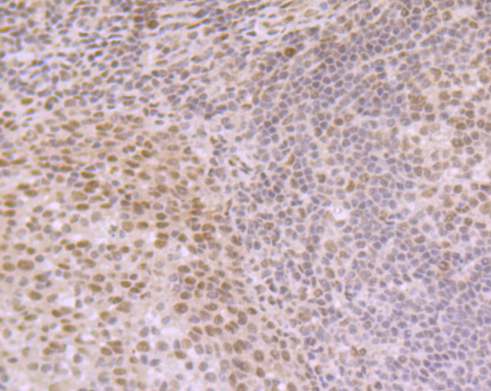
Fig5: Immunohistochemical analysis of paraffin-embedded human tonsil tissue using anti-eIF4A3 antibody. Counter stained with hematoxylin.Application
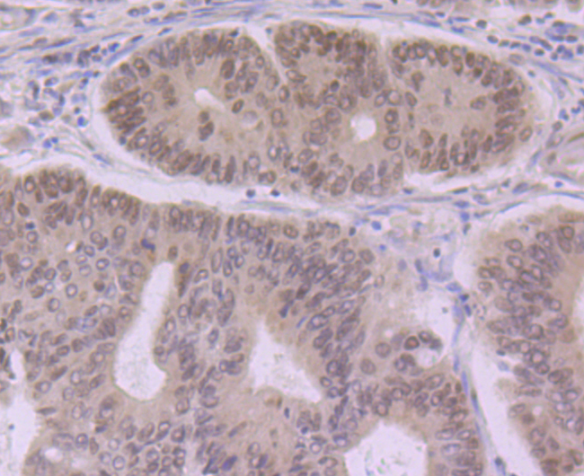
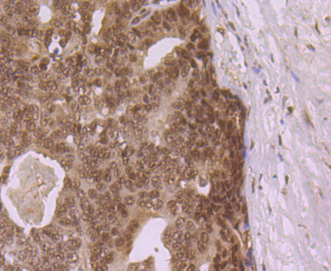
Fig6: Immunohistochemical analysis of paraffin-embedded human colon cancer tissue using anti-eIF4A3 antibody. Counter stained with hematoxylin.Application
Fig7: Immunohistochemical analysis of paraffin-embedded human prostate cancer tissue using anti-eIF4A3 antibody. Counter stained with hematoxylin.Application
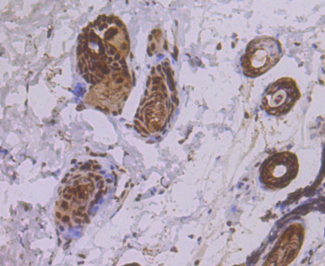
Fig8: Immunohistochemical analysis of paraffin-embedded mouse skin tissue using anti-eIF4A3 antibody. Counter stained with hematoxylin.Application
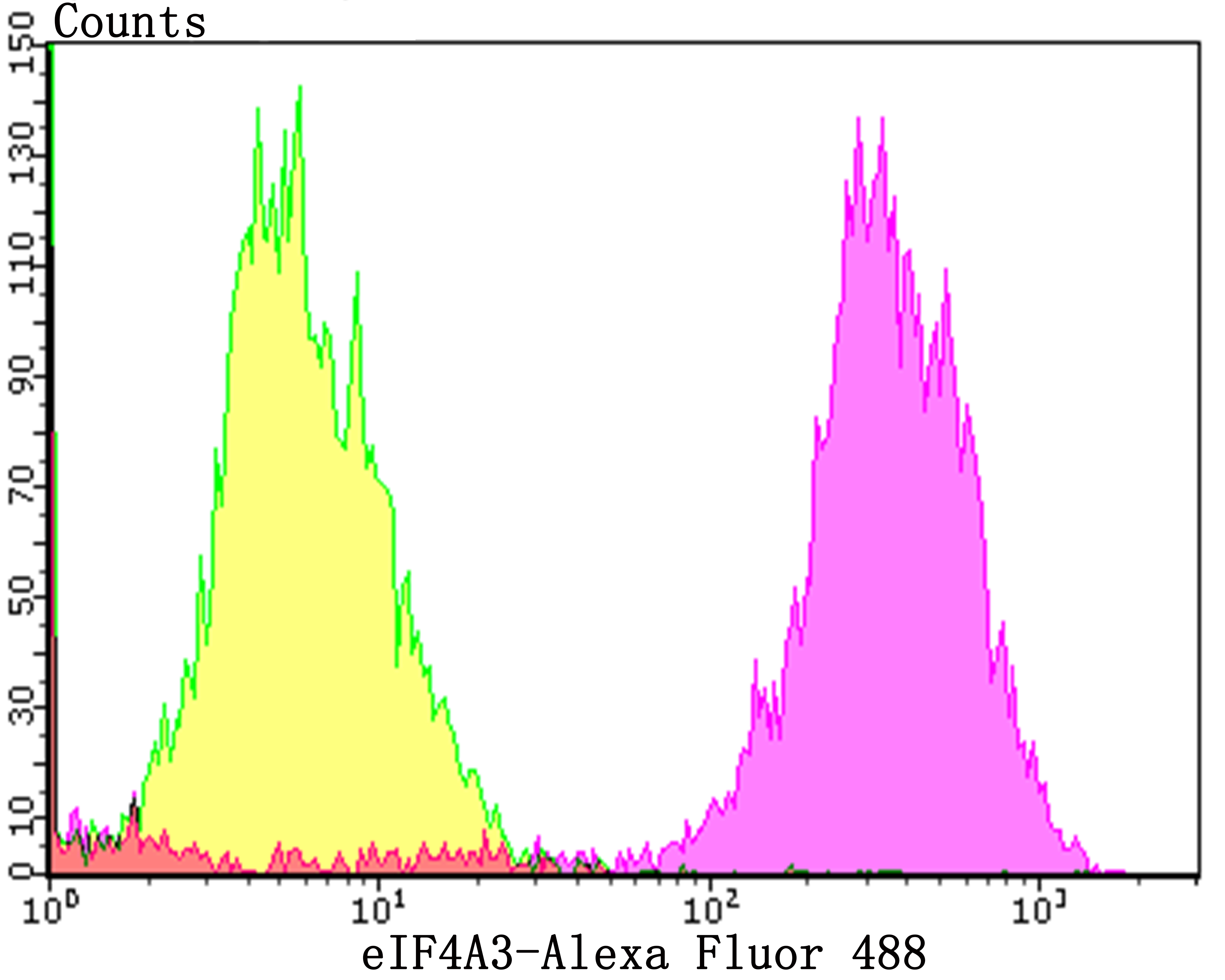
Fig9: Flow cytometric analysis of MCF-7 cells with eIF4A3 antibody at 1/50 dilution (purple) compared with an unlabelled control (cells without incubation with primary antibody; yellow). Alexa Fluor 488-conjugated goat anti-rabbit IgG was used as the secondary antibody.| Product Name | eIF4A3 [JG35-33] |
|---|---|
| Antibody Type | Primary Antibodies |
| Antigen Alias | ATP-dependent RNA helicase DDX48 antibody ATP-dependent RNA helicase eIF4A-3 antibody DDX48 antibody DEAD box protein 48 antibody eIF-4A-III antibody eIF4A-III antibody EIF4A3 antibody eIF4AIII antibody Eukaryotic initiation factor 4A-III antibody Eukaryotic initiation factor 4A-like NUK-34 antibody Eukaryotic translation initiation factor 4A isoform 3 antibody hNMP 265 antibody IF4A3_HUMAN antibody NMP 265 antibody NMP265 antibody Nuclear matrix protein 265 antibody NUK34 antibody |
| Product description | ATP-dependent RNA helicase. Involved in pre-mRNA splicing as component of the spliceosome. Core component of the splicing-dependent multiprotein exon junction complex (EJC) deposited at splice junctions on mRNAs. The EJC is a dynamic structure consisting of core proteins and several peripheral nuclear and cytoplasmic associated factors that join the complex only transiently either during EJC assembly or during subsequent mRNA metabolism. The EJC marks the position of the exon-exon junction in the mature mRNA for the gene expression machinery and the core components remain bound to spliced mRNAs throughout all stages of mRNA metabolism thereby influencing downstream processes including nuclear mRNA export, subcellular mRNA localization, translation efficiency and nonsense-mediated mRNA decay (NMD). Its RNA-dependent ATPase and RNA-helicase activities are induced by CASC3, but abolished in presence of the MAGOH-RBM8A heterodimer, thereby trapping the ATP-bound EJC core onto spliced mRNA in a stable conformation. |
| Immunogen | Recombinant protein within human eIF4A3 aa 1-150. |
| Clonality | Monoclonal |
|---|---|
| Isotype | IgG |
| Host Species | Recombinant rabbit |
| Tested Applications | WBICCIFIHCFCIP |
| WB:1:500-1:1,000 ICC:1:50-1:200 IHC:1:50-1:200 FC:1:50-1:100 |
|
| Species Reactivity | HumanMouseRat |
| Concentration | 1mg/ml |
| Alternative Names | ATP-dependent RNA helicase DDX48 antibody ATP-dependent RNA helicase eIF4A-3 antibody DDX48 antibody DEAD box protein 48 antibody eIF-4A-III antibody eIF4A-III antibody EIF4A3 antibody eIF4AIII antibody Eukaryotic initiation factor 4A-III antibody Eukaryotic initiation factor 4A-like NUK-34 antibody Eukaryotic translation initiation factor 4A isoform 3 antibody hNMP 265 antibody IF4A3_HUMAN antibody NMP 265 antibody NMP265 antibody Nuclear matrix protein 265 antibody NUK34 antibody |
|---|---|
| Molecular Weight(MW) | 47 kDa |
| Cellular Localization | Nucleus. |
| SwissProt ID | P38919 |
|---|
Application
Fig1: Western blot analysis of eIF4A3 on SiHa cell lysates using anti-eIF4A3 at 1/500 dilution.
Application
Fig2: ICC staining eIF4A3 in LOVO cells (green). The nuclear counter stain is DAPI (blue). Cells were fixed in paraformaldehyde, permeabilised with 0.25% Triton X100/PBS.
Application
Fig3: ICC staining eIF4A3 in MCF-7 cells (green). The nuclear counter stain is DAPI (blue). Cells were fixed in paraformaldehyde, permeabilised with 0.25% Triton X100/PBS.
Application
Fig4: ICC staining eIF4A3 in SiHa cells (green). The nuclear counter stain is DAPI (blue). Cells were fixed in paraformaldehyde, permeabilised with 0.25% Triton X100/PBS.
Application
Fig5: Immunohistochemical analysis of paraffin-embedded human tonsil tissue using anti-eIF4A3 antibody. Counter stained with hematoxylin.
Application
Fig6: Immunohistochemical analysis of paraffin-embedded human colon cancer tissue using anti-eIF4A3 antibody. Counter stained with hematoxylin.
Application
Fig7: Immunohistochemical analysis of paraffin-embedded human prostate cancer tissue using anti-eIF4A3 antibody. Counter stained with hematoxylin.
Application
Fig8: Immunohistochemical analysis of paraffin-embedded mouse skin tissue using anti-eIF4A3 antibody. Counter stained with hematoxylin.
Application
Fig9: Flow cytometric analysis of MCF-7 cells with eIF4A3 antibody at 1/50 dilution (purple) compared with an unlabelled control (cells without incubation with primary antibody; yellow). Alexa Fluor 488-conjugated goat anti-rabbit IgG was used as the secondary antibody.| Positive Control | SiHa, LOVO, MCF-7, SiHa, human tonsil tissue, human colon cancer tissue, human prostate cancer tissue, mouse skin tissue. |
|---|---|
| Application Notes | WB:1:500-1:1,000 ICC:1:50-1:200 IHC:1:50-1:200 FC:1:50-1:100 |
| Form | Liquid |
|---|---|
| Storage Instructions | Store at +4℃ after thawing. Aliquot store at -20℃. Avoid repeated freeze / thaw cycles. |
| Storage Buffer | 1*TBS (pH7.4), 1%BSA, 40%Glycerol. Preservative: 0.05% Sodium Azide. |


