WB
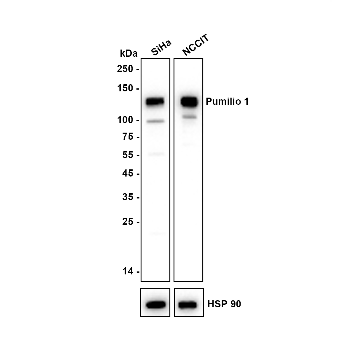
Western blot analysis of Pumilio 1 on different lysates with Rabbit anti-Pumilio 1 antibody at 1/1,000 dilution. Lane 1: SiHa cell lysate, Lane 2: NCCIT cell lysate, Lysates/proteins at 20 µg/Lane. Exposure time: 25 seconds; Blocking: 5% NFDM/TBST, 1 hour at room temperature Primary antibody: 1/1,000 in 5% NFDM/TBST, overnight at 4 ℃ Secondary antibody: Goat anti-Rabbit IgG-HRP, 1/50,000 in 5% NFDM/TBST, 1 hour at room temperature.ICC/IF
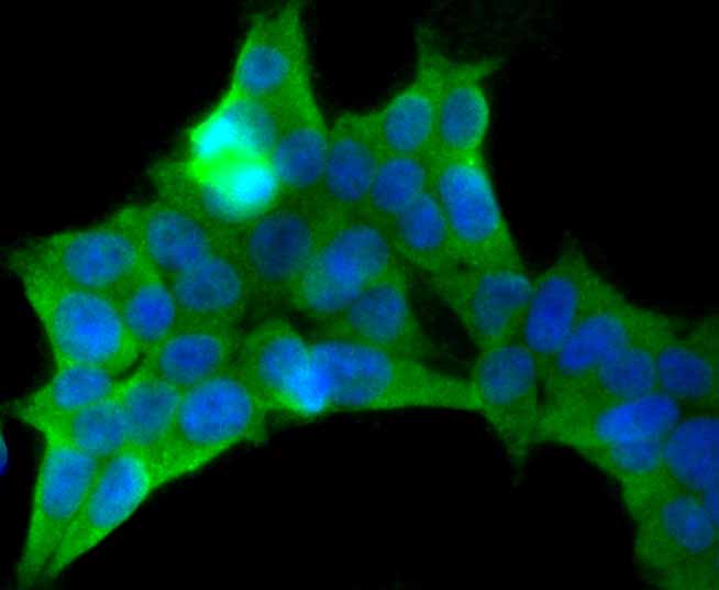
ICC staining Pumilio 1 in 293T cells (green). The nuclear counter stain is DAPI (blue). Cells were fixed in paraformaldehyde, permeabilised with 0.25% Triton X100/PBS.ICC/IF
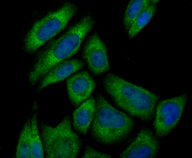
ICC staining Pumilio 1 in SiHa cells (green). The nuclear counter stain is DAPI (blue). Cells were fixed in paraformaldehyde, permeabilised with 0.25% Triton X100/PBS.IHC
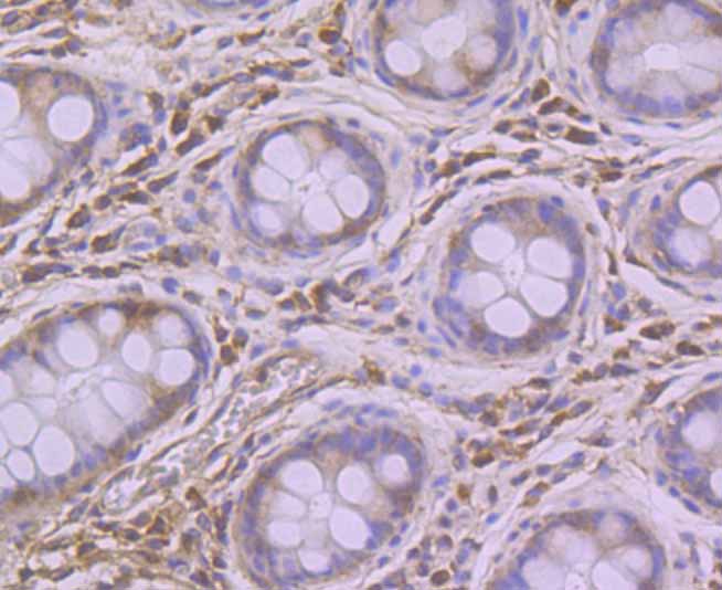
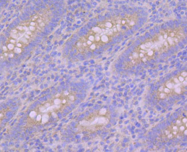
Immunohistochemical analysis of paraffin-embedded human colon tissue using anti-Pumilio 1 antibody. Counter stained with hematoxylin.IHC
Immunohistochemical analysis of paraffin-embedded human appendix tissue using anti-Pumilio 1 antibody. Counter stained with hematoxylin.IHC
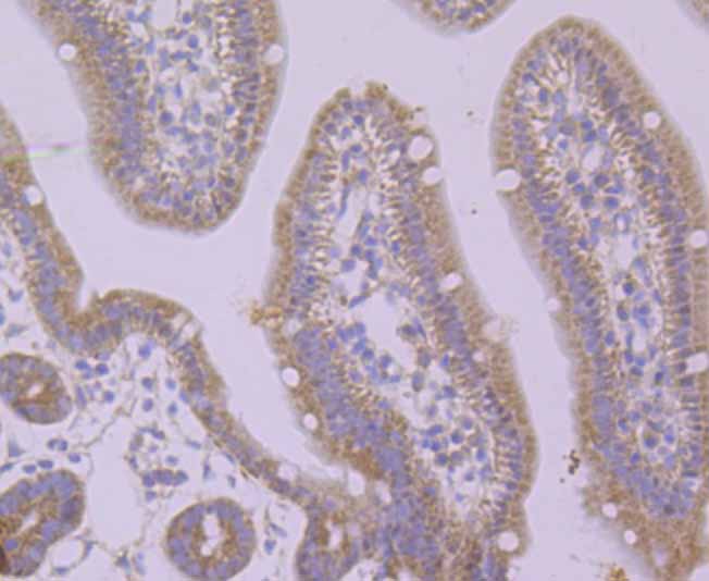
Immunohistochemical analysis of paraffin-embedded mouse small intestine tissue using anti-Pumilio 1 antibody. Counter stained with hematoxylin.IHC
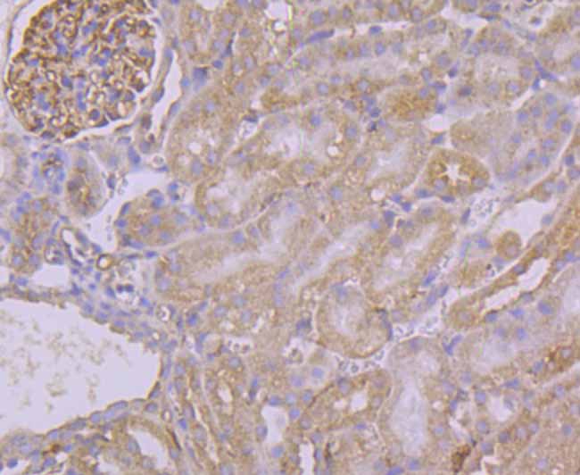
Immunohistochemical analysis of paraffin-embedded mouse kidney tissue using anti-Pumilio 1 antibody. Counter stained with hematoxylin.| Product Name | Pumilio 1 Recombinant Rabbit Monoclonal Antibody |
|---|---|
| Antibody Type | Primary Antibodies |
| Product description | Sequence-specific RNA-binding protein that acts as a post-transcriptional repressor by binding the 3'-UTR of mRNA targets. Binds to an RNA consensus sequence, the Pumilio Response Element (PRE), 5'-UGUANAUA-3', that is related to the Nanos Response Element (NRE). Mediates post-transcriptional repression of transcripts via different mechanisms: acts via direct recruitment of the CCR4-POP2-NOT deadenylase leading to translational inhibition and mRNA degradation. Also mediates deadenylation-independent repression by promoting accessibility of miRNAs. |
| Immunogen | Recombinant protein within human Pumilio 1 aa 1-200. |
| Clonality | Monoclonal |
|---|---|
| Isotype | IgG |
| Host Species | Recombinant rabbit |
| Tested Applications | ICC/IFIHCWB |
| WB:1:1000 IHC:1:50-1:200 ICC/IF:1:50-1:200 |
|
| Species Reactivity | HumanMouse |
| Concentration | 1mg/ml |
| Purification | Protein A |
| Alternative Names | HsPUM antibody KIAA0099 antibody PUM 1 antibody PUM L1 antibody Pum1 antibody PUM1_HUMAN antibody PUMH 1 antibody PUMH antibody PUMH1 antibody pumilio homolog 1 (Drosophila) antibody Pumilio homolog 1 antibody Pumilio-1 antibody Pumilio1 antibody PUML 1 antibody PUML1 antibody |
|---|---|
| Molecular Weight(MW) | 126 kDa |
| Cellular Localization | Cytoplasm. Nucleus. |
WB
Western blot analysis of Pumilio 1 on different lysates with Rabbit anti-Pumilio 1 antibody at 1/1,000 dilution. Lane 1: SiHa cell lysate, Lane 2: NCCIT cell lysate, Lysates/proteins at 20 µg/Lane. Exposure time: 25 seconds; Blocking: 5% NFDM/TBST, 1 hour at room temperature Primary antibody: 1/1,000 in 5% NFDM/TBST, overnight at 4 ℃ Secondary antibody: Goat anti-Rabbit IgG-HRP, 1/50,000 in 5% NFDM/TBST, 1 hour at room temperature.
ICC/IF
ICC staining Pumilio 1 in 293T cells (green). The nuclear counter stain is DAPI (blue). Cells were fixed in paraformaldehyde, permeabilised with 0.25% Triton X100/PBS.
ICC/IF
ICC staining Pumilio 1 in SiHa cells (green). The nuclear counter stain is DAPI (blue). Cells were fixed in paraformaldehyde, permeabilised with 0.25% Triton X100/PBS.
IHC
Immunohistochemical analysis of paraffin-embedded human colon tissue using anti-Pumilio 1 antibody. Counter stained with hematoxylin.
IHC
Immunohistochemical analysis of paraffin-embedded human appendix tissue using anti-Pumilio 1 antibody. Counter stained with hematoxylin.
IHC
Immunohistochemical analysis of paraffin-embedded mouse small intestine tissue using anti-Pumilio 1 antibody. Counter stained with hematoxylin.
IHC
Immunohistochemical analysis of paraffin-embedded mouse kidney tissue using anti-Pumilio 1 antibody. Counter stained with hematoxylin.| Application Notes | WB:1:1000 IHC:1:50-1:200 ICC/IF:1:50-1:200 |
|---|
| Form | Liquid |
|---|---|
| Storage Instructions | Store at +4℃ after thawing. Aliquot store at -20℃. Avoid repeated freeze / thaw cycles. |
| Storage Buffer | 1*TBS (pH7.4), 1%BSA, 40%Glycerol. Preservative: 0.05% Sodium Azide. |


