Application
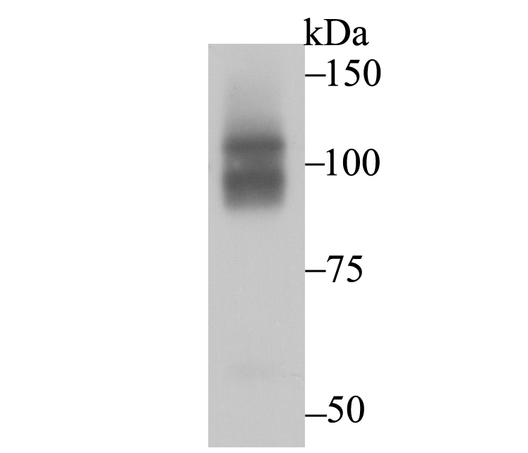
Fig1: Western blot analysis of CHD1L on HepG2 cell using anti-CHD1L antibody at 1/1,000 dilution.Application
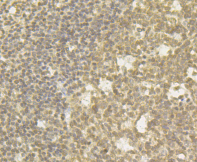
Fig2: Immunohistochemical analysis of paraffin-embedded human tonsil tissue using anti-CHD1L antibody. Counter stained with hematoxylin. The section was pre-treated using heat mediated antigen retrieval with sodium citrate buffer (pH6) for 20 mins.Application
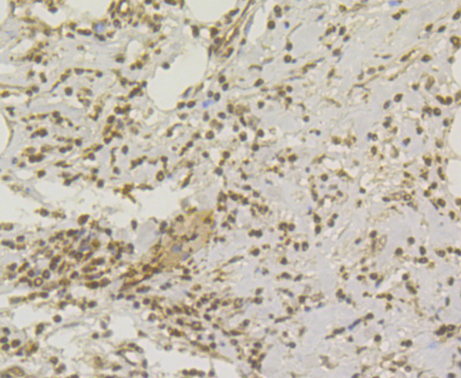
Fig3: Immunohistochemical analysis of paraffin-embedded human colon cancer tissue using anti-CHD1L antibody. Counter stained with hematoxylin. The section was pre-treated using heat mediated antigen retrieval with sodium citrate buffer (pH6) for 20 mins.Application
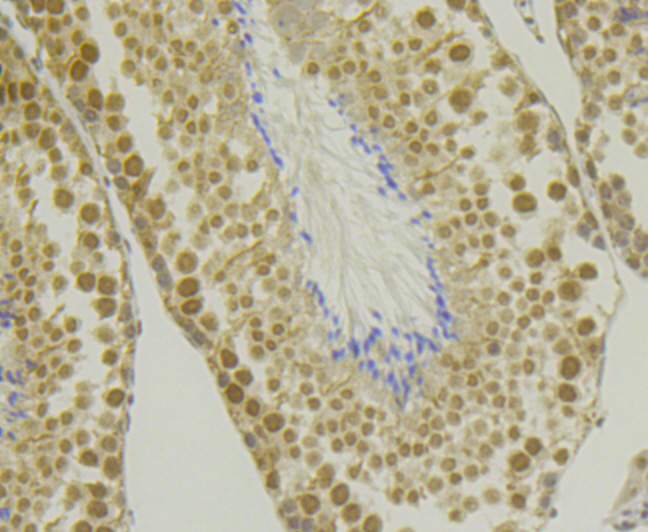
Fig4: Immunohistochemical analysis of paraffin-embedded mouse testis tissue using anti-CHD1L antibody. Counter stained with hematoxylin. The section was pre-treated using heat mediated antigen retrieval with sodium citrate buffer (pH6) for 20 mins.Application
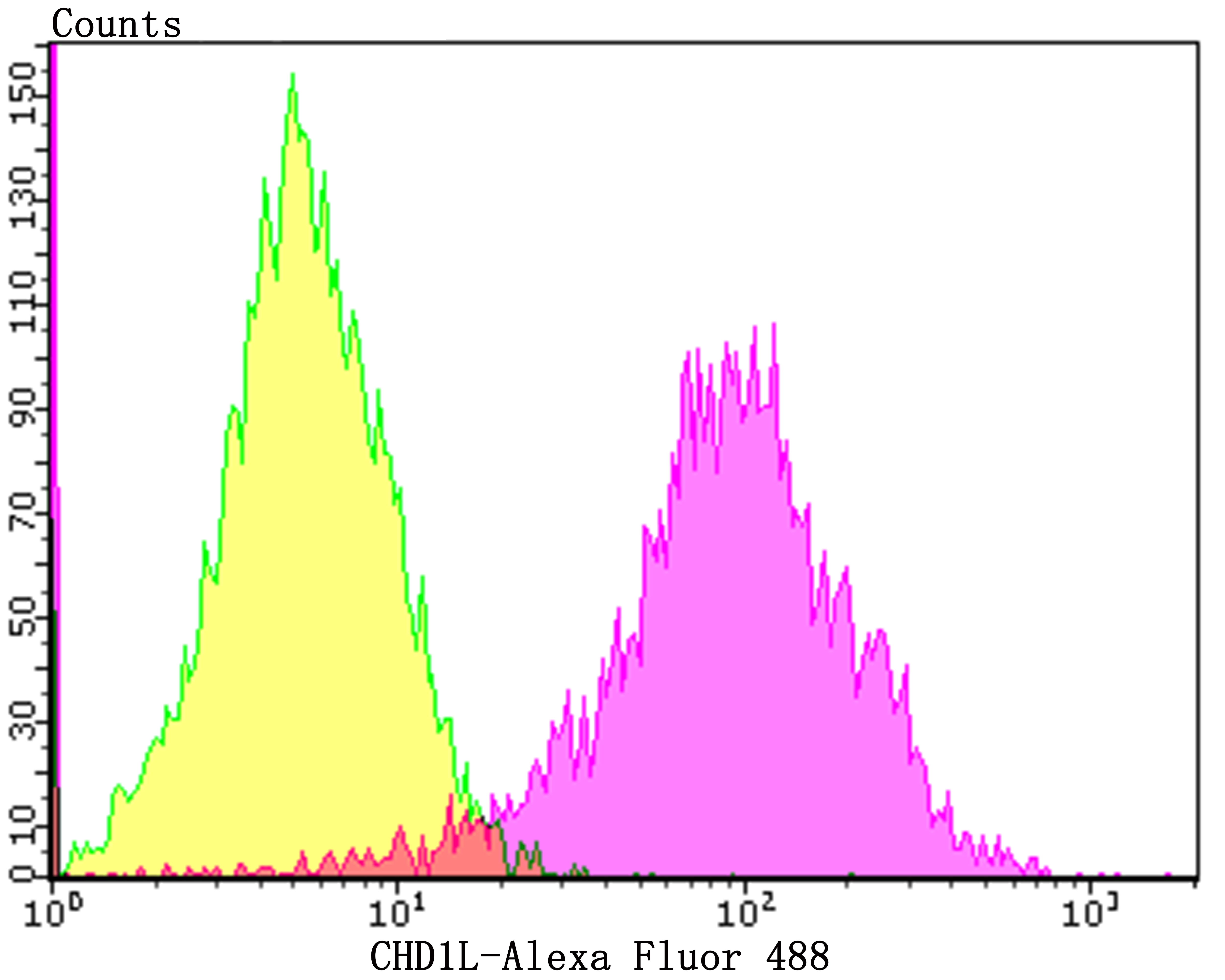
Fig5: Flow cytometric analysis of HepG2 cells with CHD1L antibody at 1/100 dilution (purple) compared with an unlabelled control (cells without incubation with primary antibody; yellow). Alexa Fluor 488-conjugated goat anti-rabbit IgG was used as the secondary antibody.| Product Name | CHD1L [JG35-44] |
|---|---|
| Antibody Type | Primary Antibodies |
| Antigen Alias | ALC1 antibody Amplified in liver cancer 1 antibody Amplified in liver cancer protein 1 antibody chd1l antibody CHD1L_HUMAN antibody CHDL antibody Chromodomain helicase DNA binding protein 1 like antibody Chromodomain-helicase-DNA-binding protein 1-like antibody FLJ22530 antibody |
| Product description | CHD1L (Chromodomain helicase DNA-binding protein 1-like) is a 897 amino acid protein encoded by the human gene CHD1L. The CHD family of proteins are ATP-dependent chromatin remodeling enzymes, which combine chromodomains, with SWI2/SNF2 ATPase/helicase motifs and DNA-binding capability. Chromodomains are protein regions of about 40-50 amino acid residues found in proteins associated with chromatin remodeling and manipulation. The domain is highly conserved among both plants and animals and is found in a large variety of proteins from many genomes. CHD1L and CHD1 share most homology within two internal helicase domains. |
| Immunogen | Recombinant protein within human CHD1L aa 600-800. |
| Clonality | Monoclonal |
|---|---|
| Isotype | IgG |
| Host Species | Recombinant rabbit |
| Tested Applications | WBIHCFC |
| WB:1:500-1:1,000 IHC:1:50-1:100 FC:1:50-1:100 |
|
| Species Reactivity | HumanMouse |
| Concentration | 1mg/ml |
| Alternative Names | ALC1 antibody Amplified in liver cancer 1 antibody Amplified in liver cancer protein 1 antibody chd1l antibody CHD1L_HUMAN antibody CHDL antibody Chromodomain helicase DNA binding protein 1 like antibody Chromodomain-helicase-DNA-binding protein 1-like antibody FLJ22530 antibody |
|---|---|
| Molecular Weight(MW) | 101/90 kDa |
| Cellular Localization | Nucleus. |
| SwissProt ID | Q86WJ1 |
|---|
Application
Fig1: Western blot analysis of CHD1L on HepG2 cell using anti-CHD1L antibody at 1/1,000 dilution.
Application
Fig2: Immunohistochemical analysis of paraffin-embedded human tonsil tissue using anti-CHD1L antibody. Counter stained with hematoxylin. The section was pre-treated using heat mediated antigen retrieval with sodium citrate buffer (pH6) for 20 mins.
Application
Fig3: Immunohistochemical analysis of paraffin-embedded human colon cancer tissue using anti-CHD1L antibody. Counter stained with hematoxylin. The section was pre-treated using heat mediated antigen retrieval with sodium citrate buffer (pH6) for 20 mins.
Application
Fig4: Immunohistochemical analysis of paraffin-embedded mouse testis tissue using anti-CHD1L antibody. Counter stained with hematoxylin. The section was pre-treated using heat mediated antigen retrieval with sodium citrate buffer (pH6) for 20 mins.
Application
Fig5: Flow cytometric analysis of HepG2 cells with CHD1L antibody at 1/100 dilution (purple) compared with an unlabelled control (cells without incubation with primary antibody; yellow). Alexa Fluor 488-conjugated goat anti-rabbit IgG was used as the secondary antibody.| Positive Control | HepG2, human tonsil tissue, human colon cancer tissue, mouse testis tissue, HepG2. |
|---|---|
| Application Notes | WB:1:500-1:1,000 IHC:1:50-1:100 FC:1:50-1:100 |
| Form | Liquid |
|---|---|
| Storage Instructions | Store at +4℃ after thawing. Aliquot store at -20℃. Avoid repeated freeze / thaw cycles. |
| Storage Buffer | 1*TBS (pH7.4), 1%BSA, 40%Glycerol. Preservative: 0.05% Sodium Azide. |


