Application
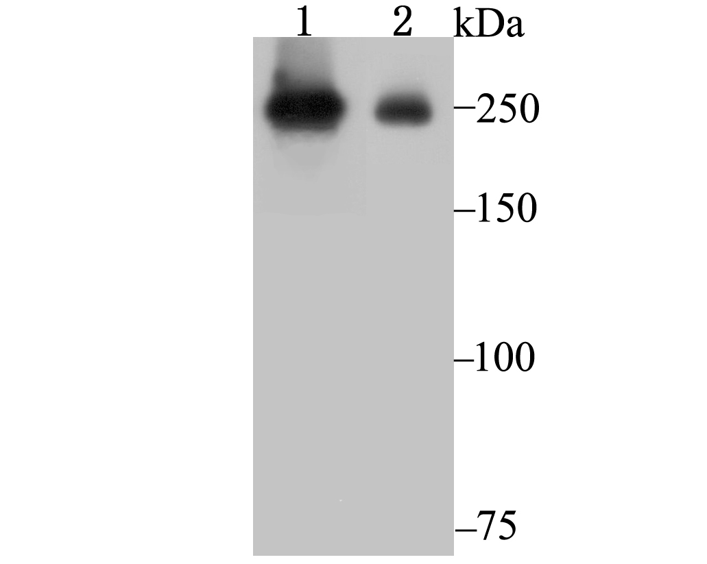
Fig1: Western blot analysis of PRPF8 on SiHa (1) and A549 (2) cell lysate using anti-PRPF8 antibody at 1/1,000 dilution.Application
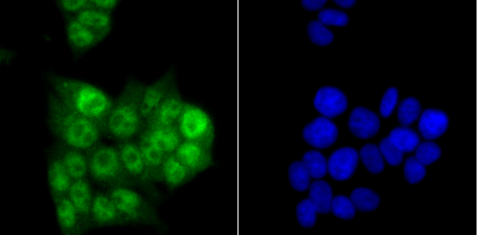
Fig2: ICC staining PRPF8 in 293T cells (green). The nuclear counter stain is DAPI (blue). Cells were fixed in paraformaldehyde, permeabilised with 0.25% Triton X100/PBS.Application
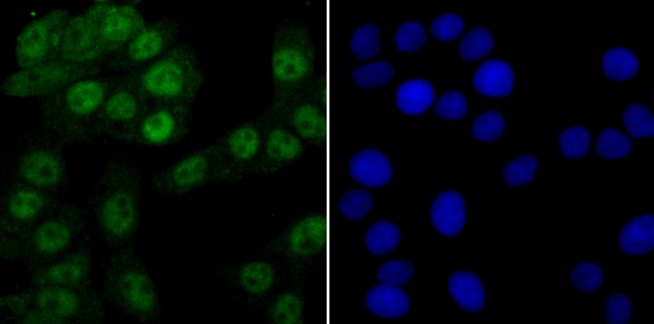
Fig3: ICC staining PRPF8 in LOVO cells (green). The nuclear counter stain is DAPI (blue). Cells were fixed in paraformaldehyde, permeabilised with 0.25% Triton X100/PBS.Application
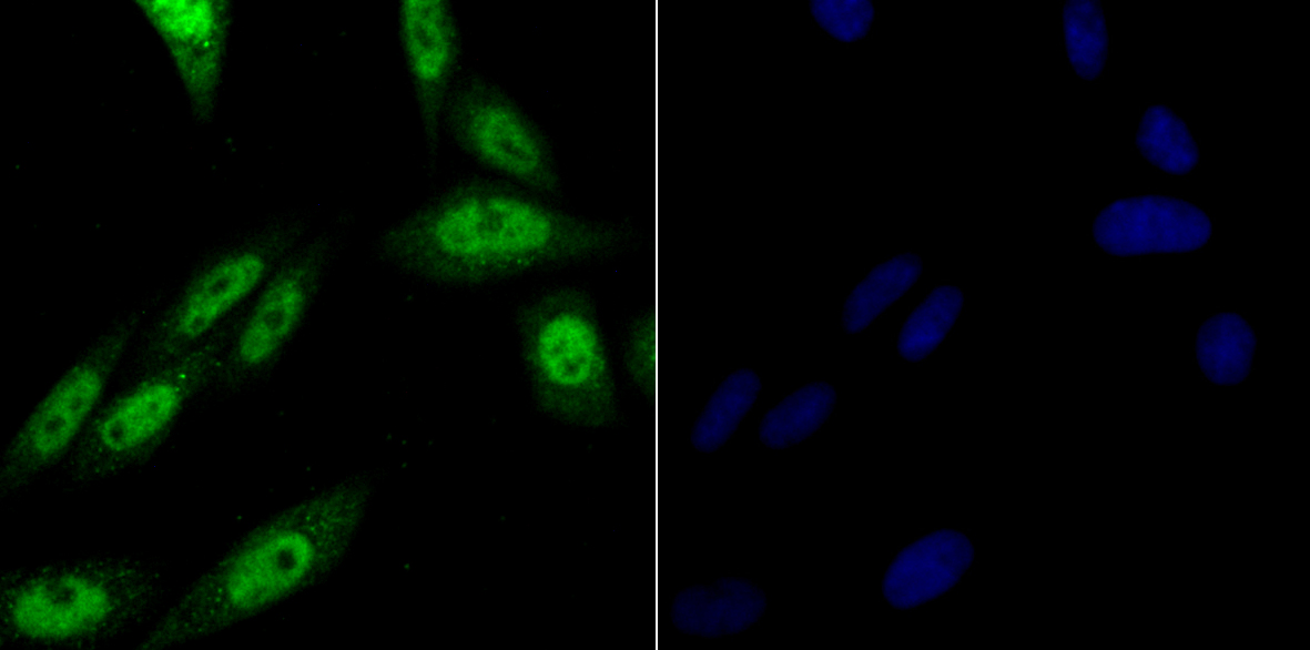
Fig4: ICC staining PRPF8 in SiHa cells (green). The nuclear counter stain is DAPI (blue). Cells were fixed in paraformaldehyde, permeabilised with 0.25% Triton X100/PBS.Application
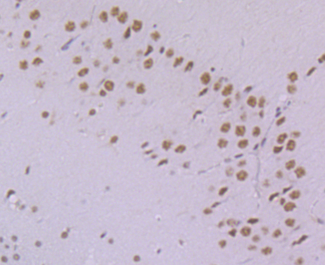
Fig5: Immunohistochemical analysis of paraffin-embedded rat brain tissue using anti-PRPF8 antibody. Counter stained with hematoxylin.Application
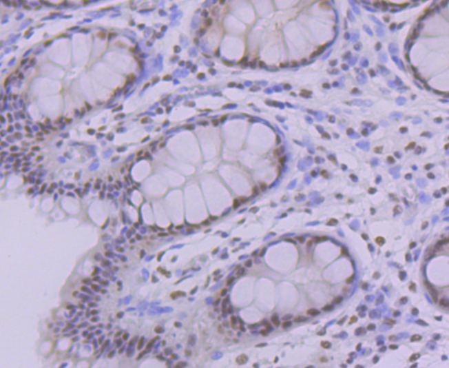
Fig6: Immunohistochemical analysis of paraffin-embedded human colon tissue using anti-PRPF8 antibody. Counter stained with hematoxylin.Application
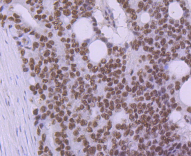
Fig7: Immunohistochemical analysis of paraffin-embedded human prostate cancer tissue using anti-PRPF8 antibody. Counter stained with hematoxylin.Application
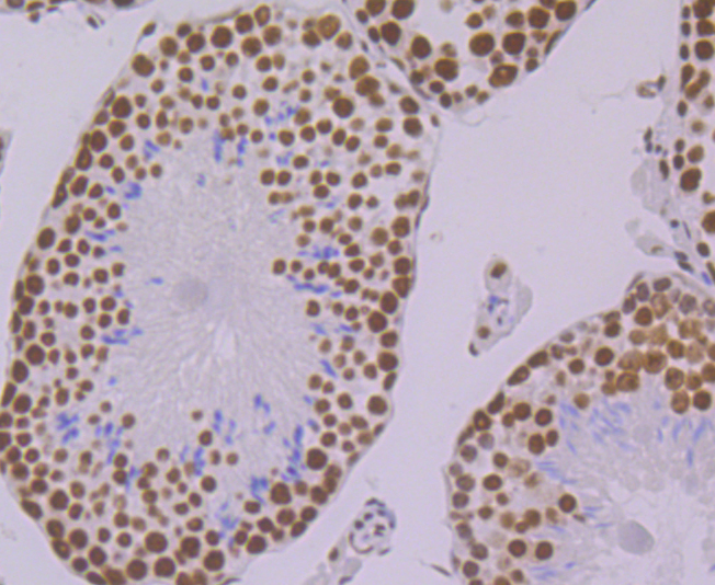
Fig8: Immunohistochemical analysis of paraffin-embedded mouse testis tissue using anti-PRPF8 antibody. Counter stained with hematoxylin.Application
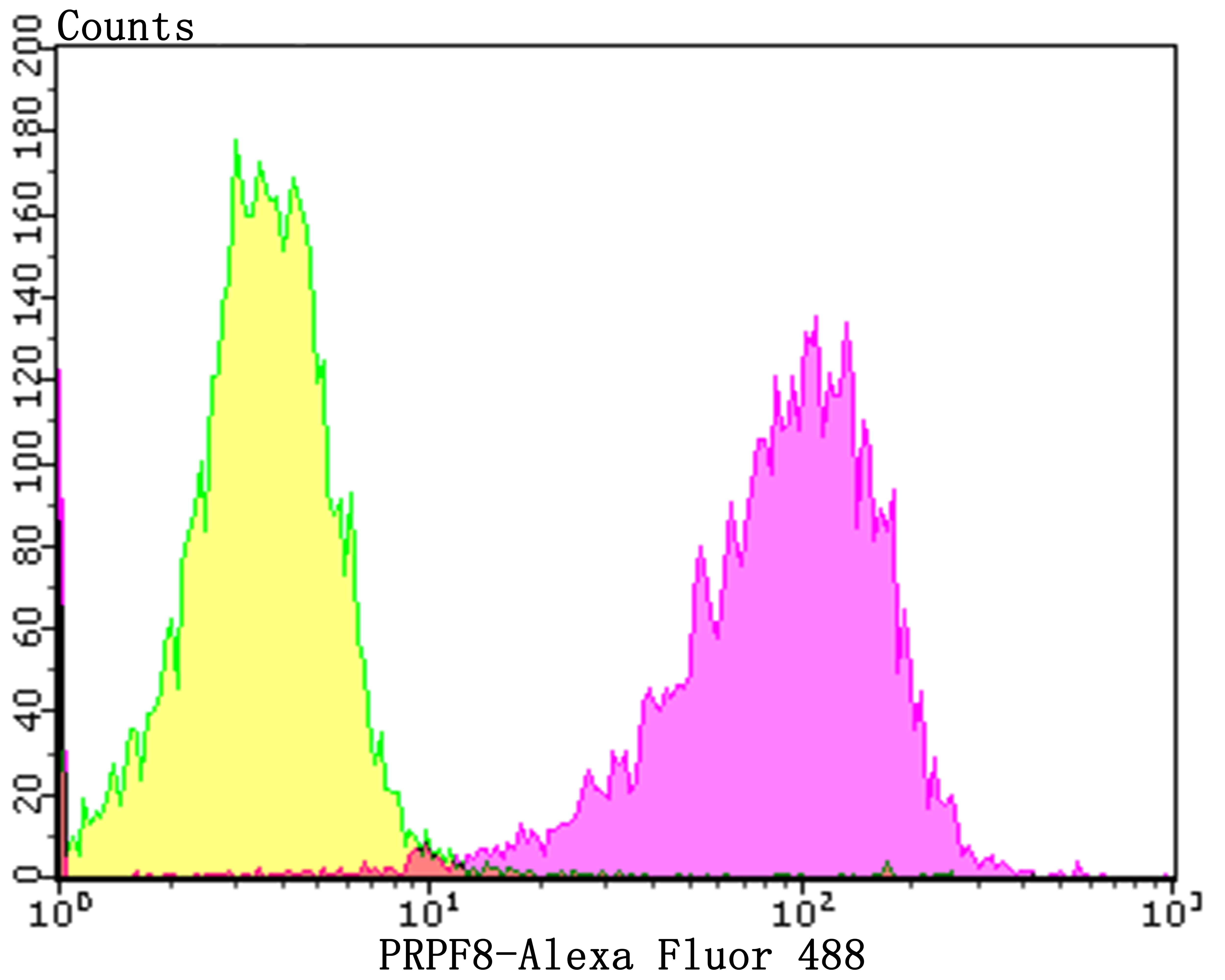
Fig9: Flow cytometric analysis of A549 cells with PRPF8 antibody at 1/100 dilution (red) compared with an unlabelled control (cells without incubation with primary antibody; black).Alexa Fluor 488-conjugated goat anti-rabbit IgG was used as the secondary antibody.| Product Name | PRPF8 [JG57-38] |
|---|---|
| Antibody Type | Primary Antibodies |
| Antigen Alias | 220 kDa U5 snRNP specific protein antibody 220 kDa U5 snRNP-specific protein antibody Apoptosis regulated protein 1 antibody Apoptosis regulated protein 2 antibody HPRP8 antibody p220 antibody Pre mRNA processing factor 8 antibody Pre mRNA-processing factor 8, S. cerevisiae, homolog of antibody Pre-mRNA-processing-splicing factor 8 antibody Precursor mRNA processing protein antibody PRP8 antibody PRP8 homolog antibody PRP8 pre mRNA processing factor 8 homolog antibody PRP8_HUMAN antibody PRPC8 antibody Prpf8 antibody Retinitis pigmentosa 13 (autosomal dominant) antibody RP13 antibody SNRNP220 antibody Splicing factor Prp8 antibody U5 snRNP specific protein antibody U5 snRNP specific protein (220 kD), ortholog of S. cerevisiae Prp8p antibody |
| Product description | PRP8, also designated pre-mRNA-processing-splicing factor 8, is a highly conserved nuclear protein and a central component of the catalytic core of the spliceosome, where it may be involved in various molecular rearrangements. PRP8, which is widely expressed, plays a role in transesterification reactions that regulate splicesome-induced pre-mRNA splicing. Specifically, PRP8 interacts with the GU dinucleotide at the 5' splice site (5'SS) and forms a specific UV-inducible cross-link. It also interacts functionally with the 3'SS, affecting the efficiency of the second catalytic step. PRP8 may play a role in the first transesterification step, as PRP8 mutations that prohibit negative regulation of PRP28 or PRP44/Brr2 subsequently block U4 activation. In addition, PRP8 interacts with a conserved region of U6 that is instrumental in the formation of the catalytic core of the spliceosome. |
| Immunogen | Recombinant protein within 1–200 of human PRPF8. |
| Clonality | Monoclonal |
|---|---|
| Isotype | IgG |
| Host Species | Recombinant rabbit |
| Tested Applications | WBICCIHCFC |
| WB:1:500-1:2,000 ICC:1:50-1:200 IHC:1:50-1:200 FC:1:50-1:100 |
|
| Species Reactivity | HumanMouseRat |
| Concentration | 1mg/ml |
| Alternative Names | 220 kDa U5 snRNP specific protein antibody
220 kDa U5 snRNP-specific protein antibody
Apoptosis regulated protein 1 antibody
Apoptosis regulated protein 2 antibody
HPRP8 antibody
p220 antibody
Pre mRNA processing factor 8 antibody
Pre mRNA-processing factor 8 S. cerevisiae homolog of antibody Pre-mRNA-processing-splicing factor 8 antibody Precursor mRNA processing protein antibody PRP8 antibody PRP8 homolog antibody PRP8 pre mRNA processing factor 8 homolog antibody PRP8_HUMAN antibody PRPC8 antibody Prpf8 antibody Retinitis pigmentosa 13 (autosomal dominant) antibody RP13 antibody SNRNP220 antibody Splicing factor Prp8 antibody U5 snRNP specific protein antibody U5 snRNP specific protein (220 kD) ortholog of S. cerevisiae Prp8p antibody |
|---|---|
| Molecular Weight(MW) | 274 kDa |
| Cellular Localization | Nucleus. Spliceosome. |
Application
Fig1: Western blot analysis of PRPF8 on SiHa (1) and A549 (2) cell lysate using anti-PRPF8 antibody at 1/1,000 dilution.
Application
Fig2: ICC staining PRPF8 in 293T cells (green). The nuclear counter stain is DAPI (blue). Cells were fixed in paraformaldehyde, permeabilised with 0.25% Triton X100/PBS.
Application
Fig3: ICC staining PRPF8 in LOVO cells (green). The nuclear counter stain is DAPI (blue). Cells were fixed in paraformaldehyde, permeabilised with 0.25% Triton X100/PBS.
Application
Fig4: ICC staining PRPF8 in SiHa cells (green). The nuclear counter stain is DAPI (blue). Cells were fixed in paraformaldehyde, permeabilised with 0.25% Triton X100/PBS.
Application
Fig5: Immunohistochemical analysis of paraffin-embedded rat brain tissue using anti-PRPF8 antibody. Counter stained with hematoxylin.
Application
Fig6: Immunohistochemical analysis of paraffin-embedded human colon tissue using anti-PRPF8 antibody. Counter stained with hematoxylin.
Application
Fig7: Immunohistochemical analysis of paraffin-embedded human prostate cancer tissue using anti-PRPF8 antibody. Counter stained with hematoxylin.
Application
Fig8: Immunohistochemical analysis of paraffin-embedded mouse testis tissue using anti-PRPF8 antibody. Counter stained with hematoxylin.
Application
Fig9: Flow cytometric analysis of A549 cells with PRPF8 antibody at 1/100 dilution (red) compared with an unlabelled control (cells without incubation with primary antibody; black).Alexa Fluor 488-conjugated goat anti-rabbit IgG was used as the secondary antibody.| Positive Control | A549, 293T, LOVO, SiHa, rat brain tissue, human colon tissue, human prostate cancer tissue, mouse testis tissue. |
|---|---|
| Application Notes | WB:1:500-1:2,000 ICC:1:50-1:200 IHC:1:50-1:200 FC:1:50-1:100 |
| Form | Liquid |
|---|---|
| Storage Instructions | Store at +4℃ after thawing. Aliquot store at -20℃. Avoid repeated freeze / thaw cycles. |
| Storage Buffer | 1*TBS (pH7.4), 1%BSA, 40%Glycerol. Preservative: 0.05% Sodium Azide. |


